The ficolins are soluble pattern recognition molecules in the lectin pathway of complement, but the spectrum and mode of interaction with pathogens are largely unknown. In this study, we investigated the binding properties of the murine serum ficolin-A towards a panel of different clinical relevant microorganisms (N = 45) and compared the binding profile with human serum ficolin-2 and ficolin-3. Ficolin-A was able to bind Gram-positive bacteria strains including E. faecalis,
L. monocytogenes and some S. aureus strains, but not to the investigated S. agalactiae (Group B streptococcus) strains. Regarding Gram-negative bacteria ficolin-A was able to bind to someE. coli and P. aeruginosa strains, but not to the investigatedSalmonellastrains. Of particular interest ficolin-A bound strongly to the pathogenicE. coli,O157:H7 and O149 strains, but it did not bind to the non-pathogenicE. coli,ATCC 25922 strain. Additionally, ficolin-A was able to bind purified LPS from these pathogenic strains. Furthermore, ficolin-A bound to a clinical isolate of the fungusA. fumigatus. In general ficolin-2 showed similar selective binding spectrum towards pathogenic microorganisms as observed for ficolin-A indicating specific pathophysiological roles of these molecules in host defence. In contrast, ficolin-3 did not bind to any of the investigated microorganisms and the anti-microbial role of ficolin-3 still remains elusive.
Citation:Hummelshøj T, Ma YJ, Munthe-Fog L, Bjarnsholt T, Moser C, et al. (2012) The Interaction Pattern of Murine Serum Ficolin-A with Microorganisms. PLoS ONE 7(5): e38196. doi:10.1371/journal.pone.0038196
Editor:Andreas Klos, Institute of Medical Microbiology and Hospital Epidemiology-Hanover Medical School, Germany
ReceivedJanuary 13, 2012;AcceptedMay 1, 2012;PublishedMay 30, 2012
Copyright:ß2012 Hummelshøj et al. This is an open-access article distributed under the terms of the Creative Commons Attribution License, which permits unrestricted use, distribution, and reproduction in any medium, provided the original author and source are credited.
Funding:This work was supported by grants from The Carlsberg Foundation, The Novo Nordisk Research Foundation, The Lundbeck Foundation, The Research Foundation of the Capital Region of Denmark and The Danish Agency for Science, Technology and Innovation. The funders had no role in study design, data collection and analysis, decision to publish, or preparation of the manuscript.
Competing Interests:The authors have declared that no competing interests exist.
* E-mail: [email protected]
Introduction
The ficolins are recognition molecules in the lectin pathway of the complement system that bind to specific structures on microorganisms and self cellular debris enabling quick and simple aggregation and opsonization of hazardous material [1,2].
The ficolins are highly oligomerized glycoproteins containing both collagen-like (CL) and fibrinogen-like (FBG) regions. As seen for mannose-binding lectin (MBL), the CL region is able to associate with the MBL/ficolin associated serine proteases (MASPs), which activate the lectin pathway of the complement system [1]. The ficolins are built up of several structural subunits each composed of three identical polypeptide chains [3–5]. Through the FBG domain they bind structures such as N-acetyl-glucosamine (GlcNAc), N-acetyl-galactosamine (GalNAc), and acetylated compounds located on the target cell [6,7].
The ficolins have been described in both vertebrates and invertebrates where each species contain several paralogues ficolins (typical 2–4). In human, three ficolins are described: ficolin-1 (M-ficolin), ficolin-2 (L-ficolin), and ficolin-3 (H-ficolin). Ficolin-1 is present in low serum concentrations whereas ficolin-2 and ficolin-3 are present in concentrations of ,5mg/ml and ,25mg/ml, respectively [8–11]. Two ficolin molecules have been identified in mice termed ficolin-A and ficolin-B. Ficolin-B is
expressed in myeloid bone marrow derived cells and ficolin-A is expressed in the liver and spleen and is present in serum [5,12]. Based on the protein sequence and exon organisation it is likely that ficolin-B is the orthologue to the human ficolin-1, whereas both the mouse ficolin-A and the human ficolin-2 seems to have evolved independently from a common ficolin-1/ficolin-B lineage [13,14]. The mouse homologue of the human ficolin-3 gene is identified as a pseudogene in mice and rats [15].
Both ficolin-1 and ficolin-2 have been shown to bind different bacteria strains such asStaphylococcus aureus, Salmonella typhimurium, Streptococcus pneumonia,Streptococcus pyogenesandStreptococcus agalactiae and are able to stimulate phagocytosis by polymorphonuclear neutrohpils and monocytes [16–18]. However, the anti-bacterial significance of ficolin-3 still remains to be determined.
In this study we investigated the binding of the murine serum ficolin-A towards a panel of different clinical relevant bacterial and fungal strains and compared this binding profile with that from human serum ficolin-2 and ficolin-3.
Results
Ficolin Quantification Assay
based on acetylated bovine serum albumin (acBSA) as ligand molecule. Recombinant ficolin-A, ficolin-2 and ficolin-3 were able to bind acBSA in a dose dependent manner and this assay was used for determination of ficolin binding to microorganisms.
Binding of Ficolins to Microorganisms
The binding of mouse A and human 2 and ficolin-3 were investigated towards a panel of different microorganisms (table 1). Binding was detected by incubating bacteria or fungus with ficolins and the residual ficolin protein in the supernatant after centrifugation was measured and the percentage of remaining ficolin was determined. A result of 100% indicates no binding to microorganisms whereas 0% indicates complete binding. Values between 100% and 75% were not considered as positive interaction between ficolins and microorganisms. Positive binding was determined to be percentage value below 75% and values below 10% were considered as very strong binding. Ficolin-A and ficolin-2 were able to bind several of the investigated microorganisms (Fig.1A and Fig.1B). Ficolin-A interacted with all theEnterococcus faecalis(EFs) strains and bound strongly to all the Listeria monocytogenes (LMs) strains (Fig.1A). It also binds to Staphylococcus epidermidis(SE) andAspergillus fumigatus(AF). Ficolin-A selectively interacts with some strains ofEscherichia coli(ECs) and Pseudomonas aeruginosa(PSs). Ficolin-2 interacted with allE. faecalis (EFs) strains as wells as theS. epidermidis(SE) andA. fumigatus(AF). As ficolin-A, ficolin-2 bound selectively to someE. coli(ECs) and P. aeruginosa (PSs) strains (Fig.1B) and to some degree to the Salmonella(ST, SD, SG) strains. At these settings, ficolin-3 did not interact with any of the bacteria strains investigated (Fig.1C). Control experiments demonstrated ficolin-3 binding to acBSA-coupled sepharose beads and not to BSA acBSA-coupled sepharose beads. A serial dilution of the bacteria was incubated with fixed concentrations of the ficolins. A dose-dependent ficolin-A binding was confirmed to E. faecalis(EF2), L. monocytogenes (LM2), E. coli (EC2),P. aeruginosa(PS2) andA.fumigutus(AF) (Fig.2A). Binding of ficolin-2 was also confirmed toE. faecalis(EF2),A. fumigatus(AF),
E. coli(EC2) andP. aeruginosa(PS2) although the dose-dependency was not as dynamic as seen for ficolin-A (Fig.2B). No binding to bacteria was detected for ficolin-3 (Fig.2C).
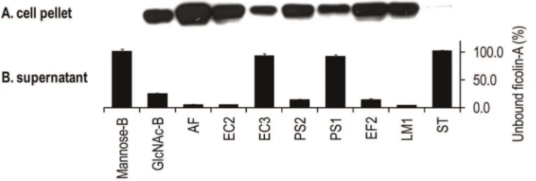
Ficolin-A was incubated with relevant microorganisms and the bacteria cells were analysed on SDS-PAGE followed by western blotting (Fig.3A). Ficolin-A binding was confirmed to E. faecalis (EF2),L. monocytogenes(LM2),E. coli(EC2) andP. aeruginosa(PS2). Weaker binding was detected to E. coli (EC3) and P. aeruginosa (PS1). No binding was detected toS. Typhimurium (ST). Control experiments showed no binding to mannan-agarose beads but positive binding to GlcNAc-agarose beads. These data were supported by analyzing the supernatant containing the unbound protein on an acBSA matrix (Fig.3B).
Binding of Serum Ficolin-A to Microorganisms
Bacteria were incubated with ficolin-A knock-out serum or wildtype serum. Ficolin-A binding was detected in the wildtype serum to A. fumigatus (AF), E. coli (EC2), P. aeruginosa (PS2), E. faecalis(EF2) andL. monocytogenes(LM2) (Fig.4). These data confirm the results obtained with recombinant ficolin-A.
Binding of Ficolin-A to LPS
Overnight bacteria culture was cleared from bacteria cells by centrifugation and the medium was sterile filtrated and incubated in different dilutions with ficolin-A and added to acBSA plates. The ficolin-A binding to the acBSA matrix was inhibited with increased concentration of bacteria growth medium fromL. mono-cytogenes(LM2),E. faecalis(EF2),E. coli(EC2) andP. aeruginosa(PS2) (Fig.5A). However, no inhibition to acBSA was observed when incubating ficolin-A with growth medium fromS. aureus(SA19),S. Typhimurium(ST) orP. aeruginosa(PS1). In order to investigate if the bacteria produce proteases that inactivate ficolin-A, and thereby could explain the observed reduced binding to acBSA, the bacteria overnight culture medium was heat inactivated, sterile filtrated and incubated with ficolin-A and measured on the acBSA plates (Fig.5B). No difference between the heat-inactivated growth
Table 1.investigated microorganisms and ligands.
ABBAbbrev. Name Gram-classification N
SA Staphylococcus aureus Positive 19
SR Streptococcus agalactiae(Group B) Positive 6
EF Enterococcus faecalis Positive 6
LM Listeria monocytogenes Positive 3
SE Staphylococcus epidermidis Positive 1
ST Salmonella Typhimurium Negative 1
SD Salmonella Dublin Negative 1
SG Salmonella Gallinarium Negative 1
EC Escherichia coli Negative 3
PS Pseudomonas aeruginosa Negative 2
KB Klebsiella pneumoniae Negative 1
AF Aspergillus fumigatus Negative 1
acBSA-B acetylated bovine serum albumin sepharose beads – –
BSA-B bolive serum albumin sepharose beads – –
PBS phosphate buffered saline – –
Mannose-B mannose agarose beads – –
GlcNAc-B N-acetyl-D-glycosamine agarose beads – –
medium and control growth medium was observed. These results indicate that the ficolin-A binding bacteria strains excrete substances such as LTA or LPS to the culture medium that inhibits the binding of ficolin-A to acBSA. LPS was purified from the investigated Gram-negative bacteria and coated on a poly-styrene plate and ficolin-A was added in a serial dilution and the binding was detected. Ficolin-A bound LPS fromE. coli(EC2) and to some degree to LPS fromP. aeruginosa(PS2) (Fig5C).
Binding of Ficolin-A toA. FumigatusUsing Flow Cytometry
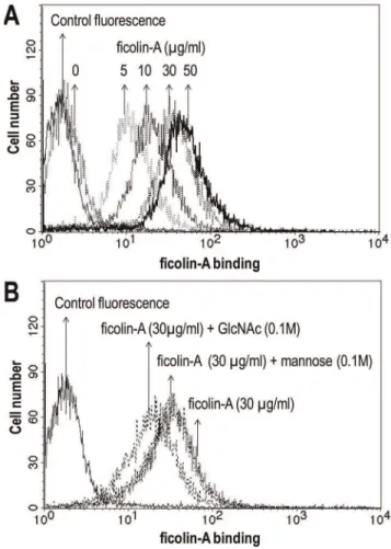
Flow cytometry was used to examine the binding of ficolin-A to A. fumigatus. The microorganism was incubated with recombinant ficolin-A and detected with polyclonal rabbit anti-mouse ficolin-A (Japan) antibody followed by FITC-conjugated anti-rabbit IgG. Fig.6A shows a significant dose-dependent binding of ficolin-A to A. fumigatus. This binding was inhibited by GlnNAc but not mannose (Fig.6B).
Binding of Ficolin-A to Microorganisms Using Confocal Laser Scanning Microscope (CLSM)
Interaction between ficolin-A and different microorganisms were assessed by CLSM. Ficolin-A bound strongly to all of the conidia cells ofA. fumigatus(Fig.7A). No fluorescence was observed when
incubating without ficolin-A (Fig.7B) or with ficolin-3 with respective antibodies (data not shown). The binding of ficolin-A to A. fumigatus was inhibited with GlcNAc but not EDTA or mannose (Fig.7C).
Ficolin-A binding was detected to E. coli (EC2), however this interaction was restricted to aggregated cells only (Fig.8A). All aggregated bacteria on the slide were positive. No binding of aggregatedE. coli(EC3) was detected (Fig.8B). As expected based on the previous results, no binding was detected toS. Typhimurium (ST) (data not shown).
Ficolin-A also bound aggregates ofE. faecalis(EF2) but not single cells (Fig.9A). No signal was detected when incubating without ficolin-A (Fig.9B). The binding appeared to be concentrated to large aggregates (Fig.9C).
Discussion
The innate immune system consists of various types of cells and molecules that specifically interact with each other to initiate a quick and effective host defence. The murine ficolin-A and human ficolin-2 and ficolin-3 are serum proteins that bind to carbohydrate structures and acetylated compounds displayed on the surfaces of both mammalian tissues and on microorganisms [19–21]. Carbohydrates may be arranged into extremely variable structures, which on some microorganisms allow the ficolins to
Figure 1. Binding of the ficolins to different microorganisms.After incubation of the microorganisms (16108cells) with 0.2mg of either
bind. The interactions between human ficolins and carbohydrates have been shown to be involved in activities such as opsonization, activation of complement and phagocytosis [2,4]. In this study we investigated the binding of the mouse ficolin-A to a number of
different clinical relevant microbial strains and compared this binding profile to human serum ficolin-2 and ficolin-3.
The results of this work demonstrate a broad binding profile of mouse ficolin-A towards both Gram-positive and Gram-negative bacterial strains. Significant ficolin-A binding was detected towards all the investigatedE. faecalis andL. monocytogenes strains whereas only some of the E. colistrains andP. aeruginosa strains reacted with ficolin-A. Ficolin-A also showed a strong binding towardsS. epidermidisand the fungusA. fumigatus. A similar binding profile was observed for human ficolin-2. However, this molecule also binds toSalmonellastrains to some degree. In contrast, ficolin-2 did not bind L. monocytogenes in the same degree as ficolin-A. Ficolin-2 has previously been shown to bind to Gram-positive bacteria such as S. pneumoniae, S. agalactiae and S. aureus, and to Gram-negative bacteria such asS. typhimuriumandE. coli[20,22]. In this study we detected binding of ficolin-2 to only a few strains ofS.aureusand the binding was apparently weaker to these strains compared to strains such asE. faecalisor some strains ofE. coli. It has been shown that ficolin-2 only bind to capsulatedS. aureus strains and not to noncapsulated strains [22]. Many bacteria, including both Gram-positive and Gram-negative, may be covered by an outer polysaccharide-containing layer termed the capsule layer. In the case of human pathogens, a large number of different capsule serotypes have been identified. For example, over 80 different capsular polysaccharides or K antigens have been described for E. coli [23,24]. Various functions have been attributed to capsules including protection from desiccation [25] and adherence to surfaces and other bacteria contributing to biofilm formation [23]. Capsules often play a role in pathogenicity [26] acting as virulence factors to protect cells from phagocytosis and complement-mediated killing. The synthesis of the capsular material depends upon the environment and whether the bacteria cultures are able shift their morphology during growth. The bacteria used in this work were grown exponential overnight in LB medium and contains likely a mixture of both capsulated and noncapsulated strains. Investigating aS. aureusbacteria culture that has been optimized for production of capsules may show a stronger interaction as described by Krarup et al. [22]. The same issue may be relevant for other bacteria strains as theGroup B Streptococcithat previously has been shown to bind ficolin-2 [16], where we could only confirm a weaker binding to these strains. However, it may be difficult to compare ‘‘strength’’ of ficolin binding to bacteria with previous published papers since the assays have been performed with different setting. Previous reports have demonstrated ficolin-3 does not appear to have a broad binding specificity towards pathogens [27]. The only bacteria that have been shown to interact with ficolin-3 areAerococcus viridiansandHafnia alvei[27,28] and the anti-microbial binding profile of the human ficolins is thereby very different. Supporting these data, the carbohydrate binding specificity of the human ficolins has been analyzed by glycan array screening demonstrating that ficolin-2 bind several different carbohydrates such as disulfated N-acetyllactosamine and tri- and tetra-saccharides containing terminal galactose or GlcNAc whereas ficolin-3 shows a very poor lectin activity [29]. Recently a mutation in theFCN3gene causing ficolin-3 deficiency has been observed in a patient with recurrent severe infections of different aetiologies [10] and ficolin-3 has also been shown to be important in tissue homeostasis [2,30]. Thus it is at present unresolved whether ficolin-3 deficiency is directly associated with increased risk of infections or may aggravate the consequences of tissue damage caused by infections.
The group ofE. faecalisbacteria appears to be a specific target for both ficolin-A and ficolin-2.E. faecalisis a opportunistic Gram-positive cocci that normally inhabits the intestinal tract but it may
Figure 2. Dose-dependent binding of the ficolins (0.2mg) to microorganisms.Ficolin-A showed strong binding toE. faecalis(EF2), A. fumigatus(AF),L. monocytogenes(LM1),E. coli(EC2) andP. aeruginosa (PS2) (A). 2 showed binding to all six microorganisms (B). Ficolin-3 did not bind any of the bacteria but a weak binding was observed to A. fumigatus (AF) (C). GlcNAc-agarose beads and acBSA-coated sepharose beads were included as positive controls. Binding is indicated with decreased percentage signals related to PBS where 100% reflects no binding. OD450nm values at 100% for the ficolin-A, ficolin-2 and ficolin-3 assays were 1.263, 2.561 and 1.338, respectively. Error bars indicate standard derivations of duplicates. The results shown are typical of three experiments.
also be responsible for surgical wound infections and urinary tract infections (for review see [31]). Ficolin-A and ficolin-2 also bind to different small-colony variants (SCVs) (EF4, EF5, EF6) in a similar manner as the normal E. faecalis counterpart (EF3). SCVs constitute slow-growing subpopulations of the bacteria with distinct phenotypes having more pathogenic characteristics. It might be speculated that mouse ficolin-A and human ficolin-2 have an important function in controlling this pathogen. Furthermore, the binding was only detected to large aggregates of this bacteria and whether the interaction is directly to the bacteria cell wall or to biofilm remains to be investigated.
The binding of the serum ficolins was investigated towards three different E. coli strains; O149 (EC1), O157:H7 (EC2) and ATCC25922 (EC3). Both ficolin-A and ficolin-2 bind to the pathogenic E. colistrains O149 and O157:H7 but not the non-pathogenic E. coliATCC25922. Supporting these data, ficolin-2 has recently been shown to bind theE. coli O104:H4 that were
responsible for the recent outbreak of food poisoning in Germany causing haemolytic-uremic syndrome (HUS) [32].
The bacterium O157:H7 has been implicated in several outbreaks of hemorrhagic colitis included fatalities caused by haemolytic uraemic syndrome [33,34] and the E. coli O149 is a pathogen that may lead to porcine postweaning diarrhea causing major problems in the pig industry worldwide [35]. The binding of ficolin-A toE. coliO157:H7 (EC2) was only detected to aggregated cells and not to single cells. E. coli strains are able to produce biofilm and the binding mechanism may be the same as observed forE. faecalis. Sterile filtrated overnight growth medium from the bacteria that associate with ficolin-A was able to inhibit the binding of ficolin-A to acBSA in a similar pattern as observed when using living bacteria. Therefore, we speculated whether the bacteria were able to produce enzymes that could inactivate the ficolin molecules. However, 96uC heat-inactivated bacteria growth medium showed a similar binding profile as observed with unheated growth medium. This strongly suggests that the reduced
Figure 3. Binding of the ficolin-A to microorganisms.After incubation of the bacteria with recombinant ficolin-A the bacteria were washed three times and analysed on SDS-PAGE followed by western blotting (A). Binding was detected to GlcNAc-agaose beads (GlcNAc-B) but not mannose-agarose beads (Mannose-B). Strong binding was observed toA. fumigatus(AF),E. coli(EC2),P. aeruginosa(PS2),E. faecalis(EF2) andL. monocytogenes (LM1). Residual concentration of ficolin-A in the supernatant was analysed on acBSA-coated polystyrene plates (B). Binding is indicated by low percentage value where 100% reflects no binding. OD450nm value at 100% was 1.799. Error bars indicate standard derivations of two experiments. doi:10.1371/journal.pone.0038196.g003
Figure 4. Binding of serum ficolin-A to microorganisms. Ficolin-A knock-out serum and wild type serum was incubated with different microorganisms. After incubation of the bacteria (16108cells) with serum diluted 1:50 in Barb-T buffer, the concentration of residual ficolin-A was
estimated on acBSA coated polystyrene plates. Binding is indicated with decreased percentage signals where 100% reflects no binding. Binding was detected toA. fumigatus(AF),E. coli(EC2),P. aeruginosa(PS2),E. faecalis(EF2) andL. monocytogenes(LM1). OD450nm at 100% is 2.731. Error bars indicate standard deviations of duplicates.
ficolin-A binding to acBSA was not due to a bacterial proteolytic digestion of ficolin-A but rather an affinity inhibition of the detection assay by secreted structures such as LPS or LTA. Ficolin-2 has been shown to bind LTA fromS. aureus,S. pyogenes
andS. agalactieaeand thereby initiate the lectin pathway-dependent C4 turnover [20]. Furthermore, porcine ficolin-a react with LPS from different bacteria such as E. coli, S. typhimurium, S. enteritidis andP.aeruginosa and LTA from theS. pyogenesandS. aureus[18]. In order to study the binding of ficolin-A to endotoxins, we purified LPS from the investigated Gram-negative bacteria and measured the binding of LPS to ficolin-A. Ficolin-A was able to bind to LPS from the pathogenicE. coliO157:H7 (EC2) but not the non-pathogenic E. coli ATCC25922 (EC3). It might be speculated that LPS is the ligand structure for ficolin-A upon immune recognition of the pathogen. However, the binding of ficolins to released LPS from the pathogenic E. coli O157:H7 (EC2) could also be an evasion mechanism of the bacteria to hamper recognition and sequestration by innate immunity mechanisms as complement deposition, opsonization and phago-cytosis.
We demonstrated a clear binding of ficolin-A towards A. fumigatus. We have previously shown that ficolin-2 binds to certainA. fumigatusstrains [36] and this interaction was confirmed with new isolates in the present study. The binding of ficolin-A to A. fumigatusseems to be located to the cell membrane of the conidia and the binding was inhibited by GlcNAc but not mannose
Figure 5. Binding of ficolin-A to bacterial components. The binding of ficolin-A (0.2mg) to acBSA was inhibited with sterile filtrated overnight bacterial growth medium from some of the bacteria (A). No differences were observed when the growth medium was heat inactivated at 96uC (B). Purified LPS was coated on polystyrene plates and serum from ficolin-B knock-out serum diluted 1:10 in Barb-T buffer was added (C). Ficolin-A binding to LPS fromE. coli(EC2) was detected in a dose-dependent manner. Error bars indicate standard deviations of duplicates. Shown is one representative experiment of three. doi:10.1371/journal.pone.0038196.g005
Figure 6. Binding of ficolin-A toA. fumigatusconidia using flow cytometry.A. fumigatus(1.46106cells) were incubated with elevated
concentration of ficolin-A (A). Bound protein was detected with polyclonal rabbit anti-mouse ficolin-A antibody.A. fumigatus(1.46106
cells) were incubated with ficolin-A (30mg/ml) in the presence of GlcNAc or mannose, respectively (B). Bound ficolin-A was detected as depicted above. As a control, A. fumigatus was incubated in the absence of ficolin-A. Results are representative of three independent experiments that yield similar results.
demonstrating that the interaction is through the FBG domain. These data may indicate that the contact between ficolin-A and the fungus is a result of a direct immune mechanism for tagging the microorganism for phagocytosis and complement deposition. The long pentraxin PTX3 has been shown to bind toA. fumigatus
and to be critical against infection in mice models [37,38]. We have previously demonstrated that ficolin-2 interacts with PTX3 [36] and that these two proteins might work synergistically against A. fumigatus in vitro. A. fumigatus has become one of the most
Figure 7. Binding of ficolin-A toA. fumigatususing confocal laser scanning fluorescence microscopy.A. fumigatus(46106cells) was
incubated with ficolin-A (5mg/ml) (A) or without ficolin-A (B). The binding was inhibited with GlcNAc but not EDTA or mannose (C). The binding was detected with a monoclonal rat anti-mouse ficolin-A antibody.
prevalent airborne fungal pathogens, causing severe and usually fatal invasive infections in immunocompromised patients [39].
Even though ficolin-A and ficolin-2 are not considered as orthologues molecules based on evolutionary neighbouring three analyses [13,14], the present results show that these two molecules might have similar properties. This suggest that ficolin-A knock-out mice models might be highly relevant for human disease model in general. However, one unresolved question that remains to be addressed is the possible role of the ficolins in viral infections and viral pathophysiology.
In conclusion, our results demonstrate that ficolin-A have a broad binding profile towards both negative and
Gram-positive strains as well fungi. The binding properties towards clinical relevant pathogens suggest that ficolin-A is an important contributors to innate host defence against pathogens, but our findings also indicate that the microbes may be able to utilize the ficolins as virulence factors. Ficolin-A appears to share several of the ficolin-2 binding properties although some differences in the binding profile were observed. Ficolin-3 however, seems to have a very restricted anti-microbial profile.
Materials and Methods
Maxisorp polystyrene microtiter plates were from NUNC (Roskilde, Denmark). PBS-buffer (10 mM Na2HPO4, 1.47 mM Figure 8. Binding of ficolin-A toE. coliusing confocal laser scanning fluorescence microscopy.Ficolin-A (5mg/ml) was incubated with E. coliEC2 (A) orE. coliEC3 (B). The bacteria (46106cells) were incubated with or without ficolin-A. Ficolin-A binding was only observed toE. coliEC2
KH2PO4, 137 mM NaCl, 2.7 mM KCl, pH = 7.4) and Barbital-buffer (4 mM C8H11N2NaO3, 145 mM NaCl, 2.6 mM CaCl2, 2.1 mM MgCl2, pH = 7.4) were from the hospital pharmacy (RegionH Apoteket, Rigshospitalet, Copenhagen, Denmark). Tween-20 was from Merck (Hohenbrunn, Germany). FITC-conjugated swine anti-rabbit IgG was purchased from DAKO (Glostrup, Denmark). Goat anti-rat IgG Alexa Fluor 488, NuPAGE 3–8% Tris-acetate gels, Tris-acetate buffer, LDS loading buffer and reducing agent were purchased from Invitrogen (Taastrup, Denmark). HRP-conjugated streptavidin, HRP-cou-pled anti-rabbit immunoglobulin, nitrocellulose membranes, ECL hyperfilm and CNBr activated sepharose beads were from GE Healthcare (Buckinghamshire, United Kingdom). SuperSignal West Femto Maximum Sensitivity Substrate and Ready-to-use TMB substrate were from Pierce Biotechnology (Rockford, IL, USA). Chloramphenicol, tryptone, yeast extract, mannose agarose beads, GlcNAc agarose beads and GlcNAc were from Sigma (Brondby, Denmark). LPS Extraction Kit was from CHEMBIO (Hertfordshire, UK). Sterile filter 0.2mm was from VWR (Herlev, Denmark). Sterile 40mM cell strainer and the BD FACSCalibur were from Becton Dickinson (Bilthoven, The Netherlands). The monoclonal antibodies were produced in house; rat anti-mouse ficolin-A (FCNA-K12), mouse anti-human ficolin-2 (FCN219), mouse human ficolin-3 (FCN334)). Polyclonal rabbit anti-mouse ficolin-A (Japan) antibody was produced in house. In selected experiments sera from ficolin-A and ficolin-B knock-out mice were used (the construction and characterisation of these mice will be reported elsewhere).
Recombinant Ficolins
Recombinant mouse ficolin-A was synthesized as previously described for ficolin-2 and ficolin-3 [3,40]. Briefly, theFCNAgene was amplified from mouse liver cDNA using the forward primer 59-ctagctcgacgcgagatgcagtggcctacgctgt-39 and the reverse primers 59-tccggaattccgttaagatgctcggattttcatctc-39. The constructs were cloned into the expression vector pED.dC and transfected into the Chinese hamster ovary (CHO) cell line CHO-DG44 (a kind gift from professor Lawrence Chasin, Columbia University, New York) using dhfr selection system. Subsequently, the recombinant ficolin-A were purified from the cell supernatant on GlcNAc agarose beads. Culture supernatants were centrifuged for 20 min at 15,000 g to obtain cleared supernatants and then loaded onto a column containing GlcNAc-agarose beads, which was been equilibrated with Barb-T buffer (Barbital-buffer with 0.05% Tween-20). The beads were washed with 100 times column volume of Barb-T buffer and the ficolins were eluted with 0.25 M GlcNAc in PBS buffer and dialyzed first into PBS containing 5 mM EDTA buffer and then pure PBS.
Coupling of acBSA and BSA to Sepharose Beads
A total of 0.1 g dry CNBr-activated Sepharose 4B beads were allowed to swell in 1 mM HCl. After washing in 1 mM HCl, the beads were incubated with 4 mg BSA or acetylated BSA for 4 h at 4uC in a buffer composed of 0.1 M NaHCO3 and 0.5 M NaCl (pH 8.3). The beads were washed three times with the same buffer and blocked with 1 M ethanolamine. The beads were washed in 0.1 M TRIS-HCl, 0.5 M NaCl (pH 8.5).
Bacteria
The following bacteria strains were examined in the assays: Staphylococcus aureus (SA1–19: 8325-4, SH1000, COL, EMRSA, SA564, 15981, USA300, Na34, m70, m124, m332, m137, Mu50, M9, ST398, S84F9, UAMS-1, P1, ATCC25923),Escherichia coli (EC1-3: O149, O157:H7, ATCC25922),Enterococcus faecalis(EF 1-6: TX0052, OG1RF, HEF5, scv1, scv2, HEF5-scv3),Listeria monocytogenes(LM1-3:10403S, ScottA, LO28), Salmo-nella Typhimurium(ST1:4/74), Salmonella dublin(ST2:3246), Salmo-nella gallinarium (ST3: G9), Pseudomonas aeruginosa (PS1-2: ATCC27853, 53381), Streptococcus agalactiae(Group B streptococ-cus) (SR1-6:33304, 33327, 33354, 54381, 33999, 33061), Staph-ylococcus epidermidis (clinical isolate A33), and Klebsiella pneumoniae (KB1:51046). All strains were grown in standard LB (Luria-Bertani broth) medium.
Preparation ofA. FumigatusConidia
The Aspergillus fumigatus strain was obtained from a clinical isolate 6871 (University of Perugia, Italia). The A. fumigatuswas grown on Sabouraud dextrose agar supplemented with chloram-phenicol by agar-streak for 4 days at 28uC. Abundant conidia were obtained under these conditions. The conidia were harvested by washing the slant culture with PBS containing 0.05% Tween-20 (PBS-T) and gently scraping the conidia from the mycelium with sterile cotton-tipped applicators. The conidia were then allowed to settle by gravity followed by filtration through sterile 40mM cell strainer to remove hyphal fragments and cell debris. After extensive washing with PBS-T, the conidia were counted and diluted to the desired concentrations. TheA. fumigatuswas heat-inactivated at 121uC for 15 min prior to use.
Consumption Assay
Bacteria were incubated overnight at 37uC, shaking at 130 rmp in 5 ml LB-medium. The overnight cultures were centrifuged at 9,000 g for 5 min and washed three times in PBS. The cell pellet was resuspended in Barb-T buffer and the bacteria concentration was estimated spectrophotometric at OD600nm, where absor-bance value 1 corresponds to approximately 16109 cells/ml. Bacteria (16108 cells) or fungus (16107 cells) were incubated shaking for 2 h at 37uC with 0.4mg/ml rficolin-A, rficolin-2 or rficolin-3 in Barb-T buffer in a total volume of 500ml. After centrifugation (9,000 g, 5 min) the supernatant was transferred to ficolin-A, ficolin-2 and ficolin-3 quantification assays described below. The percentage of unbound ficolin was estimated by dividing the signal from the sample containing the microorganisms with the signal from the control sample without microorganisms. Each test was repeated three times in duplicates. In some experiments, ficolin-A wildtype or ficolin-A knock-out mice sera (1:50) were incubated with bacteria and analysed as described above. The bacteria pellet was washed three times in Barb-T buffer and resuspended in SDS-PAGE loading buffer and analyzed by western blotting.
Ficolin-A, Ficolin-2 and Ficolin-3 Quantification Assay Microtiter plates were coated with 100ml of 5mg/ml acBSA in PBS-buffer overnight at 4uC. Plates were blocked for 1 h with Barb-T and subsequently washed three times in Barb-T. Samples were added to the wells and incubated for 2 h at 37uC. Ficolin-A
Figure 9. Binding of ficolin-A toE. faecalis(EF2) using confocal laser scanning fluorescence microscopy.TheE. faecalis(46106cells) was
incubated with ficolin-A (5mg/ml) (A) or without ficolin-A (B). Ficolin-A binding was only observed to aggregatedE. faecaliscells. 3D view of the ficolin-A binding to aggregated bacteria (C). The binding was detected with a monoclonal rat anti-mouse ficolin-A antibody.
starting from 1.56108 cells) were incubated for 2 h at 37uC, shaking with 0.4mg/ml rficolin-A, rficolin-2 or rficolin-3 in Barb-T buffer in a total volume of 500ml. After centrifugation (9,000 g, 5 min) the supernatants were transferred to ficolin-A, ficolin-2 and ficolin-3 quantification assays described above.
Western Blotting
Bacteria pellet samples were diluted in SDS-buffer with reducing agent and heated for 5 min at 90uC and applied to SDS-PAGE using 3–8% Tris-acetate gels. The separated proteins were subsequently blotted onto nitrocellulose membranes and the membranes were blocked using a 5% skim milk solution in PBS-T for 1 h at room temperature shaking. After a brief wash in PBS-T, the membranes were incubated shaking for 1 h at room temperature with 0.25mg/ml polyclonal rabbit anti-mouse ficolin-A (Japan) antibody. Following incubation, the membranes were washed thoroughly in PBS-T and subsequently incubated with 0.25mg/ml anti-rabbit-HRP. Membranes were developed on ECL hyperfilm with SuperSignal West Femto Maximum Sensi-tivity Substrate.
Sterile Filtration and Heat Inactivation of Bacteria Growth Medium
Overnight bacteria culture was centrifuged at 9,000 g for 5 min and the growth medium were sterile filtrated using 0.2mm filters and heat inactivated at 96uC for 30 min.
LPS Purification
LPS was purified from 5 ml of overnight bacteria cultures (S. typhimurium,P. aeruginosaandE.coli) using LPS Extraction Kit at standard conditions. LPS was dissolved in 50ml PBS.
LPS Assay
Purified LPS fromS. typhimurium, P. aeruginosaand E.coli were coated in polystyrene plates in 2-fold dilutions starting from 5ml/ well. Plates were blocked for 1 h with Barb-T and were subsequently washed twice in Barb-T. Ficolin-B knock-out serum diluted 1:10 in Barb-T buffer was added to the wells and incubated for 2 h at 37uC. Ficolin-A was detected with 0.25mg/ml polyclonal rabbit anti-mouse ficolin-A (Japan) antibody for 1 h at 37uC. After washing, HRP-coupled anti-rabbit immunoglobulin (0.25mg/ml) was added for 1 h at 37uC. Plates were washed and developed for 10–15 min with TMB substrate. Addition of 50ml/ well sulphuric acid terminated the enzymatic reaction. The optical density was measured at 450 nm.
cold TBS-Mg2+
/HI-FCS. Flow cytometry was performed at the BD FACSCalibur and data was analyzed by BD CellQuest Pro software. In some experiments, A. fumigatus (1.46106 cells) were incubated with mouse ficolin-A (30mg/ml) in TBS-Mg2+
/HI-FCS in the presence of GlcNAc (0.1 M) and mannose (0.1 M), respectively. Bound ficolin-A was detected as depicted above. As a control A. fumigatus was incubated in the absence of mouse ficolin-A.
Confocal Laser Scanning Fluorescence Microscopy Ficolin-A (5mg/ml) was incubated with 46106microbial cells in Barb-T buffer in a total volume of 200ml for 1 h at 37uC. After centrifugation (9,000 g, 5 min) the cell pellet was incubated with an in house produced monoclonal rat anti-mouse ficolin-A antibody (FCNA-K12) for 1 h at 37uC. After centrifugation (9,000 g, 5 min) the cell pellet was washed and incubated with anti-rat IgG Alexa Fluor 488 for 1 h at 37uC. The cells were washed and the samples were examined using a Zeiss LSM 710 confocal laser scanning microscope (CLSM) equipped with an Argon laser for excitation of the Alexa Fluor 488. The images were further processed using the IMARIS software package (Bitplane AG).
Ethics Statement
This study was conducted in accordance with Danish law for animal protection and with the European Communities Council Directive 86/609/EEC for the protection of animals used for experimental purposes. The protocol was approved and experi-ments were permitted by the local government, The Animal Experiments Inspectorate, The National Authority, Denmark (permission No. 2009/561-1616) to be executed at the De-partment of Experimental Medicine, Faculty of Health Sciences, University of Copenhagen.
Acknowledgments
The authors wish to thank Ms Sandra Faerch for excellent technical assistance.
Author Contributions
Conceived and designed the experiments: TH PG. Performed the experiments: TH YJM LMF TB CM MOS LR TF YE PG. Analyzed the data: TH YJM LMF TB CM MOS LR TF YE PG. Contributed reagents/materials/analysis tools: TH YJM LMF TB CM MOS LR TF YE PG. Wrote the paper: TH PG. Revision of the manuscript: TH YJM LMF TB CM MOS LR TF YE PG.
References
1. Endo Y, Matsushita M, Fujita T (2011) The role of ficolins in the lectin pathway of innate immunity. Int J Biochem Cell Biol 43: 705–712.
2. Honore C, Hummelshoj T, Hansen BE, Madsen HO, Eggleton P, et al. (2007) The innate immune component ficolin 3 (Hakata antigen) mediates the clearance of late apoptotic cells. Arthritis Rheum 56: 1598–1607.
3. Hummelshoj T, Thielens NM, Madsen HO, Arlaud GJ, Sim RB, et al. (2007) Molecular organization of human Ficolin-2. Mol Immunol 44: 401–411. 4. Matsushita M, Endo Y, Taira S, Sato Y, Fujita T, et al. (1996) A novel human
5. Ohashi T Erickson HP (1998) Oligemeric structure and tissue distribution of ficolins from mouse, pig and human. Arch Biochem Biophys 360: 223–232. 6. Garlatti V, Belloy N, Martin L, Lacroix M, Matsushita M, et al. (2007)
Structural insights into the innate immune recognition specificities of L- and H-ficolins. EMBO J 26: 623–633.
7. Yang L, Zhang J, Ho B, Ding JL (2011) Histidine-mediated pH-sensitive regulation of M-ficolin:GlcNAc binding activity in innate immunity examined by molecular dynamics simulations. PLoS One 6: e19647.
8. Honore C, Rorvig S, Munthe-Fog L, Hummelshoj T, Madsen HO, et al. (2008) The innate pattern recognition molecule Ficolin-1 is secreted by monocytes/ macrophages and is circulating in human plasma. Mol Immunol 45: 2782–2789. 9. Munthe-Fog L, Hummelshoj T, Hansen BE, Koch C, Madsen HO, et al. (2007) The impact of FCN2 polymorphisms and haplotypes on the Ficolin-2 serum levels. Scand J Immunol 65: 383–392.
10. Munthe-Fog L, Hummelshoj T, Honore C, Madsen HO, Permin H, et al. (2009) Immunodeficiency associated with FCN3 mutation and ficolin-3 deficiency. N Engl J Med 360: 2637–2644.
11. Sallenbach S, Thiel S, Aebi C, Otth M, Bigler S, et al. (2011) Serum concentrations of lectin-pathway components in healthy neonates, children and adults: mannan-binding lectin (MBL), M-, L-, and H-ficolin, and MBL-associated serine protease-2 (MASP-2). Pediatr Allergy Immunol 22: 424–430. 12. Liu Y, Endo Y, Homma S, Kanno K, Yaginuma H, et al. (2005) Ficolin A and
ficolin B are expressed in distinct ontogenic patterns and cell types in the mouse. Mol Immunol 42: 1265–1273.
13. Fujita T, Matsushita M, Endo Y (2004) The lectin-complement pathway-its role in innate immunity and evolution. Immunol Rev 198: 185–202.
14. Garred P, Honore C, Ma YJ, Rorvig S, Cowland JB, et al. (2010) The genetics of ficolins. J Innate Immun 2: 3–16.
15. Endo Y, Liu Y, Kanno K, Takahashi M, Matsushita M, et al. (2004) Identification of the mouse H-ficolin gene as a pseudogene and orthology between mouse ficolins A/B and human L-/M-ficolins. Genomics 84: 737–744. 16. Aoyagi Y, Adderson EE, Rubens CE, Bohnsack JF, Min JG, et al. (2007) L-ficolin/mannose-binding lectin-associated serine protease complexes bind to group B Streptococci largely through N-acetylneuraminic acid of capsular polysaccharide, and activate the complement pathway. Infect Immun 76: 179–188.
17. Kjaer TR, Hansen AG, Sorensen UB, Nielsen O, Thiel S, et al. (2011) Investigations on the pattern recognition molecule M-ficolin: quantitative aspects of bacterial binding and leukocyte association. J Leukoc Biol 90: 425–437.
18. Nahid AM Sugii S (2005) Binding of porcine ficolin-alpha to lipopolysaccharides from Gram-negative bacteria and lipoteichoic acids from Gram-positive bacteria. Dev Comp Immunol 30: 335–343.
19. Krarup A, Thiel S, Hansen A, Fujita T, Jensenius JC (2004) L-ficolin is a pattern recognition molecule specific for acetyl groups. J Biol Chem 279: 47513–47519. 20. Lynch NJ, Roscher S, Hartung T, Morath S, Matsushita M, et al. (2004) L-ficolin specifically binds to lipoteichoic acid, a cell wall constituent of gram-positive bacteria, and activates the lectin pathway of complement. J Immunol 172: 1198–1202.
21. Ma YG, Cho MY, Zhao M, Park JW, Matsushita M, et al. (2004) Human mannose-binding lectin and L-ficolin function as specific pattern recognition proteins in the lectin activation pathway of complement. J Biol Chem 279: 25307–25312.
22. Krarup A, Sorensen UB, Matsushita M, Jensenius JC, Thiel S (2005) Effect of capsulation of opportunistic pathogenic bacteria on binding of the pattern recognition molecules mannan-binding lectin, L-ficolin, and H-ficolin. Infect Immun 73: 1052–1060.
23. Roberts IS (1996) The biochemistry and genetics of capsular polysaccharide production in bacteria. Annu Rev Microbiol 50: 285–315.
24. Whitfield C (2006) Biosynthesis and assembly of capsular polysaccharides in Escherichia coli. Annu Rev Biochem 75: 39–68.
25. Ophir T Gutnick DL (1994) A role for exopolysaccharides in the protection of microorganisms from desiccation. Appl Environ Microbiol 60: 740–745. 26. Briles DE, Crain MJ, Gray BM, Forman C, Yother J (1992) Strong association
between capsular type and virulence for mice among human isolates of Streptococcus pneumoniae. Infect Immun 60: 111–116.
27. Tsujimura M, Miyazaki T, Kojima E, Sagara Y, Shiraki H, et al. (2002) Serum concentration of Hakata antigen, a member of the ficolins, is linked with inhibition of Aerococcus viridans growth. Clin Chim Acta 325: 139–146. 28. Swierzko A, Lukasiewicz J, Cedzynski M, Maciejewska A, Jachymek W, et al.
(2011) New functional ligands for ficolin-3 among lipopolysaccharides of Hafnia alvei. Glycobiology 22: 267–280.
29. Gout E, Garlatti V, Smith DF, Lacroix M, Dumestre-Perard C, et al. (2010) Carbohydrate recognition properties of human ficolins: glycan array screening reveals the sialic acid binding specificity of M-ficolin. J Biol Chem 285: 6612–6622.
30. Kuraya M, Ming Z, Liu X, Matsushita M, Fujita T (2005) Specific binding of L-ficolin and H-L-ficolin to apoptotic cells leads to complement activation. Immunobiology 209: 689–697.
31. Murray BE (1990) The life and times of the Enterococcus. Clin Microbiol Rev 3: 46–65.
32. Heidema J, Meijvis SCA, Rijkers GT (2011) Activation of the lectin pathway of complement by EHEC. BMJ 342: doi:10.1136/bmj.d3884.
33. Wachsmuth IK, Griffin PM, Wells JG (1991) Escherichia coli O157:H7, a cause of hemorrhagic colitis and hemolytic uremic syndrome. Acta Paediatr Jpn 33: 603–612.
34. Griffin PM Tauxe RV (1991) The epidemiology of infections caused by Escherichia coli O157:H7, other enterohemorrhagic E. coli, and the associated hemolytic uremic syndrome. Epidemiol Rev 13: 60–98.
35. Fairbrother JM, Nadeau E, Gyles CL (2005) Escherichia coli in postweaning diarrhea in pigs: an update on bacterial types, pathogenesis, and prevention strategies. Anim Health Res Rev 6: 17–39.
36. Ma YJ, Doni A, Hummelshoj T, Honore C, Bastone A, et al. (2009) Synergy between ficolin-2 and PTX3 boosts innate immune recognition and complement deposition. J Biol Chem 284: 28263–28275.
37. Garlanda C, Hirsch E, Bozza S, Salustri A, De AM, et al. (2002) Non-redundant role of the long pentraxin PTX3 in anti-fungal innate immune response. Nature 420: 182–186.
38. Gaziano R, Bozza S, Bellocchio S, Perruccio K, Montagnoli C, et al. (2004) Anti-Aspergillus fumigatus efficacy of pentraxin 3 alone and in combination with antifungals. Antimicrob Agents Chemother 48: 4414–4421.
39. Andriole VT (1993) Infections with Aspergillus species. Clin Infect Dis 17 Suppl 2: S481–S486.