Renata de Cassia Gonçalves(a) Dirceu Barnabé Raveli(a) Ary dos Santos Pinto(a)
(a)Department of Orthodontics, Faculdade de Odontologia de Araraquara, UNESP – Univ Estadual Paulista, Araraquara, SP, Brazil.
Corresponding author:
Renata de Cassia Gonçalves
Department of Orthodontics, Faculdade de Odontologia de Araraquara, UNESP - Univ Estadual Paulista
Rua Humaitá, 1680 CEP: 14801-903
Araraquara - São Paulo - Brazil E-mail: [email protected]
Received for publication on Nov 03, 2010 Accepted for publication on Mar 18, 2011
Effects of age and gender on upper
airway, lower airway and upper lip
growth
Abstract: The aim of the present retrospective study was to evaluate the inluence of age and gender on upper and lower airway width and upper lip length. In this study, 390 lateral cephalograms were divided into 13 age groups (ranging from 6 to 18 years) and were analyzed. The inter-group differences were analyzed using a MANOVA (Multivariate Analy-sis of the Variance), and the intragroup differences were analyzed using an ANOVA (Analysis of the Variance) and Tukey’s test. The results of the present study indicated that although the airway width and the upper lip length increased with age, the lower airway width exhibited variable growth between the ages of six and eighteen years. The airway width was signiicantly greater in females than males, whereas the upper air-way width was similar between these two genders. The lip length was signiicantly shorter in females than males. The lower airway width and upper lip length were signiicantly different between males and females, whereas the upper airway width was similar for the genders. The upper airway width and upper lip exhibited incremental growth between the ages of six and eighteen years. The upper lip closely followed the growth pattern of the upper airway width; the growth plateaued between the ages of 6 and 9 years, increased from 9 to 16 years and plateaued from 16 to 18 years.
Descriptors: Nasopharynx; Oropharynx; Lip.
Introduction
Normal respiratory activity inluences the growth of maxillofacial
structures, favoring advantageous growth and development.1 A
knowl-edge of craniofacial growth, dentofacial morphology and normal func-tioning are essential for the diagnosis, planning and stability of
orth-odontic treatments.2, 3
The position of an individual’s teeth are in equilibrium with the oral soft tissues (lip, tongue and cheek) and are affected by several functions:
respiration, deglutition, chewing and speech.4 Mouth breathing becomes
in-fections, which are amenable to treatment.5
Mouth-breathing children develop postural changes, such as a down and backward mandible posture and a down and forward positioning of the tongue. If these postural changes continue for a long period of time, especially during an individual’s active growth stages, various degrees of facial dis-harmony can develop. This facial disdis-harmony can result in long-face syndrome, an increased anterior face height, incompetent lip seals, short upper lips, and small nostrils. These disorders are frequently accompanied by dental changes, such as an anterior open bite, a posterior crossbite, maxillary dental
constriction, and protruding upper incisors.6-8
Pa-tients may also have a disordered dentofacial mor-phology that has been corrected with orthopedic
and orthodontic treatments.3
Mouth breathing is a common alteration with important dentofacial complications in children and adults, male or female, and is associated with vari-ous types of facial patterns. Several authors have re-ported an association between mouth breathing and
a vertical growth pattern,8, 9 whereas other authors
have reported a lack of a correlation between the growth pattern and an obstruction of the upper and lower pharyngeal airways in mouth-breathing
pa-tients.3,10 Several studies have identiied a gender
dif-ference between upper airway width and lower
air-way width growth.11,12 However, other studies have
reported no difference between girls and boys.3,13 In
addition, several studies have reported that pharynx
growth is continuous from infancy to adolescence.11
Other studies have assessed the importance of lip morphology in tooth positioning and facial
pro-iles.2,4,14-16
The aim of the present retrospective study was to evaluate the inluence of age and gender on the up-per and lower airway width and the upup-per lip length in a cohort of healthy subjects, who ranged from six to eighteen years of age, using lateral cephalometry.
Methodology
The lateral cephalograms of 390 untreated pa-tients (195 females and 195 males), who ranged from 6 to 18 years of age, were collected from the Orthodontic Department’s iles. Patient radiographs
from healthy Caucasian patients without a history of respiratory problems that were obtained using the Funk Orbital Device at Ribeirão Preto, SP, were included in the present study. No samples were ex-cluded because of a malocclusion (152 samples were Angle Class I, 214 were Angle Class II and 24 were Angle Class III). The exclusion criteria included poor radiograph images and images that included evidence of swallowing during the radiograph expo-sure. The present study was approved by the Univer-sity Research Ethics Committee.
The patients were divided into thirteen age groups, each of which covered a one-year age inter-val. Each group was composed of thirty individuals (15 males and 15 females).
Any patient-identifying information was re-moved from the lateral cephalograms, and these cephalograms were traced and digitized using a Nu-monics Accugrid light box by a single investigator (RCG). The linear measurements of the upper and lower airway width were recorded using a Mitutoyo Digimatic Caliper (Mitutoyo Products, Mitutoyo-Sul Americana Ltda., São Paulo, Brazil) in a simi-lar manner to McNamara’s Analysis. The upper lip length was determined using Ricketts Analysis with Dentofacial Planner Plus 2.01 software (Dentofacial Planner Software Inc., Toronto, Canada). The re-sults of these analyses are presented in Figure 1.
Statistical analysis
All of the statistical analyses were performed us-ing the SPSS software package (“Statistical Package for the Social Sciences” for Windows, version 16.0, SPSS Inc., Chicago, USA). The method error was as-sessed using the Intraclass Correlation Coeficient (ICC). The mean values of the replicate measure-ments were used for the statistical analysis.
Results
All of the results are summarized in Tables 1, 2, 3 and 4.
A MANOVA test indicated that there were no correlations between the age and gender effects on the variables of interest. There were no upper air-way width differences when the genders were com-pared; however, there were differences in this width when the age groups were compared. An ANOVA test for the upper airway data also indicated that the increase in the width of this airway was correlated with aging (Table 3). Other ANOVA tests indicated that the lower airway width varied by gender but not by age. The females in the present study exhibit-ed larger lower airway dimensions than those of the males (Table 4). Lastly, an ANOVA test indicated that the upper lip length increases by age but does not differ by gender (Table 4).
Discussion
There were no signiicant gender differences in the upper airway widths of the patients who were between 6 and 18 years of age (Table 3, Figure 2). In addition, there was no sexual dimorphism in the 2D lateral cephalometric analyses or the 3D airway
measurements of preadolescents.17 The increase in
the upper airway width relected differential growth patterns in males and females ranging from 4 to
18 years of age and from 4 to 26 years of age,13
re-spectively. The lower airway width was signiicantly wider in females than in males (Table 4, Figures 3a and 3b). These results are in agreement with
pre-vious results gathered by Ceylan and Oktay18 that
showed a signiicant difference in the oropharyn-geal measurements between genders in subjects who
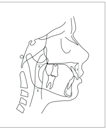
Figure 1 - Measurements on the lateral cephalograms. 1) Upper lip length, measured from the stomion superior to the anterior nasal spine. 2) Upper airway width, measured from a point on the posterior outline of the soft palate to the closest point on the posterior pharyngeal wall. 3) Lower airway width, measured from the intersection of the posterior border of the tongue and the inferior border of the mandible to the closest point on the posterior pharyngeal wall.
Table 2 - MANOVA test of the between-variable effects.
Source Dependent Variable F Sig. Observed
Power
Corrected Model
Upper Lip Length 9.819 0.000 1.000
Lower Airway Width 1.390 0.103 0.948
Upper Airway Width 10.415 0.000 1.000
Intercept
Upper Lip Length 42682.765 0.000 1.000
Lower Airway Width 3761.131 0.000 1.000
Upper Airway Width 4348.006 0.000 1.000
Age
Upper Lip Length 17.002 0.000 1.000
Lower Airway Width 1.976 0.025 0.918
Upper Airway Width 20.811 0.000 1.000
Gender
Upper Lip Length 29.048 0.000 1.000
Lower Airway Width 5.232 0.023 0.626
Upper Airway Width 0.140 0.709 0.066
Age * Gender
Upper Lip Length 1.033 0.417 0.603
Lower Airway Width 0.483 0.924 0.278
Upper Airway Width 0.875 0.573 0.515
Table 1 - Sample description.
Groups N %
G
e
nde
r Girls 195 50
Boys 195 50
Total 390 100
were 13 to 15 years of age. In contrast, the results
of research by Adamidis and Spyropoulos19 did not
indicate any gender differences in the lower airway width of patients who were an average of 9.3 years of age.
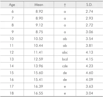
In the present study, the upper airway width increased signiicantly from six to eighteen years
of age (Table 3, Figure 2). The results of previous studies have indicated that, in growing children, the
upper airway width is dependent on age.5,7,13 The
in-crease of the upper airway width is a growth phe-nomenon that occurs because of the expansion of the wings of the sphenoid and the forward drift of the palate. The oral soft tissues, including the
ton-Table 3 - Descriptive statistics for the upper airway width.
Age Mean † S.D.
6 8.92 a 2.74
7 8.90 a 2.93
8 9.12 a 2.72
9 8.75 a 3.06
10 10.52 ab 3.54
11 10.44 ab 3.81
12 11.41 abc 4.13
13 12.59 bcd 4.15
14 13.96 cde 4.23
15 15.60 de 4.60
16 15.41 de 4.09
17 16.39 e 3.63
18 16.55 e 3.04
† Post hoc test (Tukey).
Age
Female Male
Lower airway width Upper lip length Lower airway width Upper lip length
Mean S.D. Mean † S.D. Mean S.D. Mean † S.D.
6 18.56 4.76 23.23 ab 1.56 16.37 3.72 23.99 a 2.14
7 18.31 4.12 22.41 a 1.93 16.00 4.15 23.57 a 2.28
8 17.04 4.57 22.51 a 2.01 15.34 5.80 23.95 a 1.61
9 17.70 4.32 23.01 ab 3.18 14.66 5.00 24.64 a 1.80
10 19.40 4.45 23.99 ab 3.09 18.58 5.37 25.73 abc 1.98
11 20.14 5.73 24.69 abcd 2.62 18.96 4.67 24.97 ab 1.99
12 19.90 6.46 25.69 bcd 1.06 18.06 8.74 26.26 abcd 2.38
13 18.96 5.12 26.49 cd 2.91 16.38 6.05 26.57 abcde 1.96
14 22.07 5.17 25.99 bcd 2.56 20.03 5.87 27.80 bcde 2.91
15 19.32 7.99 26.75 cd 1.70 18.55 4.30 27.97 bcde 3.54
16 18.32 7.49 26.73 cd 1.61 16.29 5.25 28.83 de 2.32
17 20.51 9.26 27.45 d 2.93 20.44 7.84 28.45 cde 2.96
18 16.89 6.04 25.85 bcd 3.40 19.70 6.43 29.45 e 3.12
† Post hoc test (Tukey). A post hoc test of the lower airway space data showed no statistically significant differences.
6 7 8 9 10 11 12 13 14 15 16 17 18 19
6 7 8 9 10 11 12 13 14 15 16 17 18 19 20
5
Age (years)
U
pper
airway
s
pace
width
(mm)
Figure 2 - Growth changes in the upper airway width, from 6 to 18 years of age. The graph shows the means and quar-tile ranges in millimeters (95% CI).
sils and adenoids, that surround the upper airway grow in proportion to the growth of the skeletal structures throughout the irst decade of life. The oral soft tissues reach their maximum size between
7 and 10 years of age.20 Then, these tissues
progres-sively decrease in size until 60 years of age.21 A
simi-lar growth pattern was identiied in this study, espe-cially an increase in the nasopharyngeal dimensions from 9 to 16 years of age.
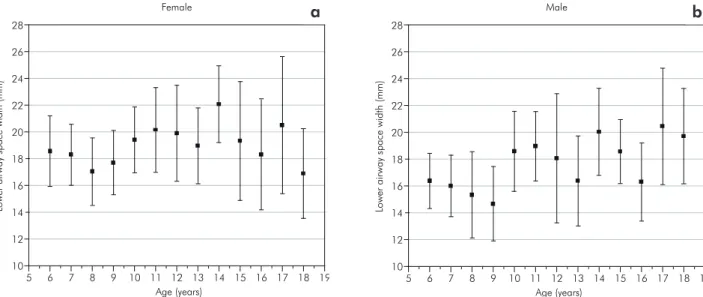
In the present study, the lower airway width dem-onstrated variable growth with no statistical differ-ences among any of the age groups (Table 4, Figures 3a and 3b). This variability may be explained by one or more of the following factors:
• the continued growth of the pharynx,
• lymphoid tissue regression,
• the presence or absence of the enlarged palatine
tonsils, and
• the positions of the tongue in relation to the
mandible and maxilla.20
A wider lower airway width may be due to a malpositioning of the tongue or to enlarged palatine
tonsils causing a mandibular prognatism.1,12
Under normal conditions, the forward transla-tion of the mandible likely contributes to the for-ward positioning of the hyoid bone, whereas the growth of the cervical vertebrae contributes to hyoid
bone descent.20,22 When there is an obstructive
con-dition, the anteroposterior dimension of the lower airway width needs to be maintained. Therefore, the mandible is moved down and backward in rela-tion to the other craniofacial structures, whereas the tongue and hyoid are not. Thus, the hyoid-associ-ated structures (the pharyngeal, cervical and dorsal muscular structures) are guided to a forward
posi-tion to avoid compromising the size of the airway5.
When these postural adaptations of the tongue, hyoid and cervical vertebrae are not suficient to maintain the size of the airway, other compensatory
mechanisms develop. For example, the lips part,8,23
the mandible moves downward to increase the
air-way space8 and the tongue assumes a lower24 or
more forward8 position within the oral cavity.
How-ever, this altered relex behavior of the lower airway width is not identical in all subjects. Consequently, different oropharyngeal widths have been observed
among individuals.5
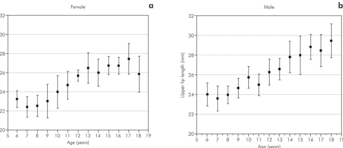
In the present study, there were no differences in the upper lip length by gender; however, there was an increase in the upper lip length by age (Table 4, Figures 4a and 4b). These results are in support of a careful analysis of the results of previous studies that suggested that gender differences of the upper lip length and age differences should be
consid-ered.14,15,16,25,26 Other authors4 have reported similar
10 24
12 22
14 26
16 28
18
6 7 8 9 10 11 12 13 14 15 16 17 18 19 20
5
Age (years)
Lo
wer
airway
s
pace
width
(mm)
Female
10 24
12 22
14 26
16 28
18
6 7 8 9 10 11 12 13 14 15 16 17 18 19 20
5
Age (years)
Lo
wer
airway
s
pace
width
(mm)
Male
Figure 3 - Growth changes in the lower airway width, from 6 to 18 years of age. The graph shows the means and quartile ranges in millimeters. a. female, b. male (95% CI).
indings of a vertical growth of the upper lip, which reaches a plateau by 15 years of age. The results of the current study indicated vertical increases of the
upper lip of 23.3 to 26.6 mm,4 20.5 to 25.0 mm15
and 19.1 to 22.5 mm.²7 In addition to its obvious
importance to the facial proile, lip morphology is
of considerable signiicance in tooth positioning.4,27
When the lips cannot perform their function proper-ly, this leads to a gradual protrusion of the anterior teeth and to the establishment of a Class II, division
1 malocclusion28. When the tongue is forward, it
causes habitual anterior mandibular posturing and,
consequently, a prognatism.1,12
The data from the present study demonstrated that the upper lip closely follows the growth pattern of the upper airway width. There is a growth pla-teau from 6 to 9 years, a progressive increase from 9 to 16 years, and another plateau from 16 to 18 years of age (Table 3, Figures 2, 4a and 4b). The form-function interaction may explain the causal associa-tion between nasal obstrucassocia-tion and facial growth
in children.10 In contrast, no correlations have been
observed between the shape and function of the up-per lip for nasal or mouth breathing, according to
previous authors.10,29 Small changes in the upper lip
length and the linear relationship prediction equa-tions indicate that patients with a short upper lip at 7 years are likely to have a short upper lip at age
18.27
Conclusions
1. The upper airway width is similar between
gen-ders but increases with age.
2. The lower airway width is signiicantly wider in
females than in males. This parameter exhibits variable growth.
3. The upper lip length is similar between males
and females but increases with age.
4. The growth of the upper lip closely follows the
growth pattern of the upper airway width. It exhibits a growth plateau from 6 to 9 years, a linear increase from 9 to 16 years and another growth plateau from 16 to 18 years of age.
20 30
22 28
24 32
26
6 7 8 9 10 11 12 13 14 15 16 17 18 Age (years)
U
pper
lip
length
(mm)
Female
19
5 20
30
22 28
24 32
26
6 7 8 9 10 11 12 13 14 15 16 17 18 19 5
Age (years)
U
pper
lip
length
(mm)
Male
Figure 4 - Growth changes in the upper lip length, from 6 to 18 years of age. The graph shows the means and quartile ranges in millimeters. a. female, b. male (95% CI).
a b
References
1. Takemoto Y, Saitoh I, Iwasaki T, Inada E, Yamada C, Iwase Y, et al. Pharyngeal airway in children with prognathism and normal occlusion. Angle Orthod. 2011 Jan;81(1):77-82.
3. Shanker S, Fields HW, Beck FM, Vig PS, Vig KWL. A longitu-dinal assessment of upper respiratory funcion and dentofacial morphology in 8- to 12-year-old children. Semin Orthod. 2004 Mar;10(1):45-53.
4. Vig PS, Cohen AM. Vertical growth of the lips: a serial cepha-lometric study. Am J Orthod 1979 Apr;75(4):405-15. 5. Tourne LP. Growth of the pharynx and its physiologic
implica-tions. Am J Orthod Dentofacial Orthop. 1991 Feb;99(2):129-39.
6. Linder-Aronson S. Respiratory function in relation to facial morphology and the dentition. Br J Orthod. 1979 Apr;6(2):59-71.
7. Preston CB, Tobias PV, Salem OH. Skeletal age and growth of the nasopharynx in the sagittal plane: A cephalometric study. Semin Orthod. 2004 Mar;10(1):16-38.
8. Subtelny JD. Oral respiration: facial maldevelopment and corrective dentofacial orthopedics. Angle Orthod. 1980 Jul;50(3):147-64.
9. de Freitas MR, Alcazar NM, Janson G, de Freitas KM, Henriques JF. Upper and lower pharyngeal airways in sub-jects with Class I and Class II malocclusions and different growth patterns. Am J Orthod Dentofacial Orthop. 2006 Dec;130(6):742-5.
10. Ambrosio AR, Trevilatto PC, Sakima T, Ignacio SA, Shi-mizu RH. Correlation between morphology and function of the upper lip: a longitudinal evaluation. Eur J Orthod. 2009 Jun;31(3):306-13.
11. Ronen O, Malhotra A, Pillar G. Influence of gender and age on upper-airway length during development. Pediatrics. 2007 Oct;120(4):e1028-34.
12. Battagel JM, Johal A, L’Estrange PR, Croft CB, Kotecha B. Changes in airway and hyoid position in response to man-dibular protrusion in subjects with obstructive sleep apnoea (OSA). Eur J Orthod. 1999 Aug;21(4):363-76.
13. Martin O, Muelas L, Vinas MJ. Nasopharyngeal cephalo-metric study of ideal occlusions. Am J Orthod Dentofacial Orthop. 2006 Oct;130(4):436 e1-9.
14. Burstone CJ. Lip posture and its significance in treatment planning. Am J Orthod. 1967 Apr;53(4):262-84.
15. Park YC, Burstone CJ. Soft-tissue profile--fallacies of hard-tissue standards in treatment planning. Am J Orthod Dento-facial Orthop. 1986 Jul;90(1):52-62.
16. Yuen SW, Hiranaka DK. A photographic study of the facial profiles of southern Chinese adolescents. Quintecensse Int. 1989 Sep;20(9):665-76.
17. Kim YJ, Hong JS, Hwang YI, Park YH. Three-dimensional analysis of pharyngeal airway in preadolescent children with different anteroposterior skeletal patterns. Am J Orthod Den-tofac. 2010 Mar;137(3):306.e1-.e11.
18. Ceylan I, Oktay H. A study on the pharyngeal size in dif-ferent skeletal patterns. Am J Orthod Dentofacial Orthop. 1995 Jul;108(1):69-75.
19. Adamidis IP, Spyropoulos MN. The effects of lymphadenoid hypertrophy on the position of the tongue, the mandible and the hyoid bone. Eur J Orthod. 1983 Nov;5(4):287-94. 20. Taylor M, Hans MG, Strohl KP, Nelson S, Broadbent
BH. Soft tissue growth of the oropharynx. Angle Orthod. 1996;66(5):393-400.
21. Vogler RC, Ii FJ,;Pilgram TK. Age-specific size of the normal adenoid pad on magnetic resonance imaging. Clin Otolaryngol Allied Sci. 2000 Oct;25(5):392-5.
22. Moss ML, Salentijn L. The primary role of functional matrices in facial growth. Am J Orthod. 1969 Jun;55(6):566-77. 23. Subtelny J. The Significance of Adenoid tissue in Orthodontia.
Angle Orthod. 1954;24(2):59-69.
24. Linder-Aronson S. Effects of adenoidectomy on dentition and nasopharynx. Trans Eur Orthod Soc. 1972:177-86. 25. Arnett GW, Jelic JS, Kim J, Cummings DR, Beress A, Worley
CM, Jr., et al. Soft tissue cephalometric analysis: diagnosis and treatment planning of dentofacial deformity. Am J Orthod Dentofacial Orthop. 1999 Sep;116(3):239-53.
26. Fernandez-Riveiro P, Suarez-Quintanilla D, Smyth-Chamosa E, Suarez-Cunqueiro M. Linear photogrammetric analysis of the soft tissue facial profile. Am J Orthod Dentofacial Orthop. 2002 Jul;122(1):59-66.
27. Nanda RS, Meng H, Kapila S, Goorhuis J. Growth chang-es in the soft tissue facial profile. Angle Orthod. 1990 Fall;60(3):177-90.
28. Ambrosio AR, Trevilatto PC, Martins LP, Santos-Pinto A, Shimizu RH. Electromyographic evaluation of the upper lip according to the breathing mode: a longitudinal study. Braz Oral Res. 2009 Oct-Dec;23(4):415-23.