Measurement of the palpebral fissure and the vermilion border of
the upper lip in newborns with gestational ages of 25 to 43 weeks
Medida da fissura palpebral e da borda vermelha do lábio superior de recém-nascidos com
idade gestacional de 25 a 43 semanas
Maria dos Anjos Mesquita1, Conceição Aparecida de Mattos Segre2
ABSTRACT
Objective: To prepare percentile curves for measurements of palpebral fissure and the greatest width of the vermilion border of the upper lip in newborns at a public maternity ward in the City of São Paulo. Methods: A descriptive cross-sectional study was carried out from August 2006 to January 2008. In the first 24 to 72 hours of life, the palpebral fissure and the greatest width of the vermilion border of the upper lip were measured in 1,964 newborns with gestational ages from 25 to 43 weeks. Percentile curves corresponding to these measurements were prepared according to gestational age. Results:
The average measurement of palpebral fissure was 1.98 cm, ranging from 0.80 to 3.00 cm, standard deviation ± 0.24 cm. The average measurement of the width of the vermilion border of the upper lip was 0.51 cm, ranging from 0.20 to 0.90 cm, standard deviation ± 0.11 cm. These measurements allowed designing the percentile curves (10th, 50th, and 90th) relative to gestational age. Conclusions:
Measurement curves of palpebral fissure and of the greatest width of the vermilion border of the upper lip may be useful to evaluate the presence of facial dimorphism in newborns.
Keywords: Infant, newborn; Measurement curve; Palpebral fissure; Lip
RESUMO
Objetivo: Elaborar curvas de percentis das medidas da fissura palpebral e da maior largura da borda vermelha do lábio superior de recém-nascidos em maternidade pública da cidade de São Paulo.
Métodos: Estudo descritivo transversal realizado de Agosto de 2006 a Janeiro de 2008. Nas primeiras 24 a 72 horas de vida, foram feitas medidas da fissura palpebral e da maior largura da borda vermelhado lábio superior de 1.964 recém-nascidos com idade gestacional de 25 a 43 semanas. Curvas de percentil correspondentes a essas medidas foram elaboradas de acordo com a idade gestacional. Resultados:
A média da medida da fenda palpebral foi de 1,98 cm, variando de 0,80 a 3,00 cm, com desvio padrão de ± 0,24 cm. A maior largura
da borda vermelha do lábio superior teve uma medida média de 0,51 cm, variando de 0,20 a 0,90 cm, com desvio padrão de ± 0,11 cm. Essas medidas permitiram a elaboração de curvas de percentil (10, 50 e 90) em relação à idade gestacional. Conclusões: As curvas elaboradas das medidas da fissura palpebral e da maior largura da borda vermelha do lábio superior podem ser úteis na avaliação da presença de dismorfias faciais de recém-nascidos.
Descritores: Recém-nascido; Curva de crescimento; Fissura
palpebral; Lábio
INTRODUCTION
Some characteristics of eyelids and lips, such as their measurements, are data that cannot be omitted from analysis in the physical examination of newborns.
Towards the 48th day of the embryo, formation of the
upper lip is completed through fusion of the two upper maxillary and the two nasomedial processes emerging from the first arch(1).
By the 6th week, small folds of ectoderm with
mesenchymal core appear just cranially and caudally to the developing cornea. These upper and lower primordial eyelids rapidly grow toward each other, meeting and fusing by the 8th week. The eyelids separate
again between the 5th and 7th months(2).
The palpebral fissure develops more quickly than the eye(3). However, the length of the palpebral fissure
directly correlates with the size of the ocular globe(4,5).
The development of the palpebral fissure does not depend solely on the development of the eye(3).
Since the eye is a neuronal organ, the length of the palpebral fissure may indirectly reflect a neurological
Study carried out at Hospital Municipal Maternidade-Escola de Vila Nova Cachoeirinha “Dr. Mário de Moraes Altenfelder Silva” – HMEVNC, São Paulo (SP), Brazil.
1 Hospital Municipal Maternidade-Escola de Vila Nova Cachoeirinha “Dr. Mário de Moraes Altenfelder Silva” – São Paulo (SP), Brazil. 2 Instituto Israelita de Ensino e Pesquisa Albert Einstein – IIEPAE, São Paulo (SP), Brazil.
Corresponding author: Maria dos Anjos Mesquita – Hospital e Maternidade Vila Nova Cachoeirinha – Avenida Deputado Emílio Carlos, 3.100 – Bairro do Limão – CEP: 02720-200 – São Paulo (SP), Brasil – E-mail: [email protected]
growth problem(4). Nevertheless, an alteration in the
measurements of the palpebral fissure may also be related to abnormalities in the development of the soft tissue around the eyes(4).
Many syndromes present with congenital
malformations in the eyelids and/or thinner upper lips, such as the fetal alcohol syndrome (FAS), Kabuki syndrome, trisomy 13, trisomy 18, among others(6). FAS,
the most frequent, is a permanent birth defect caused by maternal consumption of alcohol during pregnancy and it currently represents a major public health issue worldwide(7-9). The estimated prevalence of FAS varies
from 0.5 to 2 cases/1,000 live births(9,10). Based on
this prevalence, the Center for Disease Control and Prevention (CDC) expects there will be 1,000 to 6,000 children with FAS born every year, with approximately 4 million children born with prenatal alcohol exposure. These rates are higher than those found for other common developmental disabilities such as Down’s syndrome or spina bifida(10).
The well-known adverse effects of prenatal exposure to alcohol, however, are underdiagnosed, since healthcare professionals are unprepared to make this diagnosis and because social prejudice in pregnancy causes women to hide their alcohol-consuming habits(10,11). The diagnosis
of FAS in the newborn implies the presence of three facial characteristics: small palpebral fissure, thin upper lip, and smoothness of the philtrum, plus intrauterine growth restriction and abnormalities of the central nervous system in multiple ways(7,10). Consequently, early
identification and diagnosis of FAS in affected newborns recognized by the abovementioned characteristics are essential in order to provide health, education, and social services that may guarantee optimal well-being to these children and their families(10).
Even though some international publications describe the measurement of palpebral fissures in newborns, no reports of these measurements regarding the Brazilian population in the newborn period were found in any study. The same is true in relation to the greatest width of the vermilion border of the upper lip.
Therefore, the construction of percentile curves for the measurements of palpebral fissure and vermilion border of the upper lip in the Brazilian population must be a valid goal for neonatologists.
OBJECTIVE
To construct percentile curves for measurements of palpebral fissure length and the greatest width of the vermilion border of the upper lip in newborns at the Hospital e Maternidade-Escola Municipal Dr. Mário de Moraes Altenfelder Silva da Vila Nova Cachoeirinha (HMEVNC).
METHODS
This is a descriptive cross-sectional study carried out from August 13, 2006 to January 21, 2008 at HMEVNC, a public hospital in the City of São Paulo, located in the Northern area of the city, in Brasilândia district. This maternity hospital serves a low-income and highly mixed race population. It is a reference tertiary hospital for high-risk pregnancies, and also treats normal pregnant women belonging to this specific geographical area.
During this period, there were 7,447 live births at the hospital and 33 live children who were admitted to hospital after birth. For operational reasons, due to availability of one of the researchers (MAM), the live newborns admitted during the above-mentioned period, between 00:01 AM on Sundays and 11:59 PM on Mondays, were included. Newborns who presented with a genetic syndrome, died, were transferred to another hospital or discharged prior to their physical examination, whose mother died, who were not the first born in case of twins, or who were children of women refusing to participate in the study were excluded.
Data were collected on gestational age and measurements of the palpebral fissure and greatest width of the vermilion border of the upper lip of the newborns.
Gestational age was estimated by using the last menstrual period calculated by Naegeles’ rule(12), by
using the New Ballard Score(13) obtained between
the 6th and the 48th hour of life, or by the Capurro
method(14) carried out during the first hour of life,
when the newborn clinical condition precluded examination to be done using the New Ballard Score. The gestational age was determined through the New Ballard Score or the Capurro method when the mothers last menstrual period was unknown. Preterm newborns were defined as those whose gestational age was less than 37 weeks, term newborns as gestational age of 37 complete weeks to 41 weeks 6/7 days, and post-term newborns as gestational age equal to or above 42 weeks(15).
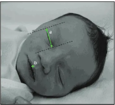
The vermilion border is the exposed mucous membrane or red portion of the upper lip, and the palpebral fissure length is the distance from the eye endocanthion to its exocanthion(16).
were taken with the patient’s eyes closed, due to the difficulty of children this age opening their eyes, and with their lips sealed (Figure 1).
6 (0.29%) were excluded from the study for the presence of syndromes, 11 (0.54%) because they died before having their physical examination, 7 (0.34%) for having been discharged from hospital before their physical examination, 1 (0.05%) due to the fact of being transferred to another hospital before the physical examination, 1 (0.05%) because the mother died before the physical examination, and 9 (0.44%) because their mothers refused to participate. Of the 2,000 (98.28%) children who remained, 36 were of twin pregnancies and the second twin was not considered. Thus the sample for analysis consisted of 1,964 newborns (96.51%).
With regard to gestational age, of the 1,964 newborns, 312 (15.90%) were premature, 1,631 (83.04%) were full term and 21 (1.06%) were post-term. The average gestational age was 38.55 weeks, ranging from 25.0 to 43 weeks and a standard deviation of 2.49 weeks. The mean birth weight was 3,048.51 g, range of 540 g to 5,680 g, and a standard deviation of 613.32 g, and 92% of the children (n = 1,807) were appropriate for gestational age.
Table 1 shows the average measurement of the palpebral fi ssure, its variation and standard deviation, as well as the measurement of the greatest width of the vermilion border of the upper lip with its variation and standard deviation.
Minimum Maximum Average Standard deviation
PF (cm) 0.80 3.00 1.98 0.24
VBUL (cm) 0.20 0.90 0.51 0.11
Table 1. Anthropometric measurements of newborn
PF: palpebral fi ssure; VBUL: vermilion border of the upper lip. Figure 1. Measurements of palpebral fi ssure and vermillion border of superior lip
“a” = palpebral fi ssure length and “b” = vermillion border width
Measuring of the palpebral fi ssure and the vermilion border of the upper lip was carried out at between 24 and 72 hours of life, so that the subcutaneous edema from labor and birth would not induce error. The newborn was lying on the crib, in supine position, in either a state of quiet sleep or quiet alert.
Data collection and anthropometric measurements of the newborns were carried out by only one of the researchers (MAM).
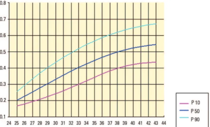
The measurements of palpebral fi ssure and greatest width of the vermilion border of the upper lip of these children, together with their gestational age, allowed the construction of the respective percentile curves for these measurements. For each curve, the 10th, 50th, and 90th percentiles were determined according to gestational age.
The newborn participation in the study was consented to after their mothers had read and signed the Parental or Legal Guardian Consent Form.
The study was approved by the HMEVNC Research Ethics Committee under protocol number 009/2006.
RESULTS
During the study period, 7,480 live children were born, with 7,447 hospital births and 33 births outside the hospital. Of this total, 2,035 were eligible to participate in the research because they were born between 00:01
AM on Sundays and 11:59 PM on Mondays. However, Figure 2. Percentile curve of measurement of palpebral fi ssure
DISCUSSION
Facial characteristics, among them the length of the palpebral fi ssure and the aspect of the upper lip, are essential components of the neonatal physical examination, and the presence of deviations from a normal pattern allows early detection of many syndromes(6). FAS, for instance, stands out for its
prevalence, and its diagnosis implies fi nding the three dysmorphic facial features: smooth philtrum, thin vermillion border, and small palpebral fi ssures(10).
Nevertheless, it is important to emphasize that each of these characteristics may differ according to race, geographical area, and can change with age(16-19).
Growth and facial anthropometry are specifi c for each population, and the sensitivity and specifi city of the evaluation will be low if the appropriate instrument for evaluation is not used. Herein lies the importance of local standardized curves for identifying congenital abnormalities(19-22).
The population comprised in this study belongs to the outskirts of a large city where miscegenation is very high, hindering the identifi cation of a “pure” representative of any race; therefore, the construction of specifi c percentile curves for the measurements of the palpebral fi ssure and the width of the upper lip according to gestational age in this population may be mandatory.
The measurement of the palpebral fi ssure has been thoroughly studied by different authors. Various studies that measured the palpebral fi ssure of newborns, covering gestational ages from 29 to 43 weeks(20,21,23-26) and fetuses
from 15.5 to 41 weeks of gestation(27) were published
by authors from the United States(23,26), Hungary(20),
China(24), Bulgaria(25), Nigeria(19, 21), and France(27).
One North American study constructed a curve of the palpebral fi ssure measurement of children from 29 weeks of gestation to 14 years of age(26). The average
measurement of the palpebral fi ssure found in this study
is comparable to that found by Denis et al.(27), who refer
a mean of 1.85 cm in fetuses aged 39 weeks of gestation, and it is also similar to the values found by Leung et al.(28),
who described a mean of 2.0 cm for White children and 2.3 cm for Black children from Cardiff.
None of the studies mentioned above, however, included the measurements of the palpebral fi ssure of children with ethnic characteristics similar to the population in this study. Furthermore, no study was found that analyzed the palpebral fi ssure measurement in the Brazilian population in order to allow comparison of data. The only Brazilian study that describes small palpebral fi ssures refers to children with SAF(29).
Astley and Clarren created an illustrated guide of the upper lip and nasal philtrum (University of Washington Lip-Philtrum Guide) that helps evaluating these structures in the children of White, North American women who consume alcoholic beverages(30). But no
study was found – whether national or international – in which the greatest width of the vermilion border of the upper lip had been measured for different gestational ages as was done in this study.
According to data found in literature, there is no signifi cant difference between the measurements of the left and right eyelids. Thus, either one of them may be measured isolatedly(20,23). In the current study only the
left palpebral fi ssure of each newborn was measured, as it was easier for the examiner to be positioned in relation to the patient. An important aspect for the measurement of the palpebral fi ssure is related to the state of contraction of the orbicular muscles of the eyelids, which leads to a reduction in length of the palpebral fi ssure, hence this measurement should be obtained when the child is relaxed(23,26). In this study,
the children were either quietly sleep or quietly alert. Some authors stated that the measurement of the palpebral fi ssure is greater in male than female newborns(24,25). However, the majority of authors assured
that there is practically no difference in the length of the palpebral fi ssure with regard to gender(20,21-23,27).
Therefore, in this study, the child sex was not considered separately in relation to this measurement.
Finally, it is worth mentioning that the palpebral fi ssure length is predominately independent of the occipitofrontal circumference in children(31), so it was
not considered for analysis in this article.
Nevertheless, this study has limitations. Only one examiner measured the variables studied, but the large number of children that were examined could compensate for error. The percentile curves for the measurements of palpebral fissure length and upper border of the superior lip according to gestational age may be appropriate for the children born in this particular municipal maternity hospital in the
city of São Paulo because of racial characteristics. However, further studies on the theme should be done and we dare say that this may be a first step towards stimulating the construction of other percentile curves in other centers, according to the characteristics of their own populations. This study did not show comparisons of these curves to others in literature, as this was not its objective.
CONCLUSION
The percentile curves for the measurements of the palpebral fissure length and the greatest width of the vermilion border of the upper lip according to gestational age, in a population of a maternity hospital in the city of São Paulo, were presented in this study.
These data can be useful in the physical evaluation of newborns in this population, as in the early diagnosis of dysmorphic characteristics and syndromic anomalies. They allow for a more precise and objective quantitative description of these structures, avoiding subjective evaluations.
REFERENCES
1. Langman J. Embriologia médica. 4a ed. São Paulo: Atheneu; 1985.
2. Larsen W. Development of the eyes. In: Larsen W, editor. Human embryology. 3a ed. Philadelphia: Churchill Livingstone; 2001. p. 379-91.
3. Denis D, Burguière O, Burillon C. A biometric study of the eye, orbit, and face in 205 normal human fetuses. Invest Ophthalmol Vis Sci. 1998;39(12)2232-8. 4. Jones KL, Hanson JW, Smith DW. Palpebral fissure size in newborn infants. J
Pediatr. 1978;92(5):787.
5. Parnell SE, Deharth DB, Wills TA, Chen SY, Hodge CW, Besheer J, et al. Maternal oral intake mouse model for fetal alcohol spectrum disorders: ocular defects as a measure of effect. Alcohol Clin Exp Res. 2006;30(10):1791-8.
6. Jones KL. Smith`s: Recognizable patterns of human malformations. 6th ed. Philadelphia: Elsevier Saunders; 2006.
7. Astley SJ, Clarren SK. Diagnosing the full spectrum of fetal alcohol-exposed individuals: introducing the 4-digit diagnostic code. Alcohol. 2000;35(4):400-10.
8. Riley EP, Guerri C, Calhoun F, Charness ME, Foroud TM, Li TK, et al. Prenatal alcohol exposure: advancing knowledge through international collaborations. Alcohol Clin Exp Res. 2003;27(1):118-35.
9. May PA, Gossage JP. Estimating the prevalence of fetal alcohol syndrome. A summary. Alcohol Res Health. 2001;25(3):159-67.
10. Bertrand J, Floyd LL, Weber MK; Fetal Alcohol Syndrome Prevention Team, Division of Birth Defects and Developmental Disabilities, National Center on Birth Defects and Developmental Disabilities, Centers for Disease Control and Prevention (CDC). Guidelines for identifying and referring persons with fetal alcohol syndrome. MMWR Recomm Rep. 2005;54(RR-11):1-14.
11. Moraes CL, Reichenheim ME. Rastreamento de uso de álcool por gestantes de serviços públicos de saúde do Rio de Janeiro. Rev Saúde Pública. 2007;41(5):695-703.
12. Watanabe EK. Evolução cronológica do concepto: duração da prenhez. In: Neme B, editor. Obstetrícia básica. São Paulo: Sarvier; 1994. p.47-9. 13. Ballard JL, Khoury JC, Wedig K, Ellerrs-Walsman BL, Lippo R. New Ballard
score, expanded to include extremely premature infants. J Pediatr. 1991;119(3):417-23.
14. Capurro H, Konichezky S, Fonseca D, Caldeyro-Barcia R. A simplified method for diagnosis of gestational age in newborn infant. J Pediatr. 1978;93(1):120-2.
15. Segre CAM. Avaliação da idade gestacional. Classificação do recém-nascido. In: Segre CAM, Costa HPF, Lippi UG, editores. Perinatologia. Fundamentos e prática. 2a ed. São Paulo: Sarvier; 2009. p. 440-7.
16. Chudley AE, Conry J, Cook JL, Loock C, Rosales T, LeBlanc N; Public Health Agency of Canada’s National Advisory Committee on Fetal Alcohol Spectrum Disorder. Fetal alcohol spectrum disorder: Canadian guidelines for diagnosis. CMAJ. 2005;172(5 Suppl):S1-S21.
17. Price KM, Gupta PK, Woodward JA, Stinnett SS, Murchison AP. Eyebrow and eyelid dimensions: an anthropometric analysis of African Americans and Caucasians. Plast Reconstr Surg. 2009;124(2):615-23.
18. Hreczko T, Farkas LG, Katic M. Clinical significance of age-related changes of the palpebral fissures between age 2 and 18 years in healthy Caucasians. Acta Chir Plast. 1990;32(4):194-204.
19. Omotade OO. Facial measurements in the newborn (towards syndrome delineation). J Med Genet. 1990;27(6):358-62.
20. Méhes K. Palpebral fissure length in newborn infants. Acta Paediatr Acad Sci Hung. 1980;21(1):55-6.
21. Adeyemo AA, Omotade OO, Olowu JA. Facial and ear dimensions in term Nigerian neonates. East Afr Med J. 1998;75(5):304-7.
22. Wu KH, Tsai FJ, Li TC, Tsai CH, Peng CT, Wang TR. Normal values of inner canthal distance, interpupillary distance and palpebral fissure length in normal Chinese children in Taiwan. Acta Paediatr Taiwan. 2000;41(1):22-7.
23. Fuchs M, Iosub S, Bingol N, Gromisch DS. Palpebral fissure size revisited. J Pediatr. 1980;96(1):77-8.
24. Fok TF, Hon KL, So HK, Wong E, Ng PC, Lee AK, et al. Craniofacial anthropometry of Hong Kong Chinese babies: the eye. Orthod Craniofac Res. 2003;6(1):48-53.
25. Madjarova LM, Madzharov MM, Farkas LG, Katic MJ. Anthropometry of soft-tissue orbits in Bulgarian newborns: norms for intercanthal and binocular widths and length of palpebral fissures in 100 boys and 100 girls. Cleft Palate Craniofac J. 1999;36(2)123-6.
26. Thomas IT, Gaitantzis YA, Frias JL. Palpebral fissure length from 29 weeks gestation to 14 years. J Pediatr. 1987;111(2):267-8.
27. Denis D, Burguière O, Burillon C. A biometric study of the eye, orbit, and face in 205 normal human fetuses. Invest Ophthalmol Vis Sci. 1998; 39(12)2232-8.
28. Leung AK, Ma KC, Siu TO, Robson WL. Palpebral fissure length. In Chinese newborn infants. Comparison with other ethnic groups. Clin Pediatr (Phila). 1990; 29(3):172-4.
29. Silva AV, Laranjeira RR, Dolnikoff M, Grinfeld H, Masur J. Alcohol consumption during pregnancy and newborn outcome: A study in Brazil. Neurobehav Toxicol Teratol.1981;3(2):169-72.
30. Astley SJ, Clarren SK. Diagnosing the full spectrum of fetal alcohol-exposed individuals: introducing the 4-digit diagnostic code. Alcohol. 2000;35(4):400-10.