Correspondence: Profa. Dra. Marcia da Silva Schmitz, Rua Pinheiro Machado, 2694 ap. 202, 97050-600 Santa Maria, RS, Brasil. Tel: +55-55-3222-4016. e-mail: [email protected]
Influence of Cervical Preflaring on Determination
of Apical File Size in Mandibular Molars:
SEM Analysis
Marcia da Silva SCHMITZ1
Roberto SANTOS2
Alexandre CAPELLI3
Marcos JACOBOVITZ3
Júlio César Emboava SPANÓ3
Jesus Djalma PÉCORA3
1Dental Course, Federal University of Santa Maria, Santa Maria, RS, Brazil
2Department of Restorative Dentistry, Dental School, University of Pernambuco, Camaragibe, PE, Brazil 3Department of Restorative Dentistry, Dental School of Ribeirão Preto,
University of São Paulo, Ribeirão Preto, SP, Brazil
This study investigated the influence of cervical preflaring with different rotary instruments on determination of the initial apical file (IAF) in mesiobuccal roots of mandibular molars. Fifty human mandibular molars whose mesial roots presented two clearly separated apical foramens (mesiobuccal and mesiolingual) were used. After standard access opening and removal of pulp tissue, the working length (WL) was determined at 1 mm short of the root apex. Five groups (n=10) were formed at random, according to the type of instrument used for cervical preflaring. In group 1, the size of the IAF was determined without preflaring of the cervical and middle root canal thirds. In groups 2 to 5, preflaring was performed with Gates-Glidden drills, ProTaperinstruments, EndoFlare instruments and LA Axxes burs, respectively. Canals were sized manually with K-files, starting with size 08 K-files, inserted passively up to the WL. File sizes were increased until a binding sensation was felt at the WL and the size of the file was recorded. The instrument corresponding to the IAF was fixed into the canal at the WL with methylcyanoacrylate. The teeth were then sectioned transversally 1 mm short of the apex, with the IAF in position. Cross-sections of the WL region were examined under scanning electron microscopy and the discrepancies between canal diameter and the diameter of IAF were calculated using the tool "rule" (FEG) of the microscope’s proprietary software. The measurements (µm) were analyzed statistically by Kruskal-Wallis and Dunn’s tests at 5% significance level. There were statistically significant differences among the groups (p<0.05). The non-flared group had the greatest discrepancy (125.30 ± 51.54) and differed significantly from all flared groups (p<0.05). Cervical preflaring with LA Axxess burs produced the least discrepancies (55.10 ± 48.31), followed by EndoFlare instruments (68.20 ± 42.44), Gattes Glidden drills (68.90 ± 42.46) and ProTaper files (77.40 ± 73.19). However, no significant differences (p>0.05) were found among the rotary instruments. In conclusion, cervical preflaring improved IAF fitting to the canals at the WL in mesiobuccal roots of maxillary first molars. The rotary instruments evaluated in this study did not differ from each other regarding the discrepancies produced between the IAF size and canal diameter at the WL.
Key Words: cervical preflaring, initial apical file, anatomic diameter.
INTRODUCTION
One of the major concerns in Endodontics is the development of instruments and techniques that are able to promote adequate disinfection and shaping of the root canal system while respecting apical anatomy. This is particularly challenging in infected curved root canals,
It is generally accepted that the operative proce-dures undertaken during endodontic treatment should be confined to the space previously occupied by the pulp tissue. Apically, the canal-dentin-cementum (CDC) limit and the diameter of this region guide the lateral extension of canal enlargement and the working length (WL) during biomechanical preparation. The anatomic diam-eter of the root canal is clinically ddiam-etermined by record-ing the size of the file that first fits to the canal walls at the WL, designated as the initial apical file (IAF). The detection of the apical constriction and determination of the IAF are thus based on the operator’s tactile sensitiv-ity. This premise relies on the assumption that the root canal is narrower in its apical third and that the file would pass without interference until reaching this constric-tion, which offers resistance to further penetration (4). Studies using optical microscopy to evaluate morphometrically the apical anatomic diameter of dif-ferent dental groups (2,5) found, on average, measure-ments corresponding to a size 25 instrument in this region. Nevertheless, it has been reported that continu-ous and progressive dentin formation within the pulp space narrows the root canal diameter, mainly at the cervical third (6). Therefore, an instrument may be equivocally chosen to initiate instrumentation due to inaccurate determination of the real anatomic diameter, in cases in which the binding sensation felt by the operator actually resulted from unappreciated engage-ment of the file at the canal entrance rather than from fitting to the WL (7). Gani and Visvisian (5) have reported that sizes 10 to 20 instruments frequently do not touch the canal walls at the CDC limit, probably because they get engaged any other point of the root canal irregularities or curvatures. Leeb (4) have also reported that the presence of dentinal projections in the middle and cervical root canal thirds hinders the free and direct access of the files to the CDC limit region.
The importance of cervical preflaring prior to IAF determination has been confirmed by different studies (6-12), which showed that it is possible to introduce a file at least one size larger up to the apical constriction after cervical preflaring. Stabholz et al. (13) have demonstrated that detection of the apical constric-tion by the operator’s tactile sensaconstric-tion is possible in 75% of the cases in which the root canals were preflared. According to Baugh and Wallace (14), most biome-chanical preparation techniques that recommend apical flaring up to a size 25 or 30 instrument, in order to
prevent the occurrence of iatrogenic events, derive from non-research supported personal opinions based on clinical experience.
Vanni et al. (7), Pécora et al. (11) and Barroso et. al (12) investigated the influence of the type of instru-ment used for cervical preflaring on the determination of IAF in maxillary central incisors, maxillary premolars and mesiobuccal canal of maxillary molars, respec-tively. These authors concluded that IAF determination by the operator’s tactile sensation is not an accurate method, especially in the absence of cervical preflaring. They also observed that the use of LA Axxess burs for cervical preflaring produced a more accurate fitting of the IAF to the anatomic diameter at the WL. The outcomes reinforce the need of studies with different groups of teeth due to the peculiar anatomy of the apical region.
The complex internal anatomy of mandibular molars, which, according to Kerekes and Tronstad (1) provides an irregular and unpredictable morphometric apical pattern, motivates the continuous development of endodontic research. The purpose of this study was to investigate the influence of cervical preflaring with different rotary instruments on determination of the IAF in mesiobuccal roots of mandibular molars.
MATERIAL AND METHODS
Fifty mesial roots of human permanent mandibu-lar momandibu-lars with fully developed roots obtained from the Tooth Bank of the Dental School of the Federal University of Santa Maria, Brazil, were used. The selected teeth presented mesial roots with two clearly separated apical foramens (mesiobuccal and mesiolingual) and root canals with curvatures between 10º and 20º, as determined by the Schneider’s method (15). The teeth were kept in 0.1% aqueous thymol solution at 9°C until use and were placed under running water to eliminate traces of thymol 48 h prior use.
until the apical foramen was reached and the file tip was visible. The real canal length was determined and the WL was established at 1 mm from root apex.
Teeth were randomly assigned to 5 groups (n=10). In Group 1, the size of the IAF was determined without previous cervical preflaring of the mesiobuccal root canal. Groups 2 to 5 had the cervical and middle thirds of the mesiobuccal root canal enlarged with sizes 1 and 2 Gates-Glidden drills (Dentsply/Maillefer), Protaper SX and S1 instruments (Dentsply/Maillefer), EndoFlare and Hero 25/.06 instruments (MicroMega, France) and titanium-nitrite treated, stainless steel LA Axxess burs (SybronEndo, Glendora, CA, USA) sizes 20/.06 and 35/ .06. All cervical preflaring procedures were performed using a TC 3,000 engine (Nouvag, TCM Endo, Goldach, Switzerland).
As the instruments used in Groups 2 and 5 do not present flexibility, the depth of penetration was deter-mined by the resistance felt at the middle root canal third and preflaring was performed at 5,000 rpm. In groups 3 and 4, the depth of penetration of the instruments was set at 5 mm short of the WL and preflaring was performed at 315 rpm. The canals were irrigated with 2 mL of 1% NaOCl at each change of instrument during preflaring, with a flush of 5 mL of this solution at the end of the procedure. A final irrigation with 10 mL of distilled water was done. The irrigating solutions were delivered with blunt tip, 31 gauge Endo-Eze irrigation needles (Ultradent Products Inc., South Jordan, UT, USA).
Root canals were negotiated using manual K-files (Dentsply/Maillefer), starting with size 08 files, ad-vanced until the WL was reached. File sizes increased progressively using clockwise and anticlockwise move-ments until an instrument firmly adjusted to the WL (IAF), as felt by the operator’s tactile sensation. The handles of the files had been painted in black to avoid identification, in such a way that the operator was unaware of the file size used until a binding sensation was felt at the WL. Instrument size was then recorded for each tooth.
The IAFs were fixed into the canals at the WL with methylcyanoacrylate. The teeth were then sec-tioned transversally 1 mm short of the apex, with the IAF in position. Cross-sections of the apical region of the mesiobuccal canal was observed with a scanning electron microscope (Philips XL-30, Philips Electric Corporation, Eindhoven, The Netherlands) at ×200 magnification and the images were recorded digitally.
Root canal and file maximum diameters were recorded for each specimen. The diameter of the root canal at the WL and the diameter of the IAF were measured for each specimen using the tool “rule” of the microscope’s proprietary software. The discrepancy between these diameters was calculated (in µm) and the
means obtained for the groups were submitted to statistical analysis.
As a non-normal data distribution was observed, statistical analysis was performed by the Kruskal-Wallis and Dunn’s tests to assess the effect of the preflaring techniques on the discrepancies between IAF diameter and root canal diameter at the WL. Signifi-cance level was set at 5%.
RESULTS
The discrepancies (in µm) (means, standard deviation and range) between the diameter of the IAF and root canal diameter at the working length for each experimental group are given on Table 1. There were statistically significant differences among the groups (p<0.05). The group without preflaring had the greatest discrepancy (mean 125.30 ± 51.54) and differed sig-nificantly (p<0.05) from all flared groups, regardless of the type of instrument (Table 1). Cervical preflaring with LA Axxess burs produced the least discrepancies (mean 55.10 ± 48.31), followed by EndoFlare instruments (68.20 ± 42.44), Gattes Glidden drills (68.90 ± 42.46) and ProTaper files (mean 77.40 ± 73.19). However, no statistically significant differences were found among the rotary instruments (p>0.05) (Table 1). Table 2 shows the ISO size of the K-files used as IAFs in the experimental groups.
Table 1. Discrepancies (in µm) (mean, standard deviation and
range) between the diameter of the initial apical file and root canal diameter at the working length, for each experimental group.
Means (±SD) Range
No preflaring 125.30a (±51.54) 79-210 Gattes Glidden 68.90b (±42.46) 14-140
ProTaper 77.40b (±73.19) 16-249
EndoFlare 68.20b (±42.44) 21-163
La Axxess 55.10b (±48.31) 22-175
DISCUSSION
The biomechanical preparation of the apical re-gion is an essential and critical operative step of the endodontic therapy, and the size of the final instrument used at the WL should be properly selected in such a way to enlarge the root canal diameter at this point.
Apical access by cervical flaring has been in-creasingly investigated (7,9-12). This procedure aims to remove cervical interferences from the root canal entrances, which represent an obstacle to free access of endodontic instruments to the apical portion of the root canals. Removal of these anatomic interferences en-hances canal shaping at the apical third. The findings of the present study showed that the removal of cervical interferences by canal preflaring with Gates Glidden drills (Group 2), ProTaper (Group 3), EndoFlare (Group 4) and LA Axxess burs (Group 5) allowed determining the IAF at the WL 1 mm short of the apical foramen with K-files of larger sizes than those used in Group I (no cervical preflaring) (Table 1). These results indicate that when the cervical third was not preflared, the determi-nation of the IAF did not reflect the real apical anatomic diameter. The non-flared group presented the greatest discrepancies between the canal size and IAF diameter at the WL, compared to the other experimental groups. These results are consistent with those of previous investigations (7,9-12).
In the present study, the groups submitted to cervical preflaring with different rotary instruments (Gates-Glidden, Endoflare, ProTaper, Endoflare and LA Axxess) showed similar mean discrepancies
be-tween the diameter of the IAF and root canal diameter at the WL. These results are consistent with those of Contreras et al. (9), who examined radiographically the position of the IAF at the WL before and after cervical preflaring of mesial canals of mandibular molars and did not find significant differences between the rotary instruments used for canal flaring. However, Vanni et al. (7), Pécora et al. (11), and Barroso et al. (12) found differences among the rotary instruments used for cervical preflaring in the different groups of teeth (maxillary central incisors, maxillary premolars and maxillary molars, respectively).
Table 2 shows that K-files of larger sizes were used in group 5, in which cervical preflaring was done with LA Axxess, in the same way as reported by Vanni et al. (7), Pécora et al. (11), and Barroso et al. (12). However, in the present study, although cervical preflaring with LA Axxess burs produced the least discrepancies, the statistical analysis did not indicate significant differ-ence among the rotary instruments. This result may be attributed to the complex anatomy of the apical region of mandibular molars (Figs. 1-5).The lack of homogeneity of the anatomic diameters of the tested specimens is supported by the findings of a previous study (1), which showed that 35% of the mandibular molars presented two canals in the mesial root 1 mm short of the root apex. At 2 mm short of the apex, this percentage dropped to 30% and to 20% at 3 mm. A study on root canal anatomy of human permanent teeth (16) showed that 10% of the mandibular molars that presented two distinct canals exiting the pulp chamber, had these canals joined in a single one in the body of the root and divided again in two canals with separated foraminal orifices close to the root apex.
Nair et al. (3) have reported that, due to the presence of isthmuses and ramifications, some areas of the mesiobuccal canal of mandibular molars are inac-cessible to the direct contact of instruments during biomechanical preparation, which favors microbial per-sistence and proliferation. Nevertheless, the use of instruments of larger sizes in the apical region during instrumentation of the mesiobuccal canal of mandibular molars has been shown to provide cleaner root canals (10,17,18).
It seems consensual that cervical preflaring should be performed prior to determination of the size of the file that first fit at the WL. The results of the present study showed that, regardless of the type of instrument, all Table 2. ISO size of the K-files used as initial apical files in the
experimental groups.
Specimen No Gates Pro Endo LA
preflaring Glidden Taper Flare Axxess
1 15 25 30 30 35
2 15 30 35 30 40
3 20 30 30 30 35
4 15 25 25 30 35
5 15 30 25 35 35
6 20 30 30 30 30
7 20 30 30 25 30
8 10 20 30 25 40
9 15 15 30 30 35
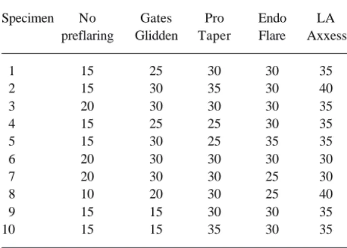
Figure 1. SEM micrograph of Group 1 (no preflaring). Cross-section at the working length (original magnification ×200).
Figure 2. SEM micrograph of Group 2 (preflaring with Gates Glidden drills). Cross-section at the working length (original magnification ×200).
flared groups presented smaller IAF discrepancy in relation to root canal diameter compared to the non-flared group. Our findings are also supportive to those of previous investigations (7,11,12,19), which demon-strated that, although cervical flaring prior to IAF determination by operator’s tactile sensation provided smaller discrepancy between the IAF and the smallest root canal diameter, in most cases, it did not accurately reflect the real canal diameter 1 mm short of the radiographic apex. Wu et al. (19) have stated that the concept of cleaning of root canals with three or four file sizes larger than the first file that fits at the WL needs to be revised due to the complex anatomy of this region. Wile some canals may be overinstrumented, causing canal transportation, apical perforations and root
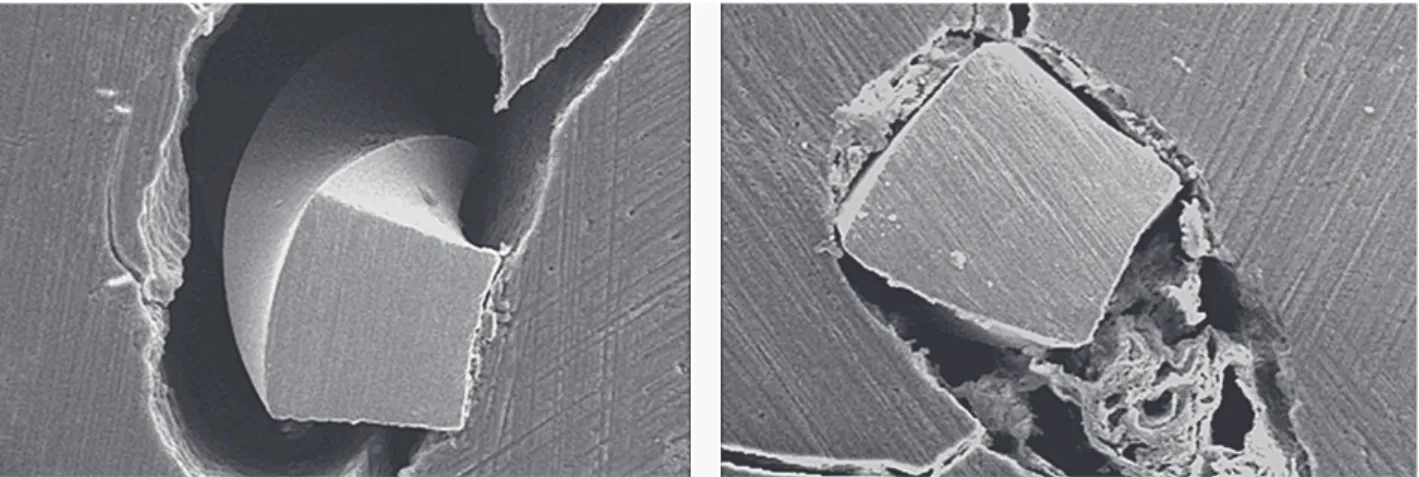
weak-Figure 4. SEM micrograph of Group 4 (preflaring with EndoFlare and Hero 25 .06 instruments). Cross-section at the working length (original magnification ×200).
Figure 5. SEM micrograph of Group 5 (preflaring with LA Axxes burs). Cross-section at the working length (original magnification ×200).
ening, others may remain underinstrumented and not properly cleaned.
Endodontic research should further develop in-struments and techniques for root canal preparation that are able to determine the real dimension of the root canals, while respecting the complexity of apical anatomy. This is expected to provide a more effective cleaning of the root canal system with no risk of causing canal deformations during instrumentation and minimizing the difficulty in the lateral extension of canal enlarge-ment. It is mandatory to change clinical decisions guided by empiric personal experiences into clinical approaches supported by scientific evidence.
1. Cervical preflaring improved the fitting of the IAF to the canals at the WL in mesiobuccal roots of maxillary first molars; 2. The non-flared group had the greatest discrepancy between the IAF diameter and canal diam-eter at the WL and differed significantly from all flared groups, regardless of the type of instrument; 3. The rotary instruments evaluated in this study (Gates-Glidden drills, EndoFlare instruments, ProTaper and LA Axxess burs) had similar performance to each other.
RESUMO
Este estudo investigou a influência do alargamento cervical feito com diferentes instrumentos rotatórios na determinação do instrumento apical inicial (IAI) das raizes mésio-vestibulares de molares inferiores. Foram utilizados 50 molares inferiores cujas raízes mesiais apresentavam dois forames apicais nitidamente separados (mésio-vestibular e mésio-lingual). Após o acesso à câmara pulpar de forma convencional e remoção do tecido pulpar, o comprimento de trabalho foi definido a 1 mm do ápice radicular. Os dentes foram divididos aleatoriamente em cinco grupos (n= 10) de acordo com o tipo de instrumento utilizado no alargamento cervical. No grupo 1, o IAI foi definido sem o prévio alargamento dos terços médio e cervical das raízes. Nos grupos 2 a 5, o terço cervical e médio do canal radicular foi alargado com as brocas de Gates-Glidden, instrumentos Pro Taper, Endo Flare e brocas LA Axxes, respectivamente. A determinação do IAI foi realizada manualmente com limas tipo K em ordem crescente de diâmetro a partir da lima 08 até se chegar ao instrumento que permitisse ao operador ter a sensação tátil do mesmo estar firmemente ajustado ao CRT. O instrumento que correspondeu ao IAI foi fixado no interior do canal radicular com cianocrilato de metila. Com o IAI posicionado, os dentes foram seccionados transversalmente até 1 mm do ápice. As seções transversais do CRT foram observadas através da microscopia eletrônica de varredura e os desajustes entre o diâmetro do canal e o diâmetro do IAI foram calculados com a função “régua” (FEG) do software do próprio microscópio. Os resultados foram avaliados estatisticamente pelo testes de Kruskal-Wallis e Dunn ao nível de significância de 5%. Houve diferenças estatisticamente significantes entre os grupos (p<0,05). O grupo sem alargamento apresentou o maior desajuste (125,30 ±51,54) e diferiu significativamente dos demais grupos (p<0,05). O alargamento cervical com as brocas LA Axxess apresentou os menores desajustes (55,10 ± 48,31), seguido de EndoFlare (68,20 ± 42,44), Gattes Glidden (68,90 ± 42,46) e limas ProTaper (77,40 ± 73,19). Contudo, não houve diferenças estatisticamente significantes entre os instrumentos rotatórios (p<0,05). Conclui-se que o alargamento cervical melhorou a adaptação do IAI aos canais no CRT das raízes mésio-vestibulares dos primeiros molares inferiores. Os instrumentos rotatórios avaliados neste estudo não apresentaram diferenças estatísticas entre si no que diz respeito aos desajustes entre as dimensões do IAI e o diâmetro do canal no CRT.
REFERENCES
1 . Kerekes K, Tronstad L. Morphometric observations on root
canals of human molars. J Endod 1977;3:114-118.
2 . Marroquin BB, El-Sayed MAA, Willershausen-Zonnchen B. Morphology of the physiological foramen: maxillary and mandibular molars. J Endod 2004;30:321-328.
3 . Nair PN, Henry S, Cano V, Vera J. Microbial status of apical root canal system of human mandibular first molars with primary apical periodontitis after “one-visit” endodontic treatment. Oral Surg Oral Med Oral Pathol Oral Radiol Endod 2005;99:231-252.
4 . Leeb J. Canal orifice enlargement as related to biomechanical preparation. J Endod 1983;9:463-470.
5 . Gani O, Visvisian C. Apical canal diameter in the first upper molar at various ages. J Endod 1999;25:689-691.
6 . Tan BT, Messer H. The effect of instrument type and preflaring on apical file size determination. Int Endod J 2002;35:752-758.
7 . Vanni JR, Santos R, Limongi O, Guerisoli DZ, Capelli A, Pécora JD. Influence of cervical preflaring on determination of apical file size in maxillary molars: SEM analysis. Braz Dent J 2005;16:181-186.
8 . Wu MK, Róris A, Barkis D, Wesselink PR. Prevalence and extent of long oval canals in the apical third. Oral Surg Oral Med Oral Pathol Oral Radiol Endod 2000;89:739-743. 9 . Contreras MA, Zinman EH, Kaplan SK. Comparison of the
first file at the apex, before and after early flaring. J Endod 2001;27:113-116.
10. Tan BT, Messer H. The quality of apical canal preparation using hand and rotatory instruments with specific criteria for enlargement based on initial apical file size. J Endod 2002;28:658-664.
11. Pécora JD, Capelli A, Guerisoli DMZ, Spanó JEC, Estrela C. Influence of cervical preflaring on apical file size determina-tion. Int Endod J 2005;38:430-435.
12. Barroso JM, Guerisoli DMZ, Capelli A, Saquy PC, Pécora JD. Influence of cervical preflaring on determination of apical file size in maxillary premolars: SEM analysis. Braz Dent J 2005;16:30-34.
13. Stabholtz A, Rotstein I, Torabinejad M. Effect of preflaring on tactile sense detection of the apical constriction. J Endod 1995;21:92-94.
14. Baugh D, Wallace J. The role of apical instrumentation in root canal treatment: a review of the literature. J Endod 2005;31:333-340.
15. Schneider SW. A comparison of canal preparations in straights and curved root canals. Oral Surg Oral Med Oral Pathol 1971;32:271-275.
16. Vertucci FJ. Root canal anatomy of the human permanent teeth. Oral Surg 1984;58:589-599.
17. Wu M, Wesselink PR. Efficacy of three techniques in clean-ing the apical portion of the curved root canals. Oral Surg Oral Med Oral Pathol 1995;79:492-496.
18. Card JC, Sigurdsson A, Orstavik D, Trope M. The effective-ness of increased apical enlargement in reducing intracanal bacteria. J Endod 2002;28:779-783.
19. Wu MK, Barkis D, Roris A, Wesselink PR. Does the first file to bind correspond to the diameter of the canal in the apical region? Int Endod J 2002;35:264-267.