ORIGINAL ARTICLE
EXERCISE AND SPORTS SCIENCES
EFFECT OF PHYSICAL TRAINING ON METABOLIC AND
BONE PROFILE IN WEANING RATS
Luciana Mendonça Arantes1 Natalia Oliveira Bertolini1 José Alexandre Leme1 Bruno Augusto Ribeiro do Vale1 Eliete Luciano1
1- UNESP – State University of São Paulo, Júlio de Mesquita Filho, Rio Claro Campus, São Paulo, Brazil.
Mailing address:
Departamento de Educação Física, Universidade Estadual Paulista “Júlio de Mesquita Filho”, Campus de Rio Claro Rua 24 A, 1.515, Bela Vista 13506-900. Rio Claro, SP, Brasil E-mail: [email protected]
ABSTRACT
Introduction: The practice of moderate-intensity exercise can reduce the risk of infections and improve metabolic aspects of the person. Objective: To investigate the effects of aerobic physical training on endocrine and metabolic aspects, bone and immune systems. Methods: Twenty Wistar rats were divided in two groups: sedentary (SG) and trained group (TG). Training program consisted
in swimming, 6 weeks, supporting a workload corresponding to 5% of body weight. At the end
of the experiment, were performed counting total and differential leukocyte count and hematocrit. After training period,were analyzed glucose, total protein, triglycerides, cholesterol, liver and muscle samples for the determination of the levels of glycogen, and determination of the tibia length and bone area. All dependent variables were analyzed by one-way analysis of variance (ANOVA) and a
significance level of P < 0.05 was used for all comparisons. Results: Hematocrit (%) analyzed showed
a significant difference, with higher values for TG (54.63 ± 1.41) than for the SG (49.5 ± 1.65). The total leukocyte count was not significantly different, as there was no difference in the differential count. Total cholesterol showed significant decrease in TG (TG = 68.27 ± 13.71 mg/dL; SG = 94.44 ± 28.09), the total protein levels also showed significant reduction (TG = 7.3 ± 0.40 g/dL; SG = 7.74 ± 0.36 g/dL) glucose levels and triglyceride showed no significant differences. The bone length showed significant difference (TG = 40±0.14 mm; SG = 42.10 ± 0.12mm). Tibial area showed a lower value for TG (1.53 ± 0.12cm²) than for SG (1.67 ± 0.18cm²); however, the difference was not statistically significant. Conclusion: It can be concluded that aerobic exercise training is able to produce some unique physiological changes in young rats. There is also the need to prescribe exercises that meet the particular maturational stage of development.
Keywords: physical training, metabolic aspects, immune system.
INTRODUCTION
In the last decade, young athletes have been on the spotlight in many sports1 which require high level of motor skill and training1. Moreover, the search for a physical appearance model set by the media also makes adolescents look for physical and/or sports activi-ties with high intensity and training volume2. Due to the dominance of cerebral impulses and lower sensation to movement exertion, children present higher motor activity than adults3, besides higher baseline metabolism mediated by the intensive growth and diffe-rentiation processes4, increasing the need for vitamins, minerals and nutrients, especially proteins5.
Extenuating training in that phase of life, may lead to predo-minance of the functional metabolism over the structural, which may harm the growth process and decrease the capacity to sup-port load6. In childhood, the bones are more flexible, less resistant to pressure and traction; the tissue of the tendons and ligaments is insufficiently resistant to traction, and the cartilage tissue and epiphyseal discs not ossified, present great risk to damage under pressure and twist forces due to the high rate of division, regulated by growth5. Thus, children and adolescents are more prone to load damage than adults, especially during puberty7. According to Mackelvieet et al.8, unilateral or maximal loads may cause, im-mediately or in the long run, tissue disorders.
Increase in bone mass during and immediately after growth is mentioned as an important preventive strategy of osteoporosis; and increase of 3 to 5% in the bone mineral density reduces the risk of fractures in 20 to 30%8. Evidence indicates that the physical activities effects on the bone mass are boosted in a period close to the maximal growth velocity peak15, improving bone mineral density of children and adolescent athletes.
Basset et al.11 concluded that once bone mineral density is incre-ased by physical exercise during early years, it reflects on adulthood, with extended benefits or decades after training interruption. However, with extenuating training, the positive effects on the bone health may be reduced or even cancelled2 usually causing fractures by stress14.
During growth, the adaptation velocity of the passive locomo-tor system (bone and cartilage tissue) is slower than the active locomotor system (muscular tissue), and is connected with the high susceptibility to overloads, needs a strict load progression with appropriate stimuli (submaximal) in children, recruiting in a multiple way the passive locomotor system which guarantees that its structures are given adequate adaptation time, avoiding hence that the load threshold is surpassed, which results in injuries 7 and positively influencing growth and its structure.
210
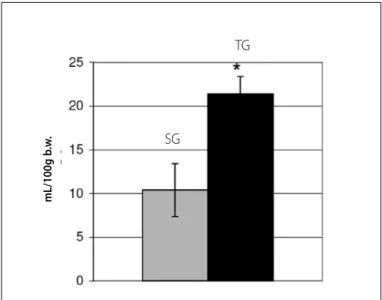
Figure 1. Mean water intake of the experimental groups.
SG
TG by insulin in the muscular, hepatic and adipose tissues through a
process aassociated with muscular contraction, with activation of the glucose transporters through insulin and exercise by distinct mechanisms17. Wistar rats submitted to a daily hour of swimming with load of 5% of body weight during 30 days, presented sig-nificant increase of muscular glycogen compared with sedentary ones18, probably due to increase of the glycogen demand imposed by exercise.
There is a great quantity of studies with adults about the effects of physical activity on the immunological system, while investigation with young athletes is scarce in the specific scientific literature. Research has shown that physical activity may cause alterations in concentration, proportion and function of blood white cells, especially in the nuclear polymorphous leukocytes, in the natural-killer cells, and in the lymphocytes, affecting also the immunoglobulins, besides other factors19.
Cortisol and catecholamine, besides active metabolites, also cause leukocyte redistribution, determining an immunosuppres-sion activity20. Despite being widely discussed, some scientists mention the existence of an ‘open window’ in a period of three to 72 hours after exercise practice, when virus and bacteria would have easier access to the organism, a fact which could be increa-sed with poor nutrition and decrease of hours of sleep21.
Practice of moderate-intensity exercises may reduce the risk to infection22; however, many studies demonstrate that practice of intense exercises of long duration inhibits the immune system23, and increases the risk to infection in the upper airways24,25.
According to Mackinnon25, physical exercise may attenuate the neutrophils function in athletes submitted to intense training; that is, the capacity to proliferate lymphocytes is increased after moderate exercise and decreased after intense exercise. Moreover, acute exercise deeply modifies in a transitory manner the number and relative distribution of many leukocytes in the blood stream, returning to baseline levels in about 24 hours after exercise.
A study conducted by Oliveira26 in rats submitted to intense physical training did not demonstrate changes in the total or di-fferential number of most of the leukocytes; however, it indicated increase in the number of monocytes in the trained group com-pared with the sedentary one, suggesting hence improvement in the immunological response in consequence to physical training. Thus, the aim of the present study was to evaluate the aerobic physical training effect on the endocrine and metabolic aspects and on the bone tissue and immunological system in weaning rats.
MATERIALS AND METHODS
Animals and their treatment
Wistar rats (Rattus norvegicusalbinus albinus, Wistar) with 30 days of life were used. The animals came from the Central Animal Facility of UNESP – Botucatu and were kept in the Animal Facility of the Laboratory of Biodynamics of the Department of Physical Education from the Institute of Biosciences – UNESP – Rio Claro. The animals were fed standard balanced chow (Purina) and were offered water ad libitum, and distributed in collective cages (five rats per cage) at controlled room temperature at 25ºC and 12h light/dark photoperiod.
Training protocol
The animals were separated in two groups (n = 10 rats per group): sedentary group (SG) and trained group (TG). The exercise protocol consisted in swimming five times per week, 1 hour/day, during six weeks, with overload of 5% of body weight of the rat, which was attached around their chests with elastic. Water tem-perature was kept between 31ºC and 32ºC for being considered neutral to the body temperature of the rats.
Evaluations pre-sacrifice
During the experimental period, weight, food and water intake of the animals were recorded for subsequent analysis. After the ex-perimental period, blood samples were collected for evaluation of the following parameters: total and differential count of leukocytes.
Evaluations post-sacrifice of animals
At the end of the experimental period, the rats from each group were kept at rest for 48 hours after the last exercise session, wi-thout previous fasting period. Sacrifice occurred by decapitation on guillotine and tissue and blood samples were removed (centrifuging at 3,000 rpm) for evaluation of many parameters: serum glucose; serum total proteins; serum triglycerides; serum total cholesterol; hematocrit; glycogen of the gastrocnemius muscle and of the liver and determination of tibial length and area.
Statistical analysis
The results were statistically evaluated by Student’s t test with significance level set at 5% (p < 0.05). The results were expressed as mean ± standard deviation.
RESULTS
Table 1 presents body weight (g). Food intake (g/100 g b.w.) and water intake (mL/100 g b.w.) values are presented in table 1 and figure 1.
Figures 1 and 2 demonstrate the food and water intake of the experimental groups.
mL/100g b
.w
.
Figure 2. Mean food intake of the experimental groups.
Figure 3. Hematocrit (%).
SG SG
TG TG
(%)
Table 1. General parameters of the sedentary group (SG) and trained group (TG) evaluated during the experimental period.
Group Body weight (g) (g/100 g b.w.)Food intake (mL/100 g b.w.)Water intake
SG 325.01 ± 76.43 18.4 ± 6.3 10.4 ±3
TG 276.5 ± 53.67 26.3 ± 4.3* 21.4 ± 2*
Values expressed in mean ± standard deviation. * indicates diference between groups (Student’s t test, p < 0.05). SG = sedentary group - TG = trained grou.
Tables 2 and figure 3 present the hematocrit behavior (%). Total leukocytes count (number of cells/mm3 blood) of sedentary and trained
rats is presented in table 2.
Table 2. Hematocrit and total leukocytes count.
Group Hematocrit (%)
Leukocytes (number of cells/mm3 blood)
SG 49.5 ± 1.65 12.280 ± 2.689
TG 54.63 ± 1.41* 13.331 ± 2.822*
Values expressed em mean ± standard deviation. * indicates diference between groups (Student’s t test, p < 0.05). SG = sedentary group TG = trained group.
Differential leukocytes count (%) of both experimental groups are presented in table 3. The serum evaluations such as: glucose (mg/ dL); cholesterol (mg/dL); proteins (g/dL) and triglycerides (mg/dL) may be observed in table 4.
Table 3.Diferential leukocytes count (%) of both experimental groups.
Group Neutrophils Lymphocytes Monocytes Eosinophils
SG 18.5 ± 5.9 77.4 ± 5 2.5 ± 1.8 1.6 ± 1
GT 18.1 ± 10.5 77.3 ± 11 3.3 ± 1.6* 1.4 ± 0.9
Values expressed in mean ± standard deviation. * indicates diference between groups (Student’s t test, p < 0.05). TG = sedentary group, TG = trained group.
Table 4. Serum evaluations obtained at the end of the experiment.
Group Glucose(mg/dL) Cholesterol(mg/dL) Proteins(g/dL) Triglycerides(mg/dL)
SG 130.22 ± 21.80
94.44 ±
28.09 7.74 ± 0.36 249.7 ± 68.66
TG 149.44 ± 22.83 68.27 ± 13.71* 7.3 ± 0.4* 284.53 ± 77.29
Values expressed in mean ± standard deviation. * indicates diference between groups (Student’s t test, p < 0.05). SG = sedentary group, TG = trained group.
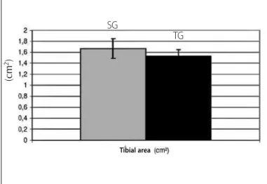
The tibial evaluations obtained at the end of the experiment such as: tibial weight (mg); tibial length (mm) and tibial area (cm²) are presented in table 5. Tibial length (mm) and tibial area (cm²) can also be observed in figures 4 and 5, respectively.
Table 5. Tibial evaluations obtained at the end of the experiment.
Group Tibial weight
(mg) Tibial length (mm)
Tibial area (cm²)
SG 662.38 ± 75.23 42.1 ± 0.12 7.74 ± 0.36
TG 703.4 ± 63.59 68.27 ± 13.71* 7.3 ± 0.4*
Values expressed in mean ± standard deviation. * indicates diference between groups (Student’s t test, p < 0.05). SG = sedentary group, TG = trained group.
Figure 4. Evaluation of the tibial length of the experimental groups.
SG
TG
(mm)
mL/100g b
.w
.
Tibial length
* indicates difference between groups (Student’s t test, p < 0.05). *indicates difference between groups (Student’s t test, p < 0.05)
212
DISCUSSION
The results indicate some specific details in the aerobic training effects in weaning rats. The records of water intake weekly followed demonstrate an adaptive compensation (table 1; figure 1), with mean water intake higher in TG (21.4 ± 2.0 mL/100 g of rat) than in SG (10.4 ± 3.0 mL/100 g of rat). Mean food intake of TG was also higher (TG = 26.3 ± 4.3 g/100 g of rat; SG = 18.4 ± 6.3 g/100 g of rat), demonstrating also food compensation due to increased energy demand by training (table 1; figure 2). Such fact can be confirmed by the absence of significant difference between groups concern-ing men body weight (table 1), related to the entire experimental period (TG = 276.50 ± 53.67 g; SG = 325.01 ± 76.43 g), which can have occurred due to the continuous and progressive increase of energy cost and reposition, with greater food intake by the trained rats27.
The hematocrit (%) analyzed presented significant difference, with higher value for TG (54.63 ± 1.41) than for SG (49.50 ± 1.65), which may represent addition to the red globules amount in response to physical training, in order to make the gas transport more efficient by the breathing process (table 2; figure 3). The to-tal leukocytes count (TG = 13.331 ± 2.822 cells/mm3 of blood; SG = 12.280 ± 2.689 cells/mm3 of blood) did not resent statistically significant difference (table 2), the same way there was no differ-ence in the differential count (table 3), indicating that there was no training influence on the immunological system concerning the parameters evaluated. Concerning some serum parameters, there were some important alterations (table 4), especially concerning rats in the developmental period under consideration. Total cho-lesterol presented relevant decrease in TG (TG = 68.27 ± 13.71 mg/ dL; SG = 94.44 ± 28.09 mg/dL), which refer back to regular aerobic training efficiency as a prevention factor for coronariopathies also in weaning rats. The total proteins levels also presented important decrease (TG = 7.30 ± 0.40 g/dL; SG = 7.74 ± 0.36 g/dL), reflecting the tissue anabolism to which the TG is submitted, with decrease of the circulating protein, which is more remarkably absorbed by the tissues28,29. Concerning the glucose levels (TG = 149.44 ± 22.83 mg/dL; SG = 130.22 ± 21.80 mg/dL) and triglycerides (TG = 284.53
± 77.29 mg/dL; SG = 249.70 ± 68.66 mg/dL), they did not present significant differences. Due to the glucose importance in the me-tabolism of the nervous system, it was already expected that the glycemic level could remain under significant alterations, since it is usually regulated within very strict thresholds. Concerning the triglycerides, the standard aerobic training programs usually do not cause any important influence on the serum levels of triglycerides29. When the hepatic glycogen (SG = 3.31 ± 0.76 mg/100 mg; SG = 3.24 ± 0.93 mg/100 mg) and muscular levels are analyzed (TG = 7.54 ± 1.93 mg/100 mg; SG = 6.03 ± 2.13 mg/100 mg), no signifi-cant differences have been found; however, observing the results for muscular glycogen, higher value for the trained group can be observed, indicating tendency to increase. More individualized load, considering for instance, the anaerobic threshold of rats, would pos-sibly present significant difference, since it is already well reported in the literature that regular training produces differences in the muscular glycogenic levels30.
Concerning aspects related to the bone tissue, when analyzing table 5, we can confirm that bone weight did not demonstrate signi-ficant difference between groups. Bone length (figure 4) presented important alteration, tibial length of TG (40.00 ± 0.14 mm) was lower than for CG (42.10 ± 0.12 mm). Tibial area (figure 5) demonstrated lower value for TG (1.53 ± 0.12 cm²) than for SG (1.67 ± 0.18 cm²); however, the difference was not statistically significant. The data suggest that the training applied to the rats was not able to bring alterations concerning bone structure, even if only considering that tibial length presented statistically significant difference. Since the rats were under a developmental period of accelerated metabolism, the data indicate that the training protocol may have negatively contributed to the bone growth of the young rats, reflecting in bone weight, which presented tendency to increase in rats submitted to physical training, and also in the tibial area, which presented tendency to reduction in the TG. It can be expected that higher bone density may have occurred as a result in the trained rats, which acquired more compact bones and hence of tibial smaller area and length, but with higher bone weight.
CONCLUSION
Regular physical training is able to produce some specific physiological alterations in young rats, especially concerning he-matological, metabolic and bone structure aspects. Therefore, the prescription of exercises which meet these developmental pe-culiarities in the maturation stage under consideration becomes necessary, since some specific training volume and intensities may be able to both positively or negatively affect those that practice regular training. The training protocol applied interfered in the he-matocrit and normal levels of proteins and cholesterol, and in the latter parameter, exercise is an important resource to fight coro-nariopathies also in young rats. Additionally, it can interfere in the bone morphology, being able to prevent normal bone growth in weaning rats.
All authors have declared there is not any potential conlict of interests concerning this article.
Figure 5. Evaluation of the tibial area of the experimental groups.
SG
TG
(cm
2)
REFERENCES
1. Guy JÁ, Micheli LJ. Strenght training for children and adolescents. J Am Acad Orthop Surg 2001;9:29-36.
2. Silva CS, Teixeira AS, Goldberg TBL. O esporte e suas implicações na saúde óssea de atletas adolescentes. Rev Bras Med Esporte 2003;9:426-31.
3. Le Boulch J. Desenvolvimento psicomotor: do nascimento até os 6 anos. 2a. ed. Porto Alegre: Art-med, 2000.
4. Bouchard CRM, Pérusse L. Genetics of fitness and physical performance. Champaigne: Human Kinetics Publishers, 1997.
5. Weineck J. Biologia do Esporte. 7a. ed. São Paulo: Manole, 2005.
6. Georgopoulos N, Markou K, Theodoropoulou A, Paraskevopoulou P, Varaki L, Kazantzi z, et al. Growth and pubertal development in elite female rhythmic gymnasts. J Clin Endocrinol Metab 1999;84:525-30. 7. Hogan KA, Gross RH. Overuse injuries in pediatric athletes. Orthop Clin North Am 2003;34:405-15. 8. Mackelvie KJ, Khan KM, Mckay HA. Is there a critical period for boneresponse to weight-bearing exercise
in children and adolescents? Br J Sports Med 2002;36:250-7.
9. Pettersson U, Nordström P, Alfredson H, Henriksson-larsen K, Lorentzon R. Effect of high impact activity on bone mass and size inadolescent females: a comparative study between two different types of sports. Calcif Tissue Int 2000;67:207-14.
10. Bass SG, Pearce M, Hendrich E, Delmas PD, Harding A, Seeman E. Exercise before puberty may confer residual benefits in bone density in adulthood: studies in active prepubertal and retired female gymnasts. J Bone Miner Res 1998;13:500-7.
11. Parker AW. Physical activity and skeletal health in children. In: Chan KM, Micheli LJ, editors. Sports and Children. Hong Kong: Williams and Wilkins, 1998;17-38.
12. Turner CH, Robling AG. Designing exercise regimens to increase bonestrength. Exerc Sport Sci Rev 2003;31:45-50.
13. Oeppen RS, Jaramillo D. Sports injuries in the young athlete. Top Magn Reson Imaging 2003;14:199-208.
14. Rodnick KJ, Haskell WL, Swislocki AL, Foley JE, Reaven GM. Improved insulin actin in muscle, liver and adipose tissue in physically trained human subjects. Am J Physiol 1987;253:E489-95.
15. Dengel DR, Hagberg JM, Pratley RE, Rogus EM, Goldberg AP. Improvements in blood pressure, glucose
metabolism, and lipoprotein lipids after aerobic exercise plus weight loss in obese, hipertensive middle-aged men. Metabolism 1998;47:1075-82.
16. Luciano E, Carneiro EM, Carvalho CR, Carvalheira JB, Peres SB, Reis MA, et al. Endurance training improves responsiveness to insulin and modulates insulin signal transduction through the phosphatidylinositol 3-Kinase/ Akt-1 pathway. Eur Endocrinol 2002;147:149-57.
17. Gomes RJ, Caetano FH, Mello MAR, Luciano E. Effects of chronic exercise on growth factors in diabetic rats. J Exerc Physiol Online 2005;8:16-23.
18. Eichner ER. Contagious infections in competitive sports. Sports Sci 1995;8:1-4. 19. Nieman DC. Immunity in athletes: current issues. Sports Sci 1998;11:1-11.
20. Nieman DC. Exercise effects on systemic immunity. Immunol Cell Biol 2000;78:496-501. 21. Nieman DC. Exercise, infection and immunity. Int J Sports Med 1994;15:S131-41.
22. Pedersen BK, Rohde T, zacho M. Immunity in athletes. J Sports Med Phys Fitness 1996;36:236-45.
23. Nieman DC, Nehlsen-Cannarella SL, Markoff PA, Balklamberton AJ, Yang H, Chritton DB, et al. The effects of moderate exercise training on natural killer cells and acute upper respiratory tract infections. Int J Sports Med 1990;11:467-73.
24. Foster C. Monitoring training in athletes with reference to overtraining syndrome. Med Sci Sports Exerc 1998;30:1164-8.
25. Mackinnon LT. Chronic exercise training effects on immune function. Med Sci Sports Exerc 2000;32:S369-76. 26. Oliveira CAM, Rogatto GP, Luciano E. Efeitos do treinamento físico de alta intensidade sobre os
leucócitos de ratos diabéticos. Rev Bras Med Esporte 2002;8:219-24.
27. Borer KT. Characteristics of growth-inducing exercise. Physiol Behav 1979;24:713-20.
28. Prado ES, Dantas EHM. Efeitos dos Exercícios Físicos aeróbios e de força nas lipo-proteínas HDL, LDL e Lipoproteína (a). Arq Bras Cardiol 2002;79:429-33.
29. Mcardle WD, Katch FI, Katch VL. Fisiologia do exercício: energia, nutrição e desempenho humano. 5a. ed. Rio de Janeiro: Guanabara Koogan, 2003.