Letters to the Editor
Radiol Bras. 2016 Jan/Fev;49(1):56–64
56
0100-3984 © Colégio Brasileiro de Radiologia e Diagnóstico por Imagem
Letters to the Editor
Tracheobronchopathia osteochondroplastica: computed tomography, bronchoscopy and histopathological findings
Traqueobroncopatia osteocondroplástica: achados tomográficos, broncoscópicos e histopatológicos
Dear Editor,
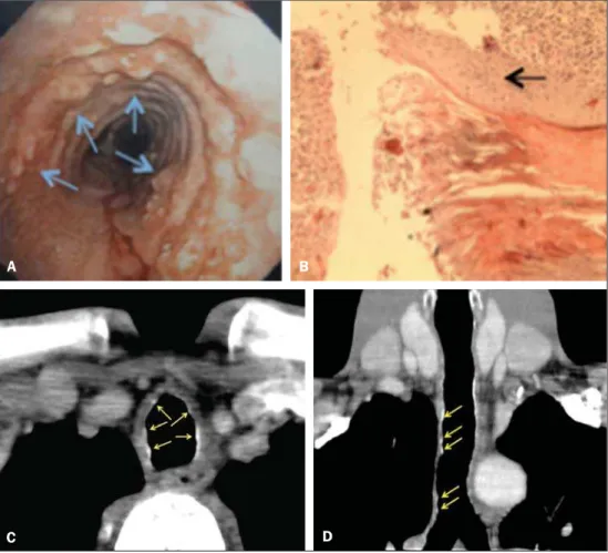
A 41-year-old man with history of recurrent airways infection since his childhood, with chronic coughing and voice hoarseness for seven years. The patient was referred to undergo laryngotra-cheobronchoscopy that revealed the presence of whitish nodular lesions on the anterolateral walls of the trachea and at the most proximal portion of the main bronchi, whose material was sent for histopathological analysis (Figures 1A and 1B). Computed tomog-raphy (CT) showed tiny, subcentimeter, submucosal, sessile nod-ules, some of them calcified, at the different levels of the trachea, with predominance in the two lower thirds of the trachea, and also in the right main bronchus. No significant luminal narrowing was observed and, typically, the posterior membranous wall of the tra-chea was spared (Figures 1C and 1D). The patient remains un-der clinical follow-up with management of symptoms.
Tracheobronchopathia osteochondroplastica is a rare chronic benign disease, with male prevalence (male:female = 3:1), and predominantly manifesting between the fifth and seventh decades of life(1,2). Association with several factors has been reported, as follows: chronic infections; chemical or medicamentous agents; degenerative tissue alterations; calcium and phosphorus metabo-lism disorders; and amyloidosis(3,4).
The disease is generally asymptomatic(1,2,5,6), and therefore, in most cases, the diagnosis is based on incidental findings at
bronchoscopy performed to investigate other diagnoses or with therapeutic purposes, or even in series of necropsy(1). In cases of symptomatic disease, cough is the main finding, present in about 66% of cases.
Generally, laryngotracheobronchoscopy raises the diagnostic suspicion and the classical finding is the presence of whitish, smooth and hard nodules, typically on the cartilaginous walls of the tra-cheal rings and of the proximal portions of the primary bronchi(7,8). The CT contributes to confirm the diagnosis(4) on the basis of its findings, namely, thickening of the inner surface of the tra-cheal cartilage with irregular, sessile nodular lesions, either calci-fied or not, focal or diffuse, sparing the posterior (membranous) trachea and leading to luminal narrowing in the affected ar-eas(1,5,6,8,9). CT is very sensitive to detect the typical calcification of the nodules, to define the extent and distribution of tracheo-bronchial stenosis, as well as to characterize complications such as atelectasis, bronchiectasis, postobstructive pneumonia(5,10).
Histopathological analysis shows that nodules correspond to submucosal osteocartilaginous growths. There are variable com-binations of fibrotic, cartilaginous, bone, hematopoietic tissue and mineralized acellular protein matrix. The epithelium lining such nodules may be normal, or present with inflammatory or meta-plastic appearance(5,8).
Some authors consider that bronchoscopic and radiological findings are sufficient to establish the diagnosis, particularly in cases where it is difficult to perform biopsy(1,5,8).
The prognosis is good in most of cases and treatment only will be requested in case of complications, principally tracheal and/ or bronchial stenosis(11).
Figure 1. A: Laryngotracheobronchoscopy: whitish nodules on the anterolateral walls of the trachea (arrows). B: Histopathology. HE, 20×. Foci of squamous metaplasia in the trachea (arrow). C,D: Axial, contrast-enhanced chest CT (C) and coronal refor-mation (D) showing micronodules, some of them calcified, on the anterolateral walls of the trachea (arrows).
A B
Letters to the Editor
Radiol Bras. 2016 Jan/Fev;49(1):56–64
57
http://dx.doi.org/10.1590/0100-3984.2014.0056 REFERENCES
1. Bioque JC, Feu N, Rubio JM, et al. Tracheobronchopathia osteochondro-plastica – clinical study and follow-up in nine cases. Journal of Bron-chology. 2001;8:78–83.
2. Pinto JA, Silva LC, Perfeito DJP, et al. Osteochondroplastic tracheo-bronchopathy: report on 02 cases and bibliographic review. Braz J Otorhinolaryngol. 2010;76:789–93.
3. Williams SM, Jones ET. General case of the day. Tracheobronchopathia osteochondroplastica. Radiographics. 1997;17:797–9.
4. Faig-Leite FS, Defaveri J. Traqueobroncopatia osteocondroplástica em portador de tumor de Klatskin: relato de caso e revisão da literatura. J Bras Patol Med Lab. 2008;44:459–62.
5. Sá JM, Almeida J, Amado J, et al. Traqueobroncopatia osteocondroplás-tica – experiência de uma unidade de broncologia. Rev Port Pneumol. 2002;VIII:329–39.
6. Webb EM, Elicker BM, Webb WR. Using CT to diagnose nonneoplas-tic tracheal abnormalities: appearance of the tracheal wall. AJR Am J Roentgenol. 2000;174:1315–21.
7. Prince JS, Duhamel DR, Levin DL, et al. Nonneoplastic lesions of the tracheobronchial wall: radiologic findings with bronchoscopic correla-tion. Radiographics. 2002;22 Spec No:S215–30.
8. Khan AM, Klapper P, Jain VR, et al. Tracheobronchopathia osteochondro-plastica: an entity diagnosed on bronchoscopy. Journal of Bronchology. 2006;13:99–101.
9. Kwong JS, Müller NL, Miller RR. Diseases of the trachea and main-stem bronchi: correlation of CT with pathological findings. Radio-graphics. 1992;12:645–57.
10. Marchiori E, Pozes AS, Souza Junior AS, et al. Alterações difusas da traquéia: aspectos na tomografia computadorizada. J Bras Pneumol. 2008;34:47–54.
11. Grenier PA, Beigelman-Aubry C, Brillet PY. Nonneoplastic tracheal and bronchial stenoses. Radiol Clin North Am. 2009;47:243–60.
Gabriela Maria Ribeiro e Ribeiro1, Marcelo Ricardo Canuto
Natal1, Eduardo Felipe Silva1, Sabrina Cardoso Freitas1,
Waldete Cabral Moraes1, Fernanda Cunha Maciel1
1. Hospital de Base do Distrito Federal (HBDF), Brasília, DF, Brazil. Mailing Address: Dra. Gabriela Maria Ribeiro e Ribeiro. Rua Gomes de Carvalho, 1005, ap. 3110, Vila Olímpia. São Paulo, SP, Brazil, 04547-004. E-mail: [email protected].
Giant pedunculated hemangioma of the liver
Hemangioma hepático gigante pedunculado
Dear Editor,
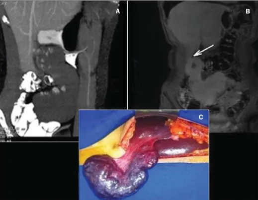
A previously healthy, 28-year-old woman presenting a palpable mass in the right hypochondrium for 3 years, evolving with local discomfort over the last 20 days. Ultrasonography (US) demon-strated an expansile mass best characterized by computed tomog-raphy (CT) and magnetic resonance imaging (MRI) which showed a well defined solid mass in continuity with the liver by a thin pedicle originating from the segment V and caudally extending towards the pelvis, measuring 18.0 × 9.4 × 5.2 cm, with features and pat-tern of enhancement suggestive of hemangioma (Figures 1A and 1B). Surgical resection was the treatment of choice because of the patient’s symptoms and the risks of torsion. The anatomo-pathological analysis confirmed the diagnosis (Figure 1C).
Hemangioma is the most common benign liver tumor(1–8), with a prevalence of 0.4–20% in necropsies(1,5–8). In most cases, hemangiomas are small, asymptomatic and incidentally found at imaging studies(1,2,5,6).
In spite of the lack of consensus about the dimensions to define a giant hemangioma, ranging from 4 to 10 cm according to the literature, it is known that the exophytic presentation, par-ticularly those pedunculated, are very rare (1–3,5,6). The first case was reported by Ellis et al. in 1985; and up to 2013, only 24 cases were described in the literature(1,4).
In almost 50% of cases, pedunculated hemangiomas are symptomatic at the diagnosis(1) and, likely any giant lesion, may determine compression of the intrahepatic biliary ducts, vascular structures or adjacent organs, manifesting with pain, early sati-ety, hemorrhage, jaundice, nausea and vomiting(1,2,5,6,8). Main complications include torsion due to a long and mobile pedicle,
Figure 1.A: Contrast-enhanced total abdominal CT (oral and intravenous contrast-enhancement), sagittal section, venous phase showing a well defined mass in the right hypochondrium/flank in continuity with the liver, presenting with a pattern of peripheral, globuliform and centripetal enhance-ment, with a thin pedicle originating from the seg-ment V. B: Coronal MRI, T1-weighted SPGR, at delayed phase showing homogenization of the lesion and identifying a pedicle contiguous with the liver parenchyma (arrow). C: Surgical speci-men of the reddish blue pedunculated lesion with cirrhotic appearance, showing pedicle contiguous with the liver parenchyma.
B