²Republication of the historical article of Diseases of the Colon & Rectum: Cutait DE, Figliolini FJ. A new method of colorectal anastomosis in
ab-dominoperineal resection. Diseases of the Colon & Rectum. 4(5):335-342, September/October 1961. Read at the meeting of the American Proctologic Society, Pittsburgh, Pennsylvania, June 21 to 24, 1961.
† Head of the group in charge of colorectal surgery (Professor Eurico da Silva Bastos). † † Assistant Professor..
* Corresponding author.
E-mail: [email protected] (A. da Luz Moreira)
2237-9363/$ - see front matter. © 2013 Elsevier Editora Ltda. All rights reserved. http://dx.doi.org/10.1016/j.jcol.2013.12.003
ABDOMINOPERINEAL rectosigmoidectomy is, at present, considered the operative method of choice for treatment of megacolon. This operation is based on the pathogenic ba-sis of dyskinesia of the distal portion of the intestine. Ac-cording to this theory, dilatation of the sigmoid lexure re-sults from lack of the propulsive function of the rectum and sometimes also of the sigmoid lexure, due to defective con-traction of its muscular ibers. The defective concon-traction is caused by agenesia of the Auerbach’s myenteric plexus in Hirschsprung’s disease and by inlammatory lesions in the acquired megacolon that cause partial or total destruction of the Auerbach’s myenteric plexus. To remedy this condi-tion, removal of the diseased, as well as the dilated portion of colon, with immediate re-establishment of bowel conti-nuity by colorectal anastomosis, seems therefore to be a ra-tional surgical procedure. This is why rectosigmoidectomy has been performed by most experienced surgeons all over the world. Nothwithstanding its effectiveness, there are some restrictions owing to the high incidence of postopera-tive complications, most of which are due to disruption, of varying degrees, of the colorectal anastomosis. In Hirsch sprung’s disease disruption is rather rare, but in acquired megacolon it is encountered in a large number of cases. In Raia and Haddad’s1 158 patients with acquired megacolon who underwent rectosigmoidectomy, this complication was encountered in 67 cases (42.4 per cent) and in our series of 222 patients it was noted in more than 30 per cent. In our
History article
A new method of colorectal anastomosis in
abdominoperineal resection
²Um novo método de anastomose colorretal em ressecção
abdominoperineal
Daher E. Cutait
†, Felipe José Figliolini
† †Department of Surgery, Medical School of the University of São Paulo, São Paulo, Brazil
experience, proximal colostomy, may reduce the hazard of disruption, but it does not prevent it.
Leakage of the anastomosis is usually accompanied by infec-tion of the presacral space. In such cases purulent discharges should be drained through a rubber tube introduced through a stab wound at the anococcygeal line or they may be drained into the peritoneal cavity. This complication may be followed by an infected perineal stercoral istula or peritonitis, respectively.
When disruption of the anastomosis produces a sinus, healing occurs spontaneously in the majority of cases. On a fair number of patients, however, it is necessary to perform correc-tive operations, such as enlargement of the perineal drainage area or istulectomy, either of which usually gives poor results. Obviously the results are worse when the disruption is larger. Stenosis, anal incontinence and permanent infection may fol-low disruption of the anastomosis, requiring the patient to continue permanently with the proximal colostomy.
Technic
Our new method of anastomosis is performed in two stages as follows:
First Stage: After dissection of the sigmoid lexure and the
rectum down to the level of the levator ani muscles and iden-tifying, with a cotton stitch, the appropriate level of division of the sigmoid lexure (Fig. 1) to be made during the perineal phase of the operation, an obturator of a sigmoidoscope is introduced through the anus and passed up to the proximal limit of the rectum (Fig. 2). The obturator is ixed at this level with a heavy silk ligature and used to apply gentle traction. This traction easily effects complete eversion or intussuscep-tion, through the anus, of the entire rectum and lower seg-ment of the sigmoid lexure (Fig. 3).
The everted rectal wall is grasped with an Allis clamp and an incision is made with scissors around the entire circumfer-ence of the rectum at a level approximately 2 to 3 cm. from the pectinate line (Figs. 4 and 5). The bowel is then pulled out through the anus as far as the level previously marked by the cotton stitch, which is situated about 6 cm. beyond the border of the everted rectum (Fig. 6). A heavy silk ligature is applied to the colon at this level and the colon is divided distal to it (Fig. 7).
The sigmoid lexure is then attached to the everted rec-tum by means of four stitches applied anteriorly, posteriorly and laterally (Fig. 8). The stitches are introduced into the se-romuscular coat of the sigmoid lexure and to the muscuIar
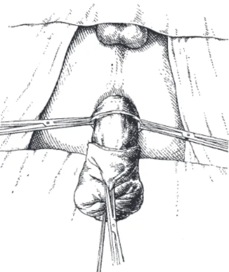
Fig. 2 - Obturator of a sigmoidoscope introduced through the anus and ixed at the proximal level of the rectum by a heavy silk ligature.
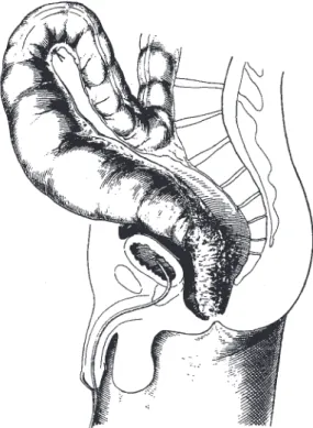
Fig. 1 - Sigmoidorectal segment dissected down to the anus in the abdominal phase of the operation. Stitch shows the level where the sigmoid is to be divided in the perineal phase of the operation.
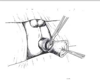
Fig. 4 - Allis clamp attached and incision of the rectal wall begun.
coat of the rectum. The two lateral stitches are then trans-ixed and attached to the perineal skin about 2.5 cm. from the pectinate line and the sigmoidorectal stump is covered with vaseline gauze (Fig. 9). This prevents retraction of this stump.
While the perineal phase of the operation is being per-formed, the assistants, who have remained at the abdomi-nal ield, reconstruct partially the pelvic peritoneal loor and close the abdominal wound. Maintenance of a com-munication between the pelvic and peritoneal cavities fa-cilitates drainage and absorption by the peritoneum of the residual blood in the pelvic cavity. The ixation skin stitches and the sigmoidal ligature are cut the next clay. The pulled-through colonic segment tllen starts to act as a temporary perineal colostomy.
Second Stage: The second stage is usually performed
seven to 10 days after the irst. The interval of seven to 10 d/tys is suficient for perfect adhesion between the mus-cular coat of the everted rectum and the serosal surface of the pulled-through portion of the colon (Figs. 10 and 11). This phase of the operation can be performed in 15 to 90 minutes and does not require anesthesia.
After the vaseline gauze dressing is removed, the colon is grasped with an Allis clamp and incised with a knife at the level of the border of the everted rectum, around its entire circumference. The incision is made as deep as the mucosa only (Fig. 12). The mucosa is dissected downward for about 0.5 cm. It is then divided with scissors aI[ around the distal limit o[ the dissection (Fig. 13). The mucosa o[ the rectum is then sutured to the mucosa of the sigmoid lex-ure with interrupted cotton sutlex-ures {Figs. 14, 15, 16 and 17). Upon completion o[ the anastomosis, the rectum is forced through the anus with the index inger (Figs. 18 and 19). The patient leaves the hospital two days later.
The rectosigmoidectomy and colorectal anastomosis just described was performed on 40 patients with acquired megacolon.
In six cases the operation was performed on patients with previous transverse colostomy performed simultane-ously with detorsion of volvulus of the sigmoid lexure.
In two cases transverse colostomy was performed si-multaneously with the rectosigmoidectomy and on the re-maining 32 cases no proximal colostomy was established. On the irst 20 patients of this series perineal drainage of the presacral space was established through a rubber tube drain. Due to the fact that in none of these cases did disrup-tion of the anastomosis occur, it was felt that this type of drainage could be abandoned. In such cases there was an accumulation of uninfected serosanguineous liquid in the presacral, space which could be drained into the peritoneal cavity. In order to facilitate this arrangement the peritoneal pelvic loor should be closed only partially.
Based on the experience acquired in the management of patients of this series, we perform, at present as a routine procedure, rectosigmoidectomy with the colorectal anasto-mosis that we have described, with- OUt establishment of a proximal colostomy and without perineal drainage of the presacral space. We feel that the results obtained by em-ploying the new technic of colorectal anastomosis justify the procedure.
Fig. 6 - Sigmoid pulled out through the anus to the level previously marked by the stitch.
Fig. 10- Appearance of the sigmoidorectal stump several days after the operation. The muscular coat of the everted rectum is irmly adherent to the serosal coat of the pulled-through portion of the bowel.
Fig. 11 - Sigmoidorectal stump on the ninth postoperative day. The mucosa of the everted rectum and the serosa of the pulled-through portion of the sigmoid are covered by a seroibrinopurulent exudate.The probe marks the pectinate line.
Fig. 12 - Incision of the sigmoid lexure at the level of the border of the everted rectum. The incision is made as deep as the mucosa and the mucosa is dissected downward for a distance of ½ cm.
Fig. 13 - Division of the mucosa of the sigmoid lexure. Fig. 9 - Transixation of the two lateral stitches to the
perineal skin.
Fig. 14 - Suture begun of the mucosa of the rectum to the mucosa of the sigmoid lexure with interrupted cotton sutures.
Fig.15 - Suture completed.
Fig. 16 - The sigmoidorectal stump, after completion of the suture (frontal view).
Fig. 17 - The sigmoidorectal stump, after completion of the suture (lateral view).
Fig.18 - Reduction of the everted rectum by forcing it in with the index inger.
Results
Disruption of the colorectal anastomosis occurred in only one case, an incidence of 2.5 per cent. This incidence of disruption is signiicantly lower than that noted in the conventional type of anastomosis (Table 1). The only disruption that occurred was in a patient who developed pseudomembranous enteritis after operation. In this case transverse colostomy had to be performed. Integrity of the suture line was demonstrated by endoscopic examination in all other cases.
patient underwent the new colon pull-through operation, but developed a serious presacral infection which required peri-neal drainage. After cessation of the drainage the patient died suddenly. Necrolz_sy revealed the presacral infection, but the actual cause of death could not be determined. Probably this was due to cardiac arrest which has been observed in some patients with Chaga’s disease.
In view of the gratifying results obtained with this new method of colorectal anastomosis, we employed it recently on ive cases requiring abdominoperineal rectosigmoidectomy for cancer of the upper rectum and rectosigmoidal area. No anastomotic leakage occurred in these patients.
Summary and conclusions
A new technic of colorectal anastomosis in abdominoperineat rectosigmoidectomy is presented.
Justiication of this technic is based on the principle of “adhesion by contact between the muscular surface of the everted rectum and the serosa surface of the pulled colon, and in the suture of the mucosa coat of the rectum to the mucosa coat of the colon.” The anastomosis is performed in two stages.
The results obtained on 40 patients with acquired mega-colon and on ive with cancer of the rectum and rectosigmoid are described.
There was a gratifying reduction in the incidence of dis-ruption of the anastomosis.
R e f e r e n c e
1. Raia, A. and J. Haddad: Complicações da retossigmoidectomia abdomino-perineal. Paper presented to the First Latin-American, Second International and Tenth Brazilian Congress of Proctology, S~o Paulo, Brazil, September 1960.
Table 1 – Disruption of colorectal anastomosis: comparative results.
Type of colorectal anastomosis
Cases, number
Number of cases of disruption of anastomosis
Percent
Primary colorectaI anastomosis
222 70 31.5
Primary-secondary 40 1 2.5
colorectal anastomosis
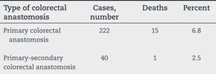
Table 2 – Mortality rate of colorectaI anastomosis: comparative results.
Type of colorectal anastomosis Cases, number Deaths Percent Primary colorectaI anastomosis
222 15 6.8
Primary-secondary 40 1 2.5
colorectal anastomosis
Infection of the presacral space was noted in three pa-tients. In the irst the infection was due to disruption of the anastomosis; in the second infection followed laceration of the vaginal wall that occurred while performing the dissec-tion incidentaI to removal of the rectum, and in the third case, infection occurred after a new pull-through procedure which was performed after the occurrence of necrosis of the intes-tine. As we have said, in only one case was infection related to imperfection of the anastomosis.
Stenosis of the anastomosis did not occur in any case. In some patients a diaphragmatic ring could be noted early in the postoperative period. However, these contractures subsid-ed in all cases in about two or three weeks. Almost all patients had anal incontinence during the early period after operation, the bowel movements numbering ive to 15 daily. This trouble, however, subsided within a few weeks.