Arq Bras Cardiol 2001; 77: 103-6.
Scorsin & Souza Cellular transplantation in heart failure
103 Clínica Cardiológyca C. Costantini - Curitiba
Mailing address: Marcio Scorsin - Clínica Cardiológyca C. Costantini - Rua Pedro Collere, 890 – Curitiba, PR - 80320-320 - e-mail: [email protected] Recebido para publicação em 12/2/01
Aceito em 28/3/01
The technique of transplanting muscle cells into the myocardium has been developed over the past few years in different animal models, with the aim of repopulating the impaired cardiac muscle with functional tissue. A clinical trial is now underway in France to assess the real benefit of this new therapy.
Heart failure is becoming a major public health pro-blem, and considering the increased longevity rates of the world population, it is expected to be the prevailing cardiac disease of the next century. This is basically caused by the progress in cardiology, which has reduced mortality from acute myocardial infarction, and has been interpreted as an “ironic failure of success”. When this disease becomes re-fractory to pharmacological therapy, available surgical treatments appear, the most radical being the replacement of the entire failing organ, as in cardiac transplantation. Howe-ver, this technique has some limitations inherent to the pro-cedure, such as organ shortage and graft vasculopathy. We must consider the fact that this treatment option cannot be proposed to all potential candidates. The intrathoracic implantation of circulatory assist devices is still primarily considered as a bridge to transplantation in the most critical-ly ill patients. Another interesting option is xenotransplan-tation, which is still confined to the experimental stage, in particular because of the difficulty in adequately addressing major safety issues.
Among the less aggressive surgical techniques pro-posed 1, the great majority attempt to change the geometry
and the dilatation of the left ventricle. To maintain cardiac output, the left ventricle increases its volume loosing its normal oval shape and acquiring a rounded aspect. Resha-ping the left ventricle could be very attractive considering the Starling law, but it does not correct the underlying disea-se that triggered the remodeling process, that is cardiomyo-cyte death, often responsible for the development of pro-gressive congestive heart failure.
Marcio Scorsin, Luiz Cesar Guarita Souza
Curitiba, PR
Cellular Transplantation for the Treatment of Heart Failure.
State of the Art
Editorial
With the objective of increasing ventricular mass, Leriche and Fontaine 2 proposed, in an experimental study
to wrap the heart with a skeletal muscle, the latissimus dorsi. Five decades later dynamic cardiomyoplasty was born 3 and
was used as an option for to restoring myocardial contracti-lity. However, it has yielded rather inconsistent results basi-cally owing to skeletal muscle atrophy.
In this setting, over these past years, cellular transplan-tation has emerged as an attractive alternate therapy for severe heart failure. This approach was encouraged by the re-cognition that normal myocardium could be successfully co-lonized by a variety of contractile cells. The engraftment of cardiomyocytes has been demonstrated by injecting cells taken from transgenic mice expressing the β-galactosidase gene 4,
which could then be identified by specific staining. Likewise, the presence of allogeneic-dystrophin-positive cells in the hearts of dogs suffering from Duchenne muscular dystrophy, which is characterized by a lack of dystrophin, has brought additional evidence for the capacity of transplanted cells to be harbored and integrated into the recipient myocardium 5 within
the cardiac tissue, as demonstrated by the formation of intercalated discs between host and recipient cells 4. Finally,
fetal cardiomyocytes have been shown to be able to survive in the bordering zone of a myocardial infarction 6 opening the
possibility of colonizing ischemic myocardium.
In a second step, different studies have assessed whe-ther cellular transplantation could effectively improve the function of ischemically diseased myocardium. A positive answer to this question has been achieved by Li and cowor-kers 7 who have shown, in a rat model of cryoinjury-induced
myocardial infarction, that the intramyocardial injection of fetal cardiomyocytes improved systolic and diastolic function up to 2 months after transplantation, as assessed by ex vivo Langendorff perfusion studies. To this end, Scorsin and coworkers 8 developed a protocol of
104
Scorsin & Souza
Cellular transplantation in heart failure
Arq Bras Cardiol 2001; 77: 103-6.
Another important issue was to determine whether functional benefits of cellular transplantation, documented in a model of regional ischemia, could be extended to the set-ting of global heart failure. To this end, in a mouse model of anthracycline-induced toxic cardiomyopathy 9, it was
possi-ble to show that injected fetal cardiomyocytes also impro-ved cardiac function 1 month after transplantation, as com-pared with control nontransplanted animals.
The advantage of fetal cardiomyocytes as cellular grafts is their capacity of entering the cell cycle and developing connections with host cells, an essential condition for func-tional improvement. Nevertheless, availability issues, ethi-cal problems related to the fetal source of these cells, and, mainly, the necessity of immunosuppressive therapy are real limitations for a potential clinical application of this allograft technique. Because of the previously mentioned drawba-cks associated with the use of fetal cells, the search for al-ternate cellular types was then refocused on the idea of a clinical use of autografts. Among the cells with the greatest therapeutic potential, bone marrow stromal cells and skele-tal myoblasts appear to be the most promising.
Bone marrow cells
Bone marrow contains multipotential progenitor cells (mesenchymal stem cells) that are in an undifferentiated state with a highly proliferative capacity. With the use of in vitro chemical induction, they can differentiate into bone, ten-don, fat, and muscle. In a very complex culture procedure 10 in
which cells were exposed to 5-azacytidine, 30% of them we-re able to change their initial stem form into cardiomyocyte-like cells, including the presence of intercalated disks and myotubes. Some studies showed 11 that stromal bone
mar-row stem cells, without in vitro muscular differentiation, transplanted in a normal myocardium could undergo “ mili-eu dependant ” differentiation and express cardiogenic phenotype. However, in a cryoinjury model of myocardial in-farction in rats 12, only previously in vitro chemically
in-duced muscular differentiated cells were able to improve function 2 months after transplantation. Despite this very interesting and promising option to restore myocardial via-bility, use of bone marrow stem cells raises two important concerns : 1) The multiplication potentiality and production of a very large number of differentiated cells in vitro has not yet been demonstrated. Cell duplication and reproduc-tion is the first condireproduc-tion to fully recolonize the diseased myocardium and thus improve ventricular function; 2) A risk is present of developing other types of tissue, especial-ly if undifferentiated stem cells are used. If the hypothesis that stromal cells can undergo phenotypic changes accor-ding to the histologic characteristic of the host tissue is legi-timate, it sounds logical to imagine that they could be diffe-rentiated into fibroblasts instead of cardiomyocytes when transplanted in a fibrotic scar, consequently reducing the therapeutic spectrum of this type of procedure.
Skeletal myoblasts (satellite cells)
The precursor of skeletal muscle fiber, the myoblast 13,
is present in adult mammals as a quiescent cell and may acti-vate, proliferate, and differentiate upon muscle injury (in vivo) or following tissue dissociation (in vitro) in culture.
Working on primary myoblast transplantations in a cry-oinjury model of myocardial infarction in dogs, Chiu et al. 14
were able to characterize the donor cells in the myocardium 14 weeks after injection. In this study, they identified myo-blasts showing a “milieu dependant” differentiation, these cells having lost their characteristic skeletal morphology, acquiring a cardiac-like phenotype. Murry et al. 15 observed
the formation of myotubes and skeletal muscle fibers within the cardiac tissue, but they were unable to identify cardiac-specific markers within the tissue formed by the injection of myoblasts. More recently, Taylor et al. 16 obtained
functio-nal improvement, as assessed by sonomicrometry, by trans-planting myoblasts in a cryoinfarcted rabbit heart model where skeletal muscle cells were found within the scar tissue following transplantation. Of note is the fact that functional improvement was only seen in those animals in which implanted cells were histologically identified, thereby providing strong evidence for a causal relationship betwe-en the presbetwe-ence of betwe-engrafted cells and the functional outco-me. In a myocardial infarction model in rats created by coro-nary artery ligation, Scorsin et al. 17 documented a
signifi-cant improvement in function, primarily manifested as a limi-tation of the postinfarction ventricular remodeling, compa-red with nontransplanted controls. Transplanted cells could be identified by a positive staining for embryonic myosin-heavy chain (which is specific for skeletal myo-blasts), and no gap-junctions were detected between trans-planted cells as expressed by the negative staining of the connexin-43, which is consistent with the study by Murry et al. Performing the same experiment 17, Scorsin et al. 17
compared the functional outcome between fetal cardiomyo-cytes and skeletal myoblasts, the latter being as effective as fetal cardiomyocytes in improving the function of the in-farcted myocardium. This was a very important datum con-sidering that the fetal cardiomyocyte remains as the stan-dard cell in terms of restoring cardiac function. The fact that myoblasts were functionally equivalent opened the way for using them as autografts in an attempt to restore myocardial viability in humans.
After many years of preclinical research studies, the French health authorities have approved the undertaking of a phase 1 trial concerning myoblast transplantation in patients with severe heart failure. The results obtained in the first clinical case 18 were very promising. After
Arq Bras Cardiol 2001; 77: 103-6.
Scorsin & Souza Cellular transplantation in heart failure
105
more resistant to fatigue. The fact that grafted myoblasts were able to convert the fiber isoform may indirectly indi-cate that they were contracting. However, if they do not form any cardiac-specific junction as shown in some expe-rimental studies 15,17, one possible hypothesis to explain
this paradox is that they probably contract in reaction to a mechanical stimulus instead of the classic electric stimula-tion through intercalated disks.
Another important point to be stressed is the ideal number of cells to be transplanted. Based on an experi-mental study in rats 19, a direct relationship was shown
between the number of cells and the improvement in func-tion. Considering the clinical case mentioned above 18, it
appears that the number of cells required to restore the viability of a medium-sized myocardial infarction (loss of 20-30% of left ventricle mass) is approximately 1 billion cells, which take 2 weeks to grow in a myogenic-specific culture medium, thus precluding the use of this therapy in an emergency setting.
Finally, looking at heart failure natural history and the severity of the remodeling process intensified over time, the optimal period for patients to undergo cellular transplanta-tion still needs to be determined. In all animal models, cellu-lar transplantation has been used with success early after the myocardial infarction, before end- stage forms of heart dysfunction appear. Perhaps one of the main advantages of this therapy is to stop the left ventricular remodeling pro-cess, when performed in a medium- dilated left ventricle, allo-wing consequently the hypertrophy of the remaining viable cardiomyocytes in association with the new engrafted ske-letal muscle to improve heart function.
In summary, it is important to emphasize the consistency of these experimental data. Regardless of the species (rat 8,
mouse 9, rabbit 16); the type of contractile cells (fetal
car-diomyocytes 7,16, skeletal myoblasts 16,17); the model (coronary
artery ligation 18, cryonecrosis 7, anthracycline-induced global
cardiomyopathy 9) and the assessment method (Langendorff
perfusion 7, sonomicrometry 16, echocardiography 6), cellular
transplantation has consistently resulted in a definite im-provement of function, which provides a strong rationale for the use of this technique in humans. The phase 1 clinical trial (in which 9 patients will be treated) is being undertaken in France with the aim of demonstrating the feasibility and the safety of the procedure. If no hazardous side effects are detected, then a large multicenter study will be initiated to assess the real place of this therapy in the heart failure thera-peutic armamentarium.
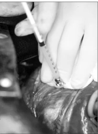
Fig. 1 - Myoblast transplantation in a patient with a 2-year-old myocardial infarction. Injection of culture medium containing150x10 6 cells/mL into the
infarcted area.
marked increase in the metabolism in the previously nonvia-ble area demonstrated by PET.
Perhaps this functional improvement needs to be cau-tiously analyzed considering that 2 associated coronary artery bypass surgeries were performed on the left side of the heart. But the eminent fact of this report is the feasibility of a large-scale production of autologous myoblasts and their successful surgical engraftment, confirmed by the new-onset metabolic activity in the previously dead zone.
Many questions still need to be clarified, however. Do skeletal myoblasts become fatigue resistant? What is their life span after transplantation? Do they form any kind of coupling with host cardiomyocytes? Do they acquire a cardiac-like phenotype? Researchers are trying to answer some of these questions. It has been demonstrated that 2 to 7 weeks after transplantation, 15 myoblasts from
fast-fiber isoforms convert into slow-twitch muscle, which is
1. Batista R, Verde J, Nery P, et al. Partial ventriculectomy to treat mid-stage heart di-sease. Ann Thorac Surg 1997; 64: 634-8.
2. Leriche R, Fontaine R. Essai expérimental de traitement de certains infarctus du myocarde et de l’anévrysme du coeur par une grffe de muscle strié. Bull Soc Nat Chir 1933; 9: 229-32.
References
3. Carpentier A, Chachques J. Myocardial stimulation with a stimulated skeletal muscle: first successfull clinical case (letter). Lancet 1985; 1: 1267. 4. Soonpaa MH, Koh GY, Klug MG, Field LJ. Formation of nascent intercalated
106
Scorsin & Souza
Cellular transplantation in heart failure
Arq Bras Cardiol 2001; 77: 103-6.
5. Koh G, Soonpaa M, Klug M, et al. Stable fetal cardiomyocyte grafts in the hearts of dystrophic mice and dogs. J Clin Invest 1995; 96: 2034-2042.
6. Scorsin M, Marotte F, Sabri A, et al. Can grafted cardiomyocytes colonize periin-farction myocardial areas? Circulation 1996; 94: II-337-II-40.
7. Li R-K, Jia Z-Q, Weisel RD, et al. Cardiomyocyte transplantation improves heart function. Ann Thorac Surg 1996; 62: 654-61.
8. Scorsin M, Hagege A, Marotte F, et al. Does transplantation of cardiomyocytes im-prove function of infarcted myocardium? Circulation 1997; 96(suppl II): 188-93. 9. Scorsin M, Hagege A, Dolizy I, et al. Can cellular transplantation improve func-tion in doxorubicin-induced heart failure? Circulafunc-tion 1998; 98: II-151-II-6. 10. Makino S, Fukuda K, Miyoshi S, et al. Cardiomyocytes can be generated from
marrow stromal cells in vitro. J Clin Invest 1999; 103: 697-705.
11. Wang J, Shum-Tim D, Galipeau J, Chedrawy E, Eliopoulos N, Chiu R. Marrow stromal cells for cellular cardiomyoplasty: feasibility and potential clinical ad-vantages. J Thorac Cardiovasc Surg 2000; 120: 999-1006.
12. Tomita S, Li R-K, Weisel R, et al. Autologous ransplantation of bone marrow cells improves dameged heart function. Circulation 1999; 100(suppl II): II-247-II56.
13. Mauro A. Satellite cell of skeletal muscle fiber. J Biochem Cytol 1961; 9: 493-5.
14. Chiu RC-J, Zibaitis A, Kao RL. Cellular cardiomyoplasty: myocardial regenera-tion with satellite cell implantaregenera-tion. Ann Thorac Surg 1995; 60: 12-18. 15. Murry C, Wiseman R, Schwartz S, Hauschka S. Skeletal myoblast transplantation
for repair of myocardial necrosis. J Clin Invest 1996; 98: 2512-23.
16. Taylor D, Atkins B, Hungspreungs P, et al. Regenerating functional myocardium: improved performance after skeletal myoblast transplantation. Nat Med 1998;4:
929-33.
17. Scorsin M, Hagege A, Vilquin J-T, et al. Comparison of the effects of fetal
cardio-myocyte and skeletal myoblast transplantation on postinfarction left ventricular function. J Thorac Cardiovasc Surg 2000; 119: 1169-75.
18. Menasché P, Hagege A, Scorsin M, et al. Myoblast transplantation for heart failure. Lancet 2001; 357: 279-80.