Comparative Transcriptome Profiling of the
Early Infection of Wheat Roots by
Gaeumannomyces graminis
var.
tritici
Lirong Yang1, Lihua Xie1, Baoguo Xue1*, Paul H. Goodwin2, Xin Quan1, Chuanlin Zheng3, Taiguo Liu3, Zhensheng Lei4, Xiaojie Yang5, Yueen Chao4, Chao Wu1
1Institute of Plant Protection Research, Henan Academy of Agricultural Sciences, Henan Key Laboratory for Control of Crop Diseases and Insect Pests, IPM Key Laboratory in Southern Part of North China for Ministry of Agriculture, Zhengzhou, P. R. China,2School of Environmental Sciences, University of Guelph, Guelph, Canada,3Institute of Plant Protection, Chinese Academy of Agricultural Sciences, Beijing, P. R. China,
4Research Centre for Wheat, Henan Academy of Agricultural Science, Zhengzhou, P. R. China,
5Economic Crop Research Institute, Henan Academy of Agricultural Science, Zhengzhou, P. R. China
Abstract
Take-all, which is caused by the fungal pathogen,Gaeumannomyces graminisvar.tritici (Ggt), is an important soil-borne root rot disease of wheat occurring worldwide. However, the genetic basis ofGgtpathogenicity remains unclear. In this study, transcriptome se-quencing forGgtin axenic culture andGgt-infected wheat roots was performed using Illu-mina paired-end sequencing. Approximately 2.62 and 7.76 Gb of clean reads were
obtained, and 87% and 63% of the total reads were mapped to theGgtgenome for RNA ex-tracted fromGgtin culture and infected roots, respectively. A total of 3,258 differentially ex-pressed genes (DEGs) were identified with 2,107 (65%) being 2-fold up-regulated and 1,151 (35%) being 2-fold down-regulated betweenGgtin culture andGgtin infected wheat roots. Annotation of these DEGs revealed that many were associated with possibleGgt pathogenicity factors, such as genes for guanine nucleotide-binding protein alpha-2 sub-unit, cellulase, pectinase, xylanase, glucosidase, aspartic protease and gentisate 1, 2-dioxygenase. Twelve DEGs were analyzed for expression by qRT-PCR, and could be gen-erally divided into those with high expression only early in infection, only late in infection and those that gradually increasing expression over time as root rot developed. This indicates that these possible pathogenicity factors may play roles during different stages of the inter-action, such as signaling, plant cell wall degradation and responses to plant defense com-pounds. This is the first study to compare the transcriptomes ofGgtgrowing saprophytically in axenic cultures to it growing parasitically in infected wheat roots. As a result, new candi-date pathogenicity factors have been identified, which can be further examined by gene knock-outs and other methods to assess their true role in the ability ofGgtto infect roots. OPEN ACCESS
Citation:Yang L, Xie L, Xue B, Goodwin PH, Quan X, Zheng C, et al. (2015) Comparative Transcriptome Profiling of the Early Infection of Wheat Roots by
Gaeumannomyces graminisvar.tritici. PLoS ONE 10(4): e0120691. doi:10.1371/journal.pone.0120691
Academic Editor:Dongsheng Zhou, Beijing Institute of Microbiology and Epidemiology, CHINA
Received:September 23, 2014
Accepted:January 25, 2015
Published:April 14, 2015
Copyright:© 2015 Yang et al. This is an open access article distributed under the terms of the Creative Commons Attribution License, which permits unrestricted use, distribution, and reproduction in any medium, provided the original author and source are credited.
Data Availability Statement:All relevant data are included within the paper and its Supporting Information. Sequence data were deposited in the NCBI SRA database under the accession number SRP050157.
Introduction
Global wheat production is severely affected by take-all, a major fungal disease that is caused byGaeumannomyces graminisvar.tritici(Ggt), which can lead to yield losses of up to 40%-60% [1].Ggtis a necrotrophic pathogen infecting wheat roots via hyphae that can survive in the soil in root debris of wheat plants.Ggthyphae penetrate root cortical cells causing a root rot and then progress into the base of the stem, disrupting water flow. The result is stunting and premature death of the plant with symptoms of white heads, empty spikes, reduced grain panicles and reduced grain weight.Ggtis highly invasive on wheat roots, but has a wide host range, including wheat, triticale, barley and rye [2,3]. Hence, considerable effort has been ex-erted to understand the mechanisms underlyingGgtpathogenicity to help reduceGgt-caused wheat losses [4–8].
The ability of a soil-borne fungus, likeGgt, to cause disease on roots is affected by many fac-tors, such as how well it is able to colonize the root surface and infect roots with the use of ex-tracellular enzymes, toxins, and effectors. InGgt, some genes coding for extracellular enzymes have been linked to pathogenicity, such as laccases [7,9], endo-β-1,4-xylanase [4],β -1,3-glucanase [5],β-1,3-exoglucanase [10] and gentisate 1,2-dioxygenase-like enzyme [10]. For an-other soil-borne fungus,Fusarium, some genes encoding for proteins of the signaling pathway, such as mitogen-activated protein kinase [11–12], vacuolar Ca2+exchanger protein [13] and plasma membrane calcium ATPase, have also been implicated in pathogenesis through recog-nition and signal transduction of extracellular signals [14]
The mechanisms of pathogenicity ofGgtto wheat roots are still not well understood as most research on wheat–Ggtinteractions has focused on biological characteristics of the disease [15], pathogen distribution [16], pathogen genetic diversity [17], and the use of antagonists [18]. One approach to elucidating the mechanisms of fungal plant pathogenicity is to conduct large scale transcipt sequencing (seq analysis) of infected plant tissues. An advantage of RNA-seq is that the level of detection of transcript abundance enables broad measurements of ex-pression levels of transcripts without prior sequence knowledge. Recent RNA-seq studies on plant-pathogen interactions includeMagnaporthe oryzaeinfection of rice [19] andFusarium graminearuminfection of barley [20]. However, there have not yet been any RNA-seq studies ofGgt-plant interactions. The present study is an RNA-seq examination of the transcriptomes ofGgtin axenic culture andGgtin infected wheat roots using the Illumina GA IIx sequencing platform. This data was then used for de novo assembly of the reads into contigs, and then mapping the reads against the contigs to identify differentially expressed genes (DEGs) be-tween the two conditions.
Materials and Methods
Plant and fungal material and infection of roots
An isolate ofGgt(GGT-007, Henan Academy of Agricultural Sciences, Zhengzhou, China) was isolated from wheat root samples, This strain was identified by morphological characteristics, pathogenicity and molecular identification according to the methods of Quan.et al. [62] Liquid cultures were grown in potato dextrose broth at 25°C with shaking at 180 rpm for 5 d to pre-pare inoculum for wheat root infection andGgtRNA extraction.
Seeds of winter wheat (Aestivum triticumcv 'Zhengmai 366') were placed in 75% ethanol for 5 s, rinsed with sterile water three times, surface sterilized with 1% AgNO3for 9 min, and then rinsed again with sterile water five times. The seeds were germinated in sterile Petri dishes (12 cm diameter) with sterile filter paper and 5 mL of sterile water. After one day, the germinated wheat seedlings were transferred to another sterile Petri dish with sterile filter paper (50 seedlings
study design, data collection and analysis, decision to publish or preparation of the manuscript.
and 10 mL sterile water added per dish). After two days, the root lengths were 2 cm to 3 cm, and the seedlings were transferred to tissue culture vessels containingGgtinoculum (5 mg mycelium in 5 mL potato dextrose broth). All plants were grown in 16 h day/8 h night at 22°C.
Sampling and experimental design
Root samples of the control group from two biological replicates were collected at 1, 2, 3, 4, 5, 7, and 9 d after inoculation. The samples were immediately frozen and then stored in liquid ni-trogen until analysis. Total RNA was extracted from these materials using RNAiso Plus (Total RNA extraction reagent) (TaKaRa, Otsu, Japan). RNA purity was verified using a NanoPhot-ometer spectrophotNanoPhot-ometer (Implen, Westlake Village, CA, USA). RNA concentration was measured using a Qubit RNA Assay Kit in a Qubit 2.0 Fluorometer (Life Technologies, Grand Island, NY, USA). RNA integrity was assessed using the RNA Nano 6000 Assay Kit of the Bioa-nalyzer 2100 system (Agilent Technologies, Wilmington, DE, USA).
Library construction and sequencing for RNA-seq
Sequencing libraries were generated using 3μg of RNA per sample with a NEBNext Ultra RNA
Library Prep Kit for Illumina (NEB, Ipswich, MA, USA) following the manufacturer’s recom-mendations, and tags were added to each sample for identification. Briefly, mRNA was purified from total RNA using poly-T oligo-attached magnetic beads. Fragmentation was performed using divalent cations under elevated temperature in NEBNext First Strand Synthesis Reaction Buffer (5×). First-strand cDNA was synthesized using random hexamer primer and M-MuLV Reverse Transcriptase (RNase H-). Second-strand cDNA was synthesized using DNA polymer-ase I and RNpolymer-ase H. Remaining overhangs were converted into blunt ends via exonuclepolymer-ase/poly- exonuclease/poly-merase activities. After the adenylation of 30
ends of DNA fragments, NEBNext Adaptor with hairpin loop structure was ligated to prepare for hybridization. To select cDNA fragments of preferentially 150 bp to 200 bp in length, the library fragments were purified with the AMPure XP system (Beckman Coulter, Beverly, MA, USA). Then, 3μL of USER Enzyme (NEB, Ipswich,
MA, USA) was used with size-selected, adaptor-ligated cDNA at 37°C for 15 min and then at 95°C for 5 min before PCR. PCR was performed with Phusion High-Fidelity DNA polymerase, Universal PCR primers, and Index (X) Primer. The PCR products were purified (AMPure XP System, Brea, CA, USA), and library quality was assessed on the Agilent Bioanalyzer
2100 system.
Clustering and sequencing and quality control
The tag-coded samples were clustered on a cBot Cluster Generation System using the TruSeq PE Cluster Kit v3-cBot-HS (Illumia, San Diego, CA, USA) according to manufacturer’s instruc-tions. After cluster generation, the library preparations were sequenced on an Illumina Hiseq 2000 platform to generate 100 bp paired-end reads were generated. Raw data (raw reads) of fastq format were initially processed through in-house PERL scripts provided by Novogene (China). In this step, the clean reads were obtained by removing adapter sequences, reads with more than 10% N, and low-quality sequences (more than 50% of the reads having aphred base sQ5. The Q20, Q30, and GC contents of the clean data were calculated. All downstream analyses were based on the quality-trimmed clean data.
Read mapping to the reference genome
dna/). Index of the reference genome was constructed using Bowtie v2.0.6 [63], and paired-end clean reads were aligned to the reference genome using TopHat v2.0.9 (http://tophat.cbcb. umd.edu/) [64] with all parameters set to their default values.
Differential expression analysis
HTSeq v0.5.4p3 [65] was used to count the read numbers that were mapped to each gene. The Reads Per Kilobase of exon model per Million mapped reads (RPKM) of each gene was calculated based on the length of the gene and read counts that were mapped to this gene. RPKM simultaneously considers the effect of sequencing depth and gene length for the read counts, and it is currently the most commonly used method for estimating gene expression levels [22].
Differential expression analysis betweenGgtin culture andGgtin infected wheat roots was performed using the DESeq R package (1.10.1) using a model based on the negative binomial distribution [66]. The resulting P-values were adjusted using the Benjamini and Hochberg’s ap-proach for controlling the false discovery rate. Genes with an adjusted P-value<0.05 found by
DESeq were assigned as differentially expressed.
Gene Ontology (GO) and Kyoto Encyclopedia of Genes and Genomes
(KEGG) enrichment analysis, novel transcript prediction, and alternative
splicing analysis
GO enrichment analysis of DEGs was implemented by the GO seq R package [67], in which gene length bias was corrected. GO terms with corrected P value less than 0.05 were considered significantly enriched by DEGs. KEGG is a database resource for understanding high-level functions and utilities of the biological system [68], such as the cell, the organism, and the eco-system, from molecular-level information, particularly large-scale molecular datasets generated by genome sequencing and other high-throughput experimental technologies (http://www. genome.jp/kegg/). KOBAS software was used to test the statistical enrichment of DEGs in KEGG pathways. The Cufflinks v2.1.1 Reference Annotation Based Transcript assembly meth-od was used to construct and identify both known and novel transcripts from TopHat align-ment results. Alternative splicing events were classified into 12 basic types by Asprofile v1.0. The number of AS events in each sample was estimated.
Quantitative real-time PCR (qRT-PCR) validation
The expression levels of 12 DEGs were determined by qRT-PCR to confirm the results of mRNA-Seq analysis. Total RNA (1μg) fromGgtculture (5 d after inoculation) andGgt
Results and Discussion
mRNA-Seq general data analyses
RNA-seq expression profiling ofGgtwas performed under two conditions:Ggtcultured in po-tato dextrose broth (PDB) and symptomaticGgt-infected wheat roots. For pure cultures ofGgt
in PDB, the fungus was grown for 5 days as that is the time period used to prepare inoculum. GrowingGgtfor longer periods in PDB does not increase the level of disease when hyphae is used to inoculated plants. The samples forGgt-infected wheat roots were chosen at 7 days post-inoculation when symptoms of root rot were prevalent but tissue damage was not so se-vere and so extraction of high quality RNA was still possible. One biological replicate with paired-end was sequenced for each condition. A total of 30,305,754 and 83,222,108 raw reads resulting in 26,172,744 and 77,645,702 clean reads were generated from the RNA ofGgtin cul-ture andGgt-infected wheat roots, respectively (Table 1). The quality of each library was simi-lar with 95.86% and 94.88% of the raw reads from theGgtculture andGgt-infected wheat roots having quality values of Q20 and error probabilities of 0.05 and 0.06, respectively. The GC contents were almost identical for theGgtandGgt-infected wheat roots (59.71% and 58.30%, respectively).
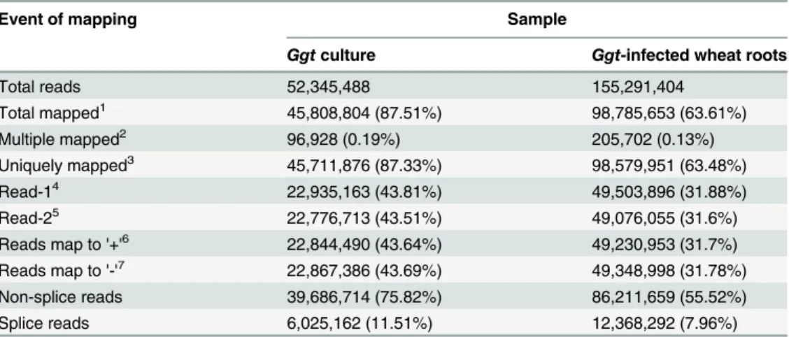
The sequenced reads were mapped to theGgtgenome. Among the transcripts from theGgt
culture andGgt-infected wheat roots, up to 87% and 63%, respectively, of the total reads (52,345,488 and 155,291,404) were uniquely mapped to theGgtgenome, whereas only small proportions (0.19% and 0.13%) were mapped to multiple locations in theGgtgenome (Table 2). Among the uniquely mapped reads, 94.5% were mapped to the genome over one exon, 5.1% over an intergenic region, and 0.4% over an intron. Reads mapped to intergenic re-gions occurred because some gene annotations were inadequate. All uniquely mapped reads were used to calculate reads per kilobase of exon model per million mapped reads (RPKM) val-ues, which were used to normalize expression levels. Alternative splicing (AS) events had oc-curred for 24,498 uniquely mapped reads from theGgtculture and 23,410 uniquely mapped reads from theGgt-infected wheat, and these were classified into 12 types using Asprofile v1.0 (S2 Table). The largest AS event groups under both conditions were TSS (alternative 5’first exon) and TTS (alternative 3’last exon) events.
Identification of differentially expressed genes (DEGs) between
Ggt
growing in culture and wheat roots
Log 2-fold DEGs betweenGgtculture andGgt-infected wheat roots were identified using DEG-Seq, and P-values were corrected by the Hochberg and Benjamini method [21].
Table 1. Transcriptome statistics of cDNA libraries from 5 day old culture ofGgtand 7 day oldGgt-infected wheat roots.
Sample name Raw reads1 Clean reads2 Cleanbases3 Error rate4(%) Q205(%) Q306(%) GC content7(%)
Ggtculture 30,305,754 26,172,744 2.62G 0.05 96.62 87.83 59.71
Ggt-infected wheat roots 83,222,108 77,645,702 7.76G 0.05 95.59 87 58.39
1The numbers of original data sequence 2The
filtered data sequence
3Qphred= - 10log 10(e)
4the sequence length multiplied by the number of sequencing 5The percentage of bases with a Phred value>20
6The percentage of bases with a Phred value>30 7The percentage of bases number of G and C
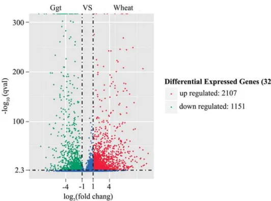
Corrected P-value of 0.005 and log2 (fold change) ±1 were set as thresholds for significant dif-ferential expression. A total of 3,258 DEGs were detected between theGgtculture and infected wheat root libraries, with 2,107 up-regulated genes and 1,151 down-regulated genes in theGgt -infected roots compared to theGgtculture (Fig 1).
The DEGs were divided into three groups according the RPKM values as per Mortazavi et al. [22]. Genes with RPKM values between 0 to 3 were considered to be expressed at a low level, 3 to 15 were at a medium level and above 15 were at a high level (Table 3). The percentage of highly expressed genes was smaller in the control than in theGgtsamples, whereas the per-centage of low-level expressed genes was larger in theGgtculture than in theGgt-infected root samples. The Pearson correlation coefficient for the replicates calculated by log10RPKM was 1 and between theGgtculture and infected samples was 0.71, indicating reliable sequencing data (S1 Fig). A comparison of the RPKM distribution of the DEGs showed that the box plots of the log10 (RPKM+1) values and distribution of the density of the log10 (RPKM+1) values over-lapped between the samples from theGgtculture andGgt-infected roots indicating that the range of of the expression values in the two samples were generally similar, although there was a considerable spread in the expression levels of the DEGs (S2 Fig).
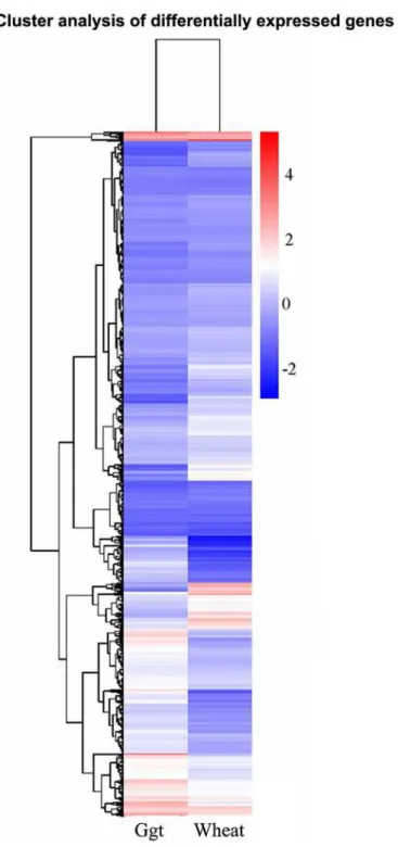
Hierarchical clustering of the DEGs according to the log10 (RPKM+1) values showed the overall gene expression pattern to be divided into several clusters based on the expression levels of the DEGs inGgtculture versusGgt-infected roots conditions (Fig 2). Only one relatively small cluster contained DEGs with very high expression levels in bothGgtculture andGgt -in-fected roots, whereas all the other DEGs formed a second cluster with multiple subclusters mostly showing medium to low expression levels under both conditions or slightly higher ex-pression under either theGgtculture orGgt-infected root conditions.
Table 2. Summary of mapping the sequenced reads to theGgtgenome from the 5 day old culture of
Ggtand 7 day oldGgt-infected wheat roots.
Event of mapping Sample
Ggtculture Ggt-infected wheat roots
Total reads 52,345,488 155,291,404
Total mapped1 45,808,804 (87.51%) 98,785,653 (63.61%)
Multiple mapped2 96,928 (0.19%) 205,702 (0.13%)
Uniquely mapped3 45,711,876 (87.33%) 98,579,951 (63.48%)
Read-14 22,935,163 (43.81%) 49,503,896 (31.88%)
Read-25 22,776,713 (43.51%) 49,076,055 (31.6%)
Reads map to '+'6 22,844,490 (43.64%) 49,230,953 (31.7%)
Reads map to '-'7 22,867,386 (43.69%) 49,348,998 (31.78%)
Non-splice reads 39,686,714 (75.82%) 86,211,659 (55.52%)
Splice reads 6,025,162 (11.51%) 12,368,292 (7.96%)
1Total number of reads mapped on theGgtgenome
2Total number of reads mapped to multiple locations inGgtgenome 3Total number of reads mapped to uniquely locations in theGgtgenome 4The two directions of the paired-end sequencing
5The two directions of the paired-end sequencing
6Total number of reads mapped to positive strand ofGgtgenome 7Total number of reads mapped to negative strand ofGgtgenome
Functional annotation and classification of DEGs
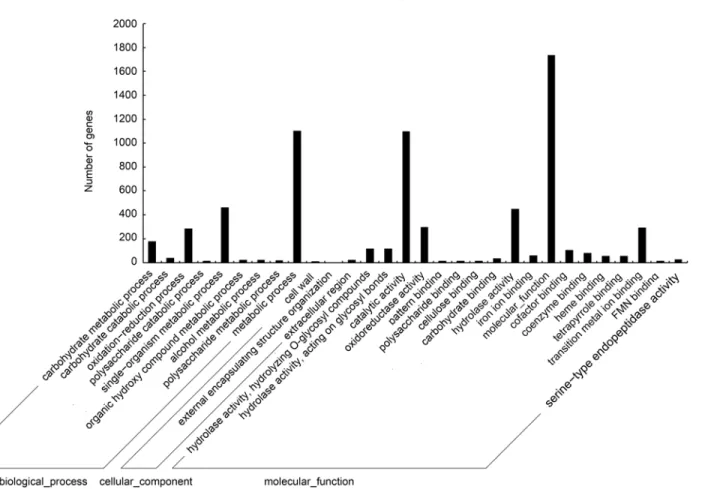
The GOseq R package was used to annotate and assign different functional GO categories to the DEGs ofGgtin infected wheat roots. The 3,258 significant DEGs between theGgtculture andGgt-infected wheat roots (q-value<0.05) belonged to 9 GO groups based on biological
process, 3 groups based on cellular component and 18 groups based on molecular function (Fig 3). For biological process, the dominant categories were metabolism process (GO: 0008152) with 1,101 DEGs, single-organism metabolism (GO: 0044710) with 461 DEGs, oxi-dation-reduction processes (GO: 0055114) with 285 DEGs and carbohydrate metabolic pro-cesses (GO: 0005975) with 176 DEGs. For cellular component, the three categories were extracellular region (GO: 0005576) with 25 DEGs, external encapsulating structure (GO: 0030312) with 14 DEGs and cell wall (GO: 0005618) with 12 DEGs. For molecular function, the largest categories were general molecular function (GO: 0008152) with 1,733 DEGs,
Table 3. Number of transcripts ofGgtculture andGgt-infected wheat roots at different expression level intervals.
Sample RPKM level
0~1 1~3 3~15 15~60 >60
Ggtculture 4068(26.90%) 2046(13.53%) 4337(28.67%) 2944(19.46%) 1730(11.44%)
Ggt-infected wheat roots 3462(22.89%) 1448(9.57%) 3755(24.83%) 4402(29.10%) 2058(13.61%)
RPKM levels were chosen based on divisions described in [22]
doi:10.1371/journal.pone.0120691.t003
Fig 1. Volcano of DEGs betweenGgtculture andGgt-infected wheat roots.Ggt:Ggt-culture, wheat:
Ggt-infected wheat roots; the y-axis corresponds to the mean expression value of log10 (p-value), and the x-axis displays the log2 fold change value. The red dots represent up-regulated DEGs, the blue dots represent down-regulated DEGs.
catalytic activity (GO: 0003824) with 1,099 DEGs, hydrolase activity (GO: 0016787) with.447 DEGs and transition metal ion binding (GO: 0046914) with 291 DEGs (Fig 3).
The biological pathways of the DEGs ofGgtwere mapped to the reference pathways in KEGG (http://www.genome.ad.jp/kegg/) [23]. The DEGs between theGgtculture andGgt
-Fig 2. Hierarchical clustering analyses of DEGs.According to the two sample’s log10 (RPKM+1), red indicates high expression of the gene, blue indicates low expression of genes. Ggt:Ggt-cultures, Wheat:Ggt -infected wheat roots.
infected wheat roots were assigned to 100 KEGG pathways (Fig 4). The pathways with the most significant representation were general metabolic pathways (mgr01100) with 242 mem-bers and biosynthesis of secondary metabolites (mgr01110) with 100 memmem-bers. These results indicate that fungal genes involved in the metabolism or biosynthesis of secondary metabolic pathways were being expressed more when the fungus was growing parasitically in wheat roots than growing saprophytically on culture medium, which may indicate an importance in the pathogenicity ofGgt.
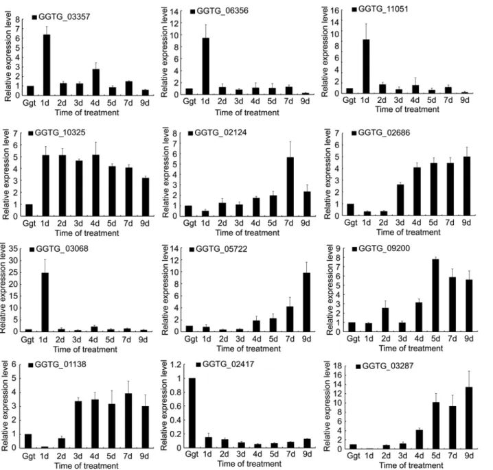
Expression of selected DEGs
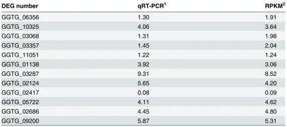
qRT-PCR of 12 selected DEGs was performed to validate the RNA-seq data. Eleven were up-regulated DEGs and one was a down-up-regulated DEG betweenGgtculture andGgt-infected wheat roots (Fig 5). A comparison of the ratio of the qRT-PCR expression and RPKM values for theGgtculture to the 7 dayGgt-infected wheat roots revealed that these were basically con-sistent indicating that the RNA-seq data were credible (Table 4).
qRT-PCR of the 11 selected up-regulated DEGs showed several patterns over the course of infection. Expression of GGTG_03357 (Adhesion and hyphal regulator 1), GGTG_06356 (Ad-enylate cyclase), GGTG_11051 (Calcium-binding protein) and GGTG_03068 (Protein scd2/
Fig 3. Functional annotation of DEGs based on gene ontology (GO) categorization.Each annotated sequence was assigned at least one GO term.
ral3) were all relatively high at 1 day of the infection followed by much lower levels for the rest of the infection comparable to that ofGgtin culture. This indicates that these DEGs may be particularly important for penetration or establishing infection. Expression of GGTG_02124 (Glucan 1,3-beta-glucosidase), GGTG_02686 (Linoleate 9S-lipoxygenase), GGTG_05722 (Xyloglucan-specific endo-beta-1,4-glucanase), GGTG_09200 (Exopolygalacturonase B), GGTG_01138 (Scytalone dehydratase) and GGTG_03287 (Endo-1,4-beta-xylanase A) were more highly expressed late in the infection indicating roles in the later stages of root coloniza-tion. However, those DEGs did not all have the same pattern of change over time with some, like GGTG_02124 (Glucan 1,3-beta-glucosidase) and GGTG_01138 (Scytalone dehydratase), generally gradually increasing over time, while others, like GGTG_03287 (Endo-1,4-beta-xyla-nase A), being only be expressed for a limited time late in infection. Expression of
GGTG_10325 (G2-specific protein kinase) was unique among the 11 selected up-regulated DEGs in that it remained relatively high throughout the infection compared to expression in culture, only slightly decreasing over time as the infection progressed. qRT-PCR of the one
Fig 4. Statistics of KEGG pathway enrichment.The y-axis corresponds to KEGG Pathway, and the x-axis shows the enrichment factor. The color of the dot represent q value, and the size of the dot represents the number of DEGs mapped to the reference pathways.
selected down-regulated DEG, GGTG_02417(Laccase-2), showed that its expression was rela-tively unchanged during infection with a slight decline in the mid-period of infection.
DEGs for signal transduction pathways
The signal transduction pathway in plant pathogenic fungi is essential for surface recognition, adaptation to the host milieu, appresorium formation, infection establishment, and invasive growth [24–33]. Several DEGs were identified from the GO annotation or KEGG analyses re-lated to Ca2+signaling, cyclic AMP- protein kinase A (cAMP-PKA), and mitogen-activated protein kinase (MAPK) pathways.
Fig 5. Relative expressions of selected DEGs inGgtculture andGgt-infected wheat roots.The x-axis shows the sample:Ggtculture, and infected wheat roots at 1 day, 2 days, 3 days, 4 days, 5 days, 7 days and 9 days post infection.
Among the up-regulated DEGs related to the Ca2+signaling pathway inGgt- infected roots versusGgtculture, there were two DEGs (GGTG_00202 and GGTG_02953) for vacuolar um ion transporter, three DEGs (GGTG_03594, GGTG_08053 and GGTG_08581) for calci-um-transporting ATPase, two DEGs (GGTG_04060 and GGTG_08412) for calcium channel protein, and one DEG (GGTG_11051) for calcium-binding protein (S3 Table). In contrast, there was only one down-regulated DEG in the infected roots related to Ca2+signaling (GGTG_02953) for vacuolar calcium ion transporter. Calcium ions are extremely important for signal transduction. Two important calcium mediators in eukaryotic cells are calmodulin and phosphatase, calcineurin. Calcineurin is required for fundamental biological events of pathogenic fungus, such as mating, morphogenesis and virulence [24–26]. Calcium transport-ers, such as vacuolar Ca2+exchanger (Vcx1), calcium-channel protein(Cch1), and plasma membrane calcium ATPase (Pmc1), are required for fungal virulence, supporting a role for cal-cium-mediated signaling in fungal pathogenesis [27]. For example, a knockout of theGgt Vcx1
significantly decreased the pathogenicity ofGgtto wheat roots [6], a knockout ofPmcA signifi-cantly reducedAspergillus fumigatusvirulence in invasive pulmonary aspergillosis of mice and a knockout ofPmcAaffected cation homeostasis and in cell wall integrity ofA.fumigatus[14]. qRT-PCR of the DEG (GGTG_11051) for calcium-binding protein showed high expression early in infection (Fig 5). Ca2+signaling may be more important forGgtin infected roots than inGgtculture because the fungus may need to identify and the change in the environment, par-ticularly early in the infection when switching from saprophytic growth in the soil to parasitic growth inside wheat roots.
Among the up-regulated DEGs related to the cAMP-PKA pathway inGgt-infected roots versusGgtin culture, there was one DEG (GGTG_02473) for guanine nucleotide-binding pro-tein alpha-2 subunit, one DEG (GGTG_06356) for adenylate cyclase, and one DEG
(GGTG_05905) for cAMP-independent regulatory protein. qRT-PCR analysis showed that GGTG_06356 expression peaked at 1 d post-infection, and then decreased to the level observed inGgtculture (Fig 5). The key components of the cAMP—PKA pathway include adenylate
Table 4. The ratio of the expression levels of 12 DGEs of 7 day oldGgt-infected wheat roots toGgt5 days in culture as determined by qRT-PCR versus RPKM values from RNA-seq.
DEG number qRT-PCR1 RPKM2
GGTG_06356 1.30 1.91
GGTG_10325 4.06 3.64
GGTG_03068 1.31 1.98
GGTG_03357 1.45 2.04
GGTG_11051 1.22 1.24
GGTG_01138 3.92 3.06
GGTG_03287 9.31 8.52
GGTG_02124 5.65 4.20
GGTG_02417 0.08 0.09
GGTG_05722 4.11 4.62
GGTG_02686 4.45 4.80
GGTG_09200 5.87 5.31
1qRT-PCR ratio was determined from the log2 of the−ΔΔCT values from theGgt-infected wheat root
sample divided by that from theGgtin culture
2RPKM ratio determined by the log2 of the RPKM value from theGgt-infected wheat roots divided by that
of theGgtculture sample.
cyclase and regulatory and catalytic subunits of protein kinase A. Both small GTPase Ras and guanine nucleotide-binding protein alpha-2 subunit function upstream from the cAMP—PKA pathway. Adenylate cyclase is activated by Gαsubunits inSchizosaccharomyces pombeand in the model filamentous fungusNeurospora crassa[28]. Once again, it appears that parasitic growth ofGgtmay involve different signaling than saprophytic growth.
For the MAPK pathway, there were 8 DEGs up-regulated for eight key enzymes in the path-way (KEGG PATH: mgr04011,http://www.genome.jp/kegg/) inGgt-infected roots versusGgt
culture. These were a DEG (GGTG_03068) for protein scd2/ral3, a DEG (GGTG_07051) for osmosensing histidine protein kinase, a DEG (GGTG_12416) for Rho guanine nucleotide ex-change factor scd1, a DEG (GGTG_07905) for cytokinesis protein sepA, a DEG (GGTG_04689) for GTP-binding protein rho5, a DEG (GGTG_05786) for MAP kinase kinase kinase mkh1, a DEG (GGTG_10157) for MAP kinase kinase kinase wis4 and a DEG (GGTG_03934) for tyro-sine-protein phosphatase pmp1. The MAPK pathway is highly conserved in eukaryotes from yeasts to humans [29,30]. In this pathway, MAP kinase kinases kinases (MAPKKK) first acti-vate MAP kinase kinases (MAPKK), which then actiacti-vate MAP kinases (MAPK). The MAPK pathway in several fungal pathogens are well known for transducing various extracellular signals in regulating cell growth, differentiation, and condition, which are important for fungal patho-genesis [30–33]. qRT-PCR for GGTG_03068 showed that its expression was high at 1 d post-in-fection and then decreased to levels similar to that inGgtcultures for the remainder of the infection (Fig 5). The scd2/ral3 protein has been shown to involved MAPK pathway in yeast during cell growth [29,31]. This indicates that theGgtscd2/ral3-honolog may be needed for ex-tracellular signal transduction in early root infection, such as during penetration. However, like the other DEGs in the signal transduction pathway ofGgt, parasitic growth in roots is a complex process that involves numerous factors, and RNA-seq only indicates its involvement. Additional studies on molecular and proteomic analysis are required to validate these predictions.
DEGs for development
Several up-regulated DEGs were also found that have previously been related to development in fungal plant pathogens, some of which are directly linked to signaling pathways. There was one DEG for adhesion and hyphal regulator 1 (GGTG_03357), one DEG for scytalone dehydratase (GGTG_01138), one DEG for linoleate 9S-lipoxygenase (GGTG_02686), one DEG for DN24 (GGTG_03133), two DEGs for cyclophilin (GGTG_01246 and GGTG_06971), two DEGs for chitin synthase (GGTG 03012 and GGTG_14037), six DEGs for hydrophobin (GGTG_03085, GGTG_02383, GGTG_06272, GGTG_07637, GGTG_04864 and GGTG_08655), and six DEGs for phosphodiesterase (GGTG_01358, GGTG_01857, GGTG_03142, GGTG_06261, GGTG_10058 and GGTG_11065). Adhesion and hyphal regulator 1 is involved in cellular pro-cesses that are mediated through an iron-independent mechanism during development [34]. Scytalone dehydratase is an enzyme involved in the synthesis of dihydroxynapthalene-derived melanin, and it has been identified as a pathogenicity determinant ofM.grisea[35]. Linoleate 9S- lipoxygenase of fungi may form specific oxylipins and participate in sporulation [36]. DN24, which is associated with nitrogen starvation, was expressed during infection of Colleto-trichum gloeosporioidesand was needed for normal hyphal development [37]. Cylcophilins are related to the signaling molecule, calcineurin, and a cyclophilin ofM.griseawas linked to path-ogenicity, appressorial development and hyphal growth [38]. Different chitin synthase genes of
pathogenicity ofM.oryzae[41]. qRT-PCR was performed for three DEGs related to fungal de-velopment. The DEG for adhesion and hyphal regulator showed high expression early in the in-fection indicating a role in penetration and inin-fection establishment. In contrast, qRT-PCR of a scytalone dehydratase DEG and a linoleate 9S- lipoxygenase DEG showed that both were highly expressed late in the infection indicating that melanin and oxylipin synthesis is needed later in the infection process.
DEGs for plant cell wall degradation
A total of 62 DEGs were related to plant cell-wall-degrading-enzymes (CWDEs) in this study (S2 Table). There were 27 DEGs for cellulase, 12 for xylanase, 1 for xyloglucanase, 21 for gluco-sidase, 2 for pectinase and 1 for aspartic protease. CWDEs are needed for initial penetration, in-vasion within the host tissue and conversion of the host tissues into nutrients [42–44]. Plant cell walls are composed of pectin, cellulose, hemicelluloses and associated proteins [43]. Polygalac-turonase can degrade pectin, which is the major sugar of the middle lamellae, resulting in rot-ting of the tissues, and an endopolygalacturonase was needed byBotrytis cinereato grow in host tissue from the inoculation site [45]. Pectinesterase catalyzes the de-esterification of pectin into pectate and methanol and is also important in the virulence ofBotrytis cinerea[44]. Cellulase hydrolyzes theβ-1, 4 glycoside bonds in the cellulose polymer and have been implicated in the virulence ofB.cinerea[46]. The hemicellulase, endo-β-1, 4-xylanase, hydrolyzes theβ-1, 4-linked polysaccharide backbone of xylan, which forms the major component of hemicellu-lose, and among the three xylanases ofB.cinerea, xyn11A is required for full virulence to toma-to [47]. There are also a variety of proteins in plant cell walls with most having structural functions cross-linking in the cell wall, although some act in plant morphogenesis and develop-ment [48]. Aspartic proteases, possibly degrading some of structural proteins thus destabilizing cell wall integrity, have been implicated in the virulence ofB.cinerea, although single and dou-ble gene knock-outs of five aspartic proteases did not affect pathogenicity [49]. qRT-PCR of two CWDEs, endo-1,4-beta-xylanase A (GGTG_03287) and exopolygalacturonase B
(GGTG_09200), showed that both had was relatively low expression in culture and during in-fection, except at 7 d post-infection (Fig 5). This indicates that thes CWDEs may only be needed late in infection byGgt, perhaps because of a need to degrade pectin and hemicellulose to obtain nutrients or to weaken the wall to allow hyphal growth in the root. If correct, then particular CWDEs may have highly specialized functions in the infection process ofGgt.
DEGs for response to plant defense compounds
Roots have a variety of defense mechanisms against fungal pathogens, such as pathogenesis-re-lated proteins and cell wall strengthening [50]. Callose is a cell wall material composed ofβ -1,3-glucan produced in response to wounding, infection by pathogens and abiotic stresses, and it provides penetration resistance against pathogens [51]. Callose degradation occurs by 1, 3-β -glucanase, which has been shown to be secreted byGgtto break down post-infectionally [9]. Lignin is composed of hydroxycinnamyl alcohols (or monolignols), coniferyl alcohol and sina-pyl alcohol and also helps to strengthen plant cell walls against microbial degradation [52]. Gentisate 1, 2-dioxygenase-like enzymes may be involved in lignin degradation by causing oxi-dative ring-opening of protocatechuate allowing lignin derived aromatic compounds to be de-graded to phenols [53]. Laccases can also participate in lignin depolymerization by oxidizing phenolic and non-phenolic components of lignin [54]. There are three laccase genes (Lac1,
defense against plant defenses related to cell wall strengthening duringGgtgrowth inside wheat roots.
Ggtinfection can induce the biosynthesis of phytoalexins in wheat [56]. The major phytoalex-ins in wheat are cyclic hydroxamic acids and 2.4-dihydroxy-7-methoxy-2H-1.4-benzoxazin-3 (4H)-one [57,58]. ATP-binding cassette (ABC) transporters are involved in limiting the sensi-tivity of the fungus to phytoalexins [59]. ABC transporter can act as efflux pumps, providing re-sistance to a variety of metabolic poisons, such as when a phytoalexin enters the hyphae [60]. There were 3 DEGs for ATP-binding cassette up-regulated in infected roots.
Conclusions
This study is the first to analyze the transcriptome ofGgtand infected wheat root using Illu-mina platform. A comparison ofGgtin culture to that in roots showed that there were 3,258 2-fold DEGs, of which twice as many were up-regulated than down-regulated. Some of these DEGs have previously been shown to be closely related toGgtpathogenicity, but many have not been previously associated withGgtand are promising candidates for further investigation. To the best our knowledge, this study is the first to use Illumina deep sequencing technology to compare the entire transcriptome ofGgtgrowing saprophytically in culture toGgtgrwoing parasitically in wheat roots. qRT-PCR of a small number of the DEGs showed that expression can be very specific to certain times during the infection, and some DEGs may play specific roles either early or late in the disease.
The use of fungal genomics and transcriptomics has started to make major contributions to identifying genes required for pathogenesis, such as those related to signaling, penetration, fun-gal nutrition and host colonization [61]. The results of this study has contributed to advancing our knowledge of this pathosystem by revealing thatGgtpreferentially expresses a considerable number of genes during parasitic growth in roots to signal changes in its environment as it col-onizes the root, to combat host resistance mechanisms related to cell wall appositions and anti-microbial compounds, as well as to degrade the plant cell walls, possibly both for obtaining nutrients and allowing for growth of its hyphae through the root tissues. Ultimately, targeting these candidate virulence genes by further analysis, such as gene disruption, will result in a bet-ter understanding of these virulence mechanisms ofGgt, which may lead to improving its con-trol resulting in higher yields of wheat and other crop hosts ofGgtworldwide
Supporting Information
S1 Fig. The Pearson correlation coefficient betweenGgtculture andGgt-infected wheat roots calculated using log10-based RPKM.Ggt:Ggtculture, Wheat:Ggt-infected wheat roots. (TIF)
S2 Fig. Comparison of the RPKM distribution for DEGs betweenGgtculture (Ggt) and
Ggt-infected wheat roots (wheat).Fig.a: RPKM distribution with the y-axis displaying log10 (RPKM+1). Fig.b: RPKM density distribution with the x-axis displaying log10 (RPKM+1). (TIF)
S1 Table. The primers used in this study.
(PDF)
S2 Table. Alternative splicing (AS) events ofGgtculture andGgt-infected wheat roots.
S3 Table. DEGs for signal transduction pathways, plant cell wall degradation and response to plant defense compounds.
(PDF)
Acknowledgments
This work was supported by grants from the National Natural Science Foundation of China (31401815) and Ministry of Science and Technology“The 12th Five-Year Plan”National Tech-nology Project in Rural Areas (Grant No. 2012BAD19B04). We thank the anonymous referees and the editor for their comments and suggestions that helped improve the manuscript.
Author Contributions
Conceived and designed the experiments: LY BX. Performed the experiments: LY LX XQ CW ZL YC. Analyzed the data: LY LX XY. Contributed reagents/materials/analysis tools: XQ CW CZ TL. Wrote the paper: PHG LY LX XY.
References
1. Gutteridge RJ, Bateman GL, Todd AD (2003) Variation in the effects of take-all disease on grain yield and quality of winter cereals in field experiments. Pest Manag Sci 59: 215–244. PMID:12587875
2. Cook RJ (2003) Take-all of wheat. Physiol Mol Plant Path 62: 73–86.
3. Guilleroux M, Osbourn A (2004) Gene expression during infection of wheat roots by the‘take-all’fungus
Gaeumannomyces graminis. Mol Plant Pathol 5: 203–216. doi:10.1111/j.1364-3703.2004.00219.x PMID:20565610
4. Dori S, Solel Z, Barash I (1995) Cell wall-degrading enzymes produced byGaeumannomyces graminis
var.triticiin vitro and in vivo. Physiol Mol Plant Path 46: 189–198.
5. Yu YT, Kang ZS, Han QM, Buchenauer H, Huang LL (2010) Immunolocalization of 1, 3-β-glucanases secreted byGaeumannomyces graminisvar.triticiin infected wheat roots. J Phytopath 158: 344–350.
6. Yang LR, Huang Y, Liang S, Xue BG, Quan X (2012) Screening wheat take-all diease mutant by Agro-bacterium-mediated genetic transformation. In: Guo ZJ (ed), Proceedings of the Annual Meeting of Chi-nese Society for Plant Pathology, Beijing: China Agricuture Press: 555.
7. Litvintseva AP, Henson JM (2002) Cloning, characterization, and transcription of three laccase genes fromGaeumannomyces graminisvar.tritici, the take-all fungus. Appl Environ Microbiol 68: 1305–
1311. PMID:11872481
8. Daval S, Lebreton L, Gazengel K, Boutin M, Guillerm-Erckelboudt AY, Sarniguet A (2011) The biocon-trol bacteriumPseudomonas fluorescensPf29Arp strain affects the pathogenesis-related gene expres-sion of the take-all fungusGaeumannomyces graminisvar.triticion wheat roots. Mol Plant Path 12: 839–854. doi:10.1111/j.1364-3703.2011.00715.xPMID:21726382
9. Edens WA, Goins TQ, Dooley D, Henson JM (1999) Purification and characterization of a secreted lac-case ofGaeumannomyces graminisvar.tritici. Appl Environ Microbiol 65: 3071–3074. PMID: 10388705
10. Pearson V (1974) Virulence and cellulolytic enzyme activity of isolates ofGaeumannomyces graminis. Trans Br Mycol Soc 63: 199–202.
11. Xu JR (2000) MAP kinases in fungal pathogens. Fungal Genet Biol 31: 137–152. PMID:11273677
12. Arbabi S, Maier RV (2002) Mitogen-activated protein kinases. Crit Care Med 30: S74–S79. PMID: 11891407
13. Kmetzsch L, Staats CC, Simon E, Fonseca FL, Oliveira DL, Sobrino L, et al. (2010) The vacuolar Ca2+
exchanger Vcx1 is involved in calcineurin-dependent Ca2+tolerance and virulence inCryptococcus
neoformans. Eukaryot Cell 9: 1798–1805. doi:10.1128/EC.00114-10PMID:20889719
14. Dinamarco TM, Freitas FZ, Almeida RS, Brown NA, Dos Reis TF, Ramalho LNZ, et al. (2012) Function-al characterization of anAspergillus fumigatuscalcium transporter (PmcA) that is essential for fungal in-fection. PloS One 7: e37591. doi:10.1371/journal.pone.0037591PMID:22649543
16. Sun JS, He YL, Wang L, Yang GQ (2010) Summarization of wheat take all and pathogenic fungi. J Henan Agricult Sci 5: 134–137.
17. Li W, Feng YX, Sun HY, Deng YY, Yu HS, Chen HG (2014) Analysis of simple sequence repeats in the
Gaeumannomyces graminisvar.triticigenome and the development of microsatellite markers. Curr Genet 5: e1432–0983.
18. Elahe B, Soheila M, Younes RD, Amir A, Mehrdad C (2011) Evaluation of some antagonistic bacteria in biological control ofGaeumannomyces graminisvartriticicausal agent of wheat take-all disease in Iran. Afr J Micro Res 5: 5165–5173.
19. Kawahara Y, Oono Y, Kanamori H, Matsumoto T, Itoh T, Minami E (2012) Simultaneous RNA-Seq analysis of a mixed transcriptome of rice and blast fungus interaction. PLoS One 7: e49423. doi:10. 1371/journal.pone.0049423PMID:23139845
20. Boddu J, Cho S, Kruger WM, Muehlbauer GJ (2006) Transcriptome Analysis of the Barley–Fusarium graminearumInteraction. Mol Plant Microbe Interact 19: 407–417. PMID:16610744
21. Hochberg Y, Benjamini Y (1990) More powerful procedures for multiple significance testing. Stati Med 9: 811–818. PMID:2218183
22. Mortazavi A, Williams BA, McCue K, Schaeffer L, Wold B (2008) Mapping and quantifying mammalian transcriptomes by RNA-Seq. Nature Methods 5: 621–628. doi:10.1038/nmeth.1226PMID:18516045
23. Kanehisa M, Goto S, Kawashima S, Okuno Y, Hattori M (2004) The KEGG resource for deciphering the genome. Nucleic Acids Res 32: D277–D280. PMID:14681412
24. Kmetzsch L, Staats CC, Cupertino JB, Fonseca FL, Rodrigues ML, Schrank A, et al. (2013) The calci-um transporter Pmc1 provides Ca2+tolerance and influences the progression of murine cryptococcal in-fection. FEBS J 280: 4853–4864. doi:10.1111/febs.12458PMID:23895559
25. Marchi V, Sorin A, Wei Y, Rao R (1999) Induction of vacuolar Ca2+-ATPase and H+/Ca2+exchange
ac-tivity in yeast mutants lacking Pmr1, the Golgi Ca2+-ATPase. FEBS Lett 454: 181–186. PMID: 10431803
26. Pinchai N, Juvvadi PR, Fortwendel JR, Perfect BZ, Rogg LE, Asfaw YG, et al. (2010) TheAspergillus fumigatusP-type Golgi apparatus Ca2+/Mn2+ATPase PmrA is involved in cation homeostasis and cell wall integrity but is not essential for pathogenesis. Eukaryot Cell 9: 472–476. doi: 10.1128/EC.00378-09PMID:20097742
27. Kurnellas MP, Nicot A, Shull GE, Elkabes S (2005) Plasma membrane calcium ATPase deficiency causes neuronal pathology in the spinal cord: a potential mechanism for neurodegeneration in multiple-sclerosis and spinal cord injury. FASEB J, 15: 298–300.
28. Kays AM, Rowley PS, Baasiri RA, Borkovich KA (2000) Regulation of conidiation and adenylyl cyclase levels by the Gαprotein GNA-3 inNeurospora crassa. Mol Cell Biol 20: 7693–7705. PMID:11003665
29. Herskowitz I (1995) MAP kinase pathways in yeast: for mating and more. Cell 80: 187–197. PMID: 7834739
30. Schaeffer HJ, Weber MJ (1999) Mitogen-activated protein kinases: specific messages from ubiquitous messengers. Mol Cell Biol 19: 2435–2444. PMID:10082509
31. Zhao X, Mehrabi R, Xu JR (2007) Mitogen-activated protein kinase pathways and fungal pathogenesis. Eukaryot Cell 6: 1701–1714. PMID:17715363
32. Kramer B, Thines E, Foster AJ (2009) MAP kinase signalling pathway components and targets con-served between the distantly related plant pathogenic fungiMycosphaerella graminicolaand Magna-porthe grisea. Fungal Genet Biol 46: 667–681. doi:10.1016/j.fgb.2009.06.001PMID:19520179
33. Dickman MB, Yarden O (1999) Serine/threonine protein kinases and phosphatases in filamentious fungi. Fungal Genet Biol 26: 99–117. PMID:10328981
34. Askew C, Sellam A, Epp E, Mallick J, Hogues H, Mullick A, et al. (2011) The zinc cluster transcription factor Ahr1p directs Mcm1p regulation ofCandida albicansadhesion. Mol Microbiol. 79: 940–53. doi: 10.1111/j.1365-2958.2010.07504.xPMID:21299649
35. Lundqvist T, Rice J, Hodge CN, Basarab GS, Pierce J, Lindqvist Y (1994) Crystal structure of scytalone dehydratasea disease determinant of the rice pathogen,Magnaporthe grisea. Structure 2: 937–44. PMID:7866745
36. Wennman A, Oliw EH (2013) Secretion of two novel enzymes, manganese 9S-lipoxygenase and epoxy alcohol synthase, by the rice pathogenMagnaporthe salvinii. J Lipid Res 54: 762–75. doi:10.1194/jlr. M033787PMID:23233731
37. Stephenson SA, Stephens CM, Maclean DJ, Manners JM (2005) CgDN24: a gene involved in hyphal development in the fungal phytopathogenColletotrichum gloeosporioides. Microbiol Res 160:389–
38. Viaud MC, Balhadère PV, Talbot NJ (2002) AMagnaporthe griseacyclophilin acts as a virulence deter-minant during plant infection. Plant Cell 14:917–30. PMID:11971145
39. Kong LA, Yang J, Li GT, Qi LL, Zhang YJ, Wang CF, et al. (2012) Different chitin synthase genes are re-quired for various developmental and plant infection processes in the rice blast fungusMagnaporthe oryzae. PLoS Path 8: e1002526.
40. Kim S, Ahn IP, Rho HS, Lee YH (2005) MHP1 aMagnaporthe griseahydrophobin gene, is required for fungal development and plant colonization. Mol Microbiol 57:1224–37. PMID:16101997
41. Zhang H, Liu K, Zhang X, Tang W, Wang J, Guo M, et al. (2011) Two phosphodiesterase genes,pdel
andpdeh, regulate development and pathogenicity by modulating intracellular cyclic AMP levels in
Magnaporthe oryzae. Plos One 6: e17241. doi:10.1371/journal.pone.0017241PMID:21386978
42. Weste G (1970) Extra-cellular enzyme production by various isolates ofOphiobolus graminisandO.
graminisvar.avenae. J Phytopath 67: 189–204.
43. Kang ZS, Huang LL, Buchenauer H (2000) Cytochemistry of cell wall component alteration on wheat roots infected byGaumonomyces graminisvartritici. J Plant Dis Prot 107: 337–351.
44. Valette-Collet O, Cimerman A, Reignault P, Levis C, Boccara M (2003) Disruption ofBotrytis cinerea
pectin methylesterase geneBcpme1reduces virulence on several host plants. Mol Plant Microbe Inter-act 16: 360–367. PMID:12744465
45. ten Have A, Mulder W, Visser J, van Kan JAL (1998) The endopolygalacturonase genebcpg1is re-quired for full virulence ofBotrytis cinerea. Mol Plant Microbe Interact 11:1009–1016. PMID:9768518
46. Urbanek H, Zalewska-Sobczak J (1984) Multiplicity of cell wall degrading glycosidic hydrolases pro-duced by apple infectingBotrytis cinerea. J Phytopath 110: 261–271.
47. Brito N, Espino JJ, González C (2006) The endo-β-1, 4-xylanase Xyn11A is required for virulence in
Botrytis cinerea. Mol Plant Microbe Interact 19: 25–32. PMID:16404950
48. Cassab GI (1998) Plant cell wall proteins. Annu Rev Plant Physiol Plant Mol Biol. 49: 281–309. PMID: 15012236
49. ten Have A, Espino JJ, Dekkers E, Van Sluyter SC, Brito N, Kay J, et al. (2010) TheBotrytis cinerea as-partic proteinase family. Fung Genet Biol 47: 53–65. doi:10.1016/j.fgb.2009.10.008PMID:19853057
50. Okubara PA, Paulitz TC (2005) Root defense responses to fungal pathogens: a molecular perspective. Plant Soil 274: 215–226.
51. Hückelhoven R (2007) Cell wall—associated mechanisms of disease resistance and susceptibility. Annu Rev Phytopath 45: 101–127 PMID:17352660
52. Vanholme R, Demedts B, Morreel K, Ralph J, Boerjan W (2010) Lignin biosynthesis and structure. Plant Physiol 153: 895–905. doi:10.1104/pp.110.155119PMID:20472751
53. Barry KP, Taylor EA (2013) Characterizing the promiscuity of LigAB, a lignin catabolite degrading extra-diol dioxygenase fromSphingomonas paucimobilisSYK-6. Biochem 52: 6724–6736. doi:10.1021/ bi400665tPMID:23977959
54. Bourbonnais R, Paice MG (1990). Oxidation of non-phenolic substrates: An expanded role for laccase in lignin biodegradation. FFEBS Lett 267: 99–102. PMID:2365094
55. Litvintseva AP, Henson JM (2002) Cloning, characterization, and transcription of three laccase genes fromGaeumannomyces graminisvar.tritici, the take-all fungus. Appl Environ Microbiol 68: 1305–
1311. PMID:11872481
56. Guilleroux M, Osbourn A (2004) Gene expression during infection of wheat roots by the‘take-all’fungus
Gaeumannomyces graminis. Mol Plant Pathol 5: 203–216. doi:10.1111/j.1364-3703.2004.00219.x PMID:20565610
57. Niemeyer HM, Copaja SV, Barría BN (1992) The triticeae as sources of hydroxamic acids, secondary metabolites in wheat conferring resistance against aphids, Hereditas 116: 295–299.
58. Schalchli H, Pardo F, Hormazabal E, Palma R, Guerrero J, Bensch E (2012) Antifungal activity of wheat root exudate extracts onGaeumannomyces graminisvar.triticigrowth. Soil Sci Plant Nut 12: 329–337.
59. Schoonbeek HJ, Raaijmakers JM, De Waard MA (2002) Fungal ABC transporters and microbial inter-actions in natural environments. Mol Plant Microbe Interact 15: 1165–1172. PMID:12423022
60. de Waard MA (1997) Significance of ABC transporters in fungicide sensitivity and resistance. Pestic Sci. 51: 271–275.
61. Van De Wouw AP, Howlett BJ (2011) Fungal pathogenicity genes in the age of 'omics'. Mol Plant Pathol 12: 507–14. doi:10.1111/j.1364-3703.2010.00680.xPMID:21535355
63. Langmead B, Salzberg SL. (2012) Fast gapped-read alignment with Bowtie 2. Nature Methods 9: 357–
359. doi:10.1038/nmeth.1923PMID:22388286
64. Trapnell C, Roberts A, Goff L, Pertea G, Kim D, Kelley DR, et al. (2012) Differential gene and transcript expression analysis of RNA-seq experiments with TopHat and Cufflinks, Nature Prot 7: 562–578.
65. Anders S (2010) HTSeq: Analysing high-throughput sequencing data with Python. doi:10.1101/ 002824
66. Wang L, Feng Z, Wang X, Wang X, Zhang X (2010) DEGseq: an R package for identifying differentially expressed genes from RNA-seq data. Bioinformatics 26: 136–138. doi:10.1093/bioinformatics/btp612 PMID:19855105
67. Young MD, Wakefield MJ, Smyth GK, Oshlack A (2010) Gene ontology analysis for RNA-seq: account-ing for selection bias. Genome Biol 11: R14. doi:10.1186/gb-2010-11-2-r14PMID:20132535
68. Kanehisa M, Araki M, Gotol S, Hattori M, Hirakawa M, Itoh1 M, et al. (2008) KEGG for linking genomes to life and the environment. Nucleic Acids Res 36: 480–484.