Nrf2 and HSF-1 Pathway Activation via
Hydroquinone-Based Proelectrophilic Small
Molecules Is Regulated by Electrochemical
Oxidation Potential
Takumi Satoh
1,2, Romain Stalder
3, Scott R. McKercher
1,
Robert E. Williamson
3, Gregory P. Roth
3,y, and Stuart A. Lipton
1Abstract
Activation of the Kelch-like ECH-associated protein 1/nuclear factor (erythroid-derived 2)-like 2 and heat-shock protein 90/heat-shock factor-1 signal-transduction pathways plays a central role in combatting cellular oxidative damage and related endoplasmic reticulum stress. Electrophilic compounds have been shown to be activators of these transcription-mediated responses throughS-alkylation of specific regulatory proteins. Previously, we reported that a prototype compound (D1, a small molecule representing a proelectrophilic,para-hydroquinone species) exhibited neuroprotective action by activating both of these pathways. We hypothesized that the para-hydroquinone moiety was critical for this activation because it enhanced transcription of these neuroprotective pathways to a greater degree than that of the correspondingortho -hydro-quinone isomer. This notion was based on the differential oxidation potentials of the isomers for the transformation of the hydroquinone to the active, electrophilic quinone species. Here, to further test this hypothesis, we synthesized a pair ofpara -and ortho-hydroquinone-based proelectrophilic compounds and measured their redox potentials using analytical cyclic voltammetry. The redox potential was then compared with functional biological activity, and thepara-hydroquinones demon-strated a superior neuroprotective profile.
Keywords
Nrf2, HSF-1, heat-shock proteins, phase 2 antioxidant enzymes
Introduction
Living tissues and their associated cells maintain a deli-cate balance between reductive and oxidative processes to survive. Perturbation of this homeostatic redox balance is thought to significantly contribute to various disorders, including Alzheimer’s and Parkinson’s diseases (Hara and Snyder, 2007; Satoh and Lipton, 2007; Bredesen, 2008; Kim et al., 2008; Nakamura and Lipton, 2009). Recently, considerable attention has focused on electro-philic and proelectroelectro-philic drugs (PEDs) as well as their related analogues because of their ability to activate cel-lular defense systems (Satoh et al., 2006; Calabrese et al., 2010; Groeger and Freeman, 2010; Satoh et al., 2013). PEDs such as carnosic acid (CA; Figure 1) are natural products found in herbs such as rosemary and sage (Aruoma et al., 1992; Satoh et al., 2008a, 2008b).
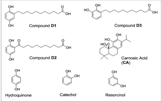
In addition, related natural product-inspired hydroqui-none-based synthetic compounds (i.e., D1 and D3; Figure 2) have recently been reported by our team and were shown to be bioactive (Satoh et al., 2011).
1Sanford-Burnham Neuroscience and Aging Research Center, La Jolla, CA,
USA
2Department of Anti-Aging Food Research, School of Bioscience and
Biotechnology, Tokyo University of Technology, Hachiouji, Japan
3Sanford-Burnham Medical Research Institute at Lake Nona, Orlando, FL,
USA
yDeceased, August 2, 2014
Corresponding Author:
Takumi Satoh, Sanford-Burnham Neuroscience and Aging Research Center, 10901 North Torrey Pines Road, La Jolla, CA 92037-1005, USA. Email: [email protected]
ASN Neuro July-August 2015: 1–13
!The Author(s) 2015 Reprints and permissions:
sagepub.co.uk/journalsPermissions.nav DOI: 10.1177/1759091415593294 asn.sagepub.com
Creative Commons CC-BY: This article is distributed under the terms of the Creative Commons Attribution 3.0 License
Importantly, CA and D1 (Figure 1), examples of PEDs, themselves are not electrophilic until they are activated at a site of tissue injury undergoing oxidative stress (Satoh et al., 2011).
PED activation under conditions of oxidative stress occurs because quinone formation is influenced by the cellular redox state, and in particular the Cu2þ/Cuþ recycling system (Wang et al., 2010; Satoh et al., 2013).
Figure 1. Chemical structures of the proelectrophiles evaluated in this study. The present study highlights compounds D1 (a) and D3 (b). CA (d) was evaluated as a neuroprotective compound in prior studies and is used here as a reference compound (Satoh et al., 2008a, 2008b). The compound notated as D2 (c) served as an inactive negative control for D1 in previous studies (Satoh et al., 2011). Note that D1 and D2 arepara-hydroquinone isomers, while D3 and the natural product CA areortho-hydroquinone isomers. CA¼carnosic acid.
The redox-active transition metal copper can catalyze oxidative activation of a number of phenolic compounds via Cu2þ/Cuþcycling (Bensasson et al., 2008; Satoh et al., 2009; Wang et al., 2010; Johansson, 2012). Under normal conditions, PED hydroquinones are very slowly oxidized to the quinone form, whereas this rate is greatly increased in the presence of Cu2þ/Cuþrecycling (Wang et al., 2010; Satoh et al., 2013). In addition, after conversion to the active form, PEDs display superiority to classical antioxi-dant molecules because of their sustained action and amplification via the nuclear factor (erythroid-derived 2)-like 2 (Nrf2) and heat-shock factor-1 (HSF-1) tran-scription-mediated signaling pathways (Satoh et al., 2013). These dynamic processes afford the potential for generating a pathologically activated therapeutic (or PAT) drug (Lipton, 2004, 2006, 2007). Importantly, we have also shown that several PEDs display an excellent absorption, distribution, metabolism, excretion, toxicity, and pharmacokinetic profile in the central nervous system, making them strong drug candidates (Satoh et al., 2008a; Rezaie et al., 2012; Satoh et al., 2013).
First, we focused on the Kelch-like ECH-associated protein 1 (Keap1)/Nrf2 pathway as one of the targets of PEDs for neuroprotection (Satoh et al., 2008b, 2011, 2013). After conversion to electrophilic quinones, PEDs activate the Keap1/Nrf2 pathway (Satoh and Lipton, 2007; Satoh et al., 2008b, 2013). We have shown that it is possible to take advantage of this intracellular mech-anism of electrophilic activation to develop a novel strat-egy for drug development against neurodegenerative diseases. As an illustration, CA activates the Nrf2 tran-scriptional pathway and protects various cells and organs against redox stress (Takahashi et al., 2009; Kosaka et al., 2010; Mimura et al., 2010; Tamaki et al., 2010; Maruoka et al., 2011; Satoh et al., 2011; Rezaie et al., 2012; Yanagitai et al., 2012). We have developed a potential strategy for clinical translation of this work based on the following chemical principles (Satoh and Lipton, 2007; Satoh et al., 2013).
1. Novel PEDs and related analogues are better tolerated than electrophiles, in part because electrophilic com-pounds can deplete glutathione in healthy, unstressed cells (Bensasson et al., 2008; Satoh et al., 2009; Wang et al., 2010; Johansson, 2012).
2. PEDs combat the oxidative stress that converts them to electrophiles through activation of the Nrf2 pathway (Bensasson et al., 2008; Satoh et al., 2009; Wang et al., 2010; Johansson, 2012).
Second, we focused on the heat-shock protein (HSP)90/HSF-1 pathway as one of the targets of PEDs for neuroprotection (Satoh et al., 2011, 2013). HSP90/ HSF-1 (Zou et al., 1998; Morimoto, 2008; Takii et al., 2010), like Keap1/Nrf2 (Talalay, 2000; Itoh et al., 2004;
Zhang et al., 2011), provides cell protection through acti-vation of endogenous gene networks involved in antioxi-dant response element (ARE) defense (Satoh and Lipton, 2007; Satoh et al., 2013). Several activators of Nrf2 via reaction with Keap1 also covalently bind to cysteine resi-dues of HSP90 to activate HSF-1 and other HSPs (Satoh et al., 2011; Zhang et al., 2011) through binding to AREs and heat-shock factor response elements (HSE), respect-ively, to provide neuroprotection against oxidative and nitrosative insults (Satoh et al., 2011; Zhang et al., 2011). Thus,S-alkylation of critical cysteine residues by electrophilic compounds can activate both of these tran-scriptional pathways, representing what has been called an electrophilic counterattack response (Satoh et al., 2006; Satoh and Lipton, 2007; Groeger and Freeman, 2010; Satoh et al., 2013). Because the capacity of neurons to protect themselves from redox insults can be easily overwhelmed, such backup systems are important homeostatic regulation processes that insure the continu-ation of normal neuronal signaling (Satoh et al., 2006; Satoh and Lipton, 2007; Groeger and Freeman, 2010; Satoh et al., 2013). D1 is a novel proelectrophilic com-pound that activates both the Nrf2 and HSF-1 pathways and can thus protect against both oxidative and endo-plasmic reticulum (ER) stress (Satoh et al., 2011; Zhang et al., 2011).
Importantly, potential clinical relevance for the use of PEDsin vivohas been obtained in models of age-related macular degeneration (AMD; Rezaie et al., 2012; Satoh et al., 2013) and cerebrovascular disease (stroke; Satoh et al., 2008b). While antioxidants were generally believed to have some effect in preventing AMD, their actions are fairly marginal (Prasad, 2009; Cano et al., 2010; Ramkumar et al., 2010). The use of antioxidants is not very effective because tissue penetrance is not reliable and often the action is not sustained. Similarly, while tissue plasminogen activator can dissolve clots, no effective neu-roprotective treatment has been proven for stroke damage in the brain (Satoh et al., 2008a, 2008b). Most importantly, the novel PEDs, such as D1 described in this article, increase the chances of producing clinically toler-ated therapeutics because they are only changed to the active state by oxidative insult at the site of impending injury (Satoh et al., 2013).
Nakamura and Lipton, 2009). Glutamate may be directly toxic to cultured neuronal cells via two different processes (Tan et al., 1998; Sagara et al., 2002). The first pathway is mediated by glutamate receptors. The second pathway is activated by a reduction in intracellular glutathione levels, leading to an imbalance in the homeostasis of the cell’s redox state (Tan et al., 1998; Sagara et al., 2002). This second pathway can be blocked by the add-ition of antioxidants. In particular, glutamate toxicity of HT22 cells has been used to model oxidative stress-induced cell death in hippocampal neurons (Tan et al., 1998; Sagara et al., 2002).
The objective of this study was to determine which isomer (para-vs.ortho-hydroquinone) could provide max-imal activation of the Nrf2/ARE and HSF-1/HSE path-ways. We sought to combine chemical and biological perspectives on this issue to define the best chemical struc-ture of an electrophilic core for use as a PED. To test the
para-hydroquinone electrophilic core of D1 versus an iso-mericortho-analogue, we synthesized compound D3 (the
ortho-hydroquinone variant of D1) having the same chem-ical scaffold (Figure 1). We then compared thepara- and
ortho-forms in terms of chemical and biological actions by monitoring oxidation potential, transcriptional activa-tion, induction of phase 2 enzymes and HSPs, as well as protection against oxidative and ER stress.
Materials and Methods
Chemicals and Antibodies
Antibodies and reagents were obtained as follows: anti-heme oxygenase-1 (HO-1) polyclonal rabbit IgG (OSA-150, Assay Design, Ann Arbor, MI), anti-NADPH quinone oxidoreductase1 (NQO1) polyclonal rabbit IgG (2618-1, Epitomics, Cambridge, MA), anti-HSP70 monoclonal mouse IgG (200-301-A27, Rockland, Pottstown, PA), IRDye 800CW goat anti-rabbit (green fluorescent; LI-COR, Lincolon, NE, catalogue number 926-32211), and IRDye 680LT goat anti-mouse (red fluorescent; LI-COR, Lincolon, NE, catalogue number 926–68020). Other reagents including dimethylsulfoxide (DMSO), sodium glutamate, fluores-cein diacetate (FDA), hydrogen peroxide (HP), tunica-mycin (TM), and Hoechst 33 258 stain were obtained from Sigma (St Louis, MO). The chemical synthesis of D1 has been described previously (Satoh et al., 2011). Synthesis of analogues D1 and D3 are described later.
General Procedure for the Synthesis of Compounds,
D1 and D3
To a 20-ml microwave vial was added methyl 10-bromo-decanoate (1.0 equiv.) and triphenylphosphine (2.0 equiv.). The mixture was placed under an argon
atmosphere and subjected to heating via microwave irradiation at 100C for 45 min. The resulting mixture
was then cooled to room temperature, followed by add-ition of anhydrous tetrahydrofuran (THF; 10 ml) and mild heating and vortexing until a solution was formed. To the resulting phosphonium salt solution was then added 1.0 M sodium bis-trimethylsilylamide (1.0 equiv.) dropwise over a 1-min period. This mixture was stirred at 0C in an ice/brine bath for 45 min followed by addition
of the corresponding ortho- or para -dimethoxybenzalde-hydes (1.48 M in THF). The reaction was allowed to pro-ceed, with stirring, at room temperature over 16 hr. The reaction product was then concentrated and reconstituted in 20 ml of ethyl acetate (EtOAc)/water (50/50 v/v), after which the layers were separated. The aqueous layer was extracted with EtOAc (38 ml) and combined with the organic layer, and the mixture was then dried over MgSO4. The product was subsequently filtered,
concen-trated, and subjected to SiO2 flash column
chromatog-raphy (Biotage SP4 system, Uppsala, Sweden) using 5% to 18% EtOAc to afford the olefinic esters as a colorless oils (46%–68%). These intermediates were used directly in the next step.
To a 5-ml round-bottom flask containing the mixed olefinic ester intermediates was added anhydrous metha-nol (0.1 M) followed by addition of 10% Pd/C (0.1 equiv.). The reaction was placed under a hydrogen atmosphere and stirred vigorously for 16 hr. After such time, the mixture was filtered through a plug of celite/ SiO2and concentrated to yield the crude aliphatic ester
in quantitative yield. To the crude ester were added equal amounts of THF and 2 M LiOH(aq) solution (4 equiv.),
and this mixture was then stirred at 40C overnight. The
reaction product was concentrated, dissolved in EtOAc, and then acidified (to pH 1–2) via addition of 1 N HCl. The layers were separated, and the acidic aqueous layer was extracted three times with EtOAc. The combined extracts were dried over Na2SO4, filtered through a
SiO2 solid phase extraction cartridge, and concentrated
to yield the crude acids affording colorless oils. The oils were diluted through addition of anhydrous CH2Cl2and
cooled to78C in a dry ice/acetone bath. To this solu-tion was added 1 M BBr3(2.1 equiv.) in CH2Cl2, and this
mixture was allowed to gradually return to room tem-perature over a period of 4 hr. Thereafter, the tempera-ture was reduced to 0C. The reaction was quenched with
deionized water and extracted three times with EtOAc. The combined extracts were dried over Na2SO4, filtered
Compound D1. 1H-nuclear magnetic resonance (NMR; 500 MHz, deuterated methanol, CD3OD) d ppm: 6.57 (d,
J¼8.5 Hz, 1H), 6.52 (d,J¼2.9 Hz, 1H), 6.43 (dd,J¼8.5, 3.0 Hz, 1H), 2.542.46 (m, 2H), 2.26 (t,J¼7.4 Hz, 2H), 1.641.51 (m, 4H), 1.391.25 (m, 12H). 13C-NMR (125 MHz, deuterated methanol, CD3OD) d ppm: 178.3, 151.1, 149.2, 131.5, 117.7, 116.7, 114.0, 35.4, 31.3, 31.1, 30.8, 30.7, 30.7, 30.7, 30.5, 30.4, 26.4. High-resolution mass spectrometry calculatedm/z for C17H26O4[M-H]¼ 293.1758;m/zfound 293.1756.
Compound D3. 1H NMR (500 MHz, deuterated methanol, CD3OD) d ppm: 6.626.52 (m, 3H), 2.612.52 (m, 2H), 2.26 (t, J¼7.5 Hz, 2H), 1.641.53 (m, 4H), 1.371.25 (m, 12H). 13C NMR (125 MHz, deuterated methanol, CD3OD) d ppm: 178.5, 146.0, 144.4, 130.9, 122.1, 120.2, 113.8, 35.6, 31.2, 31.1, 30.8, 30.7, 30.7, 30.7, 30.5, 30.4, 26.4. High-resolution mass spectrometry calculatedm/z for C17H26O4[M-H]¼293.1758; m/zfound 293.1757.
Cyclic Voltammetry Experimental Protocol
All experiments were performed using an electrochemical analyzer (CH Instruments, Austin, TX, model 600 E), glassware (cells), hardware, and electrodes (glassy carbon working, platinum counter, Ag/AgCl reference). All glassware was cleaned with concentrated nitric acid and rinsed with deionized water prior to experimentation. Prior to and between cyclic voltammetry (CV) scans, the glassy carbon electrode was polished with 0.05-micron alumina, the platinum wire auxiliary electrode was cleaned in 0.1 M H2SO4, and the Ag/AgCl reference
elec-trode was stored in 1.0 M KCl. All substrates were pre-pared as 1 mM solutions in 0.1 M tetrabutylammonium perchlorate in anhydrous acetonitrile and degassed under argon for 10 min prior to and between CV scans. Anodic and cathodic potentials were recorded after at least 10 cycles to insure reproducibility, except for resorcinol, which was recorded for the first cycle due to rapid current decrease upon subsequent cycles (Nasr et al., 2005; Astudillo et al., 2007; Nematollahi and Mohammadi-Behzad, 2009).
Cell Culture (ARPE-19 Cells)
To study the biochemistry and molecular biology of oxi-dative stress in retina, several investigators have used
in vitroculture of ARPE-19 cells. For example, exposure of ARPE-19 cells to HP is known to induce apoptosis and has thus been used as anin vitromodel of retinal degen-eration triggered by oxidative stress (Satoh et al., 2011). The cells were maintained in 10-cm dishes containing Dulbecco’s modified Eagle’s medium (DMEM) supple-mented with 10% fetal calf serum. The cells were intro-duced into wells of a 24-well plate at a density of 1105
cells/cm2and incubated for 24 hr. The medium was then changed to serum-free medium containing the designated concentrations of the test compounds, and the cultures were incubated for 24 hr. Then, HP or TM was added, and the cells were incubated for 4 hr or 24 hr, respectively. Finally, the cells were stained with FDA (1mM) and
Hoechst 33258 (5mg/ml) and observed by epifluorescence
microscopy.
Reverse Transcription-Polymerase Chain Reaction
For reverse transcription-polymerase chain reaction (RT-PCR) analysis, total RNA was obtained by use of TRIZOL Reagent (Invitrogen, Waltham, MA) from ARPE-19 cells that had been incubated with vehicle (DMSO), D1 (5mM), or D3 (5mM) in serum-free
medium for 24 hr (Satoh et al., 2000, 2003; Sasaki et al., 2011, 2013). Total RNA (1mg) from each source was
exposed to Superscript III (Invitrogen) in the presence of RNasin (20 U), random hexamers (2.5mM), dNTPs, and the supplied reverse transcription buffer. The reac-tion (20ml) was allowed to continue for 15 min at 42C. A
volume of 1/100th of this mixture from each source was then subjected to PCR conducted with the appropriate primer sets. At completion of the PCR, 10ml of the PCR
products were mixed with 2ml of loading buffer, and this
mixture was electrophoresed in 1.5% agarose gel in the presence of 0.5mg/ml ethidium bromide. The amplified
DNA fragments were visualized with UV detection. RT-PCR analysis was performed as described previ-ously (Satoh et al., 2000, 2003; Sasaki et al., 2011, 2013) with the following primers (number of PCR cycles and size of PCR product in parentheses):
50-TGA CTG ACT ACC TCA TGA AG-30(F) and
50-TTG CCA ATG GTG ATG ACC TG-30 (R) for b-actin
(22 cycles, 202 bp);
50-GAG TTG CAG CTG CTG AG-30 (F) and
50-GCA TGC CTG CAT TCA CAT G-30 (R) for ho-1 (24
cycles, 233 bp);
50-CTC CAT GTA CTC TCT GCA AG -30 (F) and
50-GTG GTG TCT CAT GAG TGT GC -30(R) for nqo1 (30 cycles, 203 bp); and
50-AGA TTC ATG ACG TCG TCC TG-30(F) and
50-GGA TGC CAT TAG CAT CAA TG-30(R) for hsp70 (28
cycles, 468 bp).
Western Blot Analysis
ARPE-19 cells were incubated for 24 hr with vehicle (DMSO), D1 (5mM), or D3 (5mM) in 10-cm dishes
#78503, Thermo Scientific, Waltham, MA) supplemented with a protease inhibition cocktail (Complete Protease Inhibitor Cocktail Tablets; catalog #11836170001, Roche, Waltham, MA). Total cell lysates (10mg each)
were separated by sodium dodecyl sulfate-polyacrylamide gel electrophoresis and then transferred onto polyvinyli-dene fluoride membranes. HO-1, NQO1, HSP70, and gly-ceraldehyde 3-phosphate dehydrogenase (GAPDH) were detected with specific antibodies, and the signals were detected using peroxidase-conjugated secondary antibo-dies. The protein signals were enhanced by use of a chemiluminescence assay (ECL Western blotting; Amersham Pharmacia, Piscataway, NJ).
Luciferase Assay
We used pGL-GSTYa ARE core-luciferase (Satoh et al., 2008, 2011) and ptK-hHSP70-luc (Takii et al., 2010) for assessment of transcriptional activation via ARE and HSE, respectively. ARPE-19 cells were seeded at a dens-ity of 1105cells/cm2into wells of a 48-well plate con-taining 1mg of plasmid DNA plus Lipofectamine 2000
(Invitrogen), and then incubated for 5 hr in PBS contain-ing 1mg of plasmid DNA plus Lipofectamine 2000
(Invitrogen). The cells were then washed in PBS and incu-bated for another 24 hr with vehicle, D1 (5 or 10mM), or
D3 (5 or 10mM). Firefly luciferase activity in cell lysates
was measured with a luminometer for reporter gene assays (Promega, Madison, WI). Transfection efficiency was normalized tob-galactosidase activity assessed by co-transfection with pSV-b-gal (Promega). For reporter gene assays, cells were transfected with 1mg of the reporter
construct (pGL-GSTYa ARE core-luciferase or ptK-hHSP70-luc) and 0.2mg pSV-b-gal for 1 hr. Cells were
then washed in PBS and incubated in serum-free culture medium for another 24 hr with vehicle, D1 (5 or 10mM),
or D3 (5 or 10mM). Firefly luciferase activity andb
-galac-tosidase activity in cell lysates were measured by using a luciferase system and b-galactosidase enzyme assay system, respectively (Promega).
Culture of HT22 Cells
Mouse hippocampal HT22 cells were maintained in 10-cm dishes containing DMEM supplemented with 10% fetal calf serum (Satoh et al., 2000, 2003; Sasaki et al., 2011, 2013). The cells were seeded onto 24-well plates at a density of 8104 cells per well in 500ml of serum-containing DMEM. One hour after seeding, the cultures were incubated for 1 hr with vehicle (DMSO) or various concentrations of D1 or D3. One hour later, the cells were exposed to 5 mM glutamate for 24 hr to induce oxidative damage. Subsequently, viability of the HT22 cells was determined using 3-(4,5-dimethylthiazol-2-yl)-2,5-diphenyl tetrazolium bromide (MTT) reduction,
as described elsewhere (Satoh et al., 2000, 2003; Sasaki et al., 2011, 2013). Cell death was also measured by the lactate dehydrogenase (LDH) cytotoxicity detection kit (TAKARA, Otsu, Shiga, Japan), which quantitatively measures the release of LDH into the medium following cell lysis or cell death. This assay was used as described by the manufacturer.
Statistical Analysis
Results are presented as the meanSD. Analysis of vari-ance with an appropriate post hoc test was performed for multiple comparisons and a Student’sttest for compari-son of two samples. A p value.05 was considered significant.
Results
Redox State of Compounds
The redox behavior of D1 and D3 was analyzed using CV in solution. Their voltammograms were compared with that of 1,2-dihydroxybenzene (catechol), 1,3-dihydroxy-benzene (resorcinol) and 1,4-dihydroxy1,3-dihydroxy-benzene (hydro-quinone), which were measured as control compounds in parallel to that of D1 and D3 (Nasr et al., 2005; Astudillo et al., 2007; Nematollahi and Mohammadi-Behzad, 2009). The CVs of catechol and hydroquinone were recorded in the0.5 V to 1.5 V potential range versus Ag/AgCl, where one anodic peak (oxidation to quinone form) and one cathodic peak (reduction back to the hydrogenated form) are observed. Table 1 sum-marizes the potential values for all compounds. Redox processes in 0.1 M tetrabutylammonium perchlorate in acetonitrile electrolyte are not reversible because the anodic and cathodic peaks are separated by over 600 mV. This was expected and consistent with values reported in the literature for these substrates/electrolyte combinations, and in particular hydroquinone is
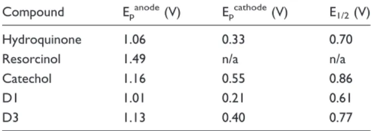
Table 1. Anodic (Oxidation) and Cathodic (Reduction) Peak Potentials and Corresponding Half-Wave Potentials From the CVs of D1, D3, and the Unsubstituted Control Compounds Shown in Figure 1.
Compound Epanode(V) Epcathode(V) E1/2(V)
Hydroquinone 1.06 0.33 0.70
Resorcinol 1.49 n/a n/a
Catechol 1.16 0.55 0.86
D1 1.01 0.21 0.61
D3 1.13 0.40 0.77
Note. CVs recorded using 0.1 M (Bu)4NClO4in acetonitrile with 100 mV/s
confirmed to oxidize more readily than catechol by 0.1 V. The CVs of resorcinol show only one oxidation peak at 0.3 V to 0.4 V higher potential than the other two isomers, which is consistent with the lack of electron delocaliza-tion on the nonconjugated meta-substitution pattern of the molecule (Nasr et al., 2005; Astudillo et al., 2007; Nematollahi and Mohammadi-Behzad, 2009). The CVs of the oxidation processes of D1 and D3 are shown in Figure 2. The anodic peak for D1 is found at 1.01 V, and that of D3 is recorded at 1.13 V. With cathodic peaks at 0.21 V and 0.40 V, respectively, the half-wave potentials for D1 and D3 are at 0.61 V and 0.77 V. This is consistent with the values found for the hydroquinone and catechol model compounds (Table 1 and Figure 1) and confirms that all redox processes involving the oxidation of D1 occur at 0.1 V to 0.2 V lower potentials than D3, that is, D1 is oxidized more readily than D3.
Activation of the Nrf2 and HSF-1 Pathways
We hypothesized that D1 activated Nrf2 and HSF-1 more than D3 based on the redox potential and ease of oxidation to the cysteine-reactive quinone form. To con-firm this notion, we performed luciferase assays in ARPE-19 cells transfected with plasmid DNAs under the transcriptional control of the ARE or HSE (Figure 3). D1 (5 mM) significantly activated both
tran-scriptional elements, indicating that D1 can stimulate both the ARE and HSE systems. In contrast, theortho -isomer D3 (5mM) activated the Nrf2/ARE pathway to a
lesser extent than that of the para-isomer. At 10mM,
D1 was still more potent than D3. D3 at 10mM but not
5mM activated the HSE transcriptional element. These
observations are consistent with the notion that the para-isomer (D1) activates the Keap1/Nrf2 and
Figure 3. Transcriptional activation of ARE and HSE. Retinal pigment epithelial ARPE-19 cells were plated at 1105cells/cm2, incubated for 24 hr, and then transfected with DNAs (ARE- or HSE-luciferase construct). After a 5-hr incubation in serum-containing medium, the medium was changed to serum-free medium containing vehicle (DMSO) versus D1 or D3 (5mM or 10mM). Cell lysates were obtained after
HSP90/HSF-1 pathways more effectively than the
ortho-isomer (D3).
Induction of Phase 2 Enzymes
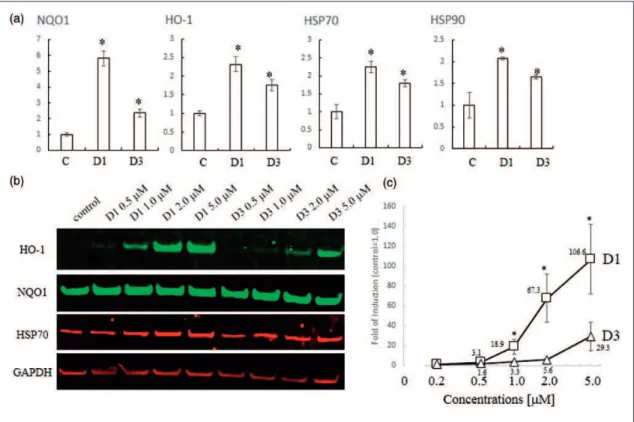
We hypothesized that the biological activity of com-pounds D1 and D3 would be closely related to their abil-ity to activate the Nrf2/ARE pathway. The previous DNA microarray study showed that compound D1 induced expression of HSPs in addition to phase 2 enzymes. Incubation with D1 resulted in activation of Nrf2 and HSF-1 transcriptional elements, thus inducing phase 2 enzymes and HSPs, respectively (Satoh et al., 2011). In this manner, D1 protected neuronal cells from both oxidative and ER-related stress. First, in this article, to compare the induction of the genes encoding phase 2 enzymes and HSPs by 5mM D1 and D3, we performed an
RT-PCR analysis using primers for the ho-1 and nqo1
phase 2 genes, and for hsp70 and hsp90 (Figure 4(a)).
D1 significantly induced each of these genes, although the magnitude of induction varied from gene to gene. Induction by D3 was significantly weaker than that by D1. Next, we confirmed the induction of HO-1, NQO1, and HSP70 at the protein level by performing immuno-blot analysis (Figure 4(b)). D1 induced HO-1 in a dose-dependent manner, whereas D3 did so only weakly, as confirmed by quantitative analysis (Figure 4(c)). While D1 and D3 induced the expression of NQO1 and HSP70 proteins, the basal levels of expression were already high in this cell line. Taken together, these data suggest that D1 induced both phase 2 enzymes and HSPs both more potently and efficaciously than D3.
Protective Effects in ARPE-19 Cells
An important biological attribute of PEDs is that they offer protection from oxidative stress (Satoh and Lipton, 2007; Satoh et al., 2013). Thus, we examined whether D1
Figure 4. Induction of phase 2 enzymes and HSPs. (a) PCR analysis of phase 2 and HSP genes induced by D1 or D3. Total RNA was extracted from ARPE cells treated with 5mM D1 or D3 for 24 hr in serum-free medium. RT-PCR was performed using cDNA template with
the specific primers listed in the Materials and Methods section mRNA levels were quantified by densitometry of qPCR band intensity after 28 cycles (n¼3). All genes were normalized tob-actin expression. *Significantly different (p<.05) from control. (b) Western blot analysis of dose-dependent induction of HO-1, NQO1, HSP70, and GAPDH proteins by the indicated concentrations of D1 or D3. Cell lysates were prepared, and 10mg protein/lane of protein was subjected to sodium dodecyl sulfate-polyacrylamide gel electrophoresis, after which
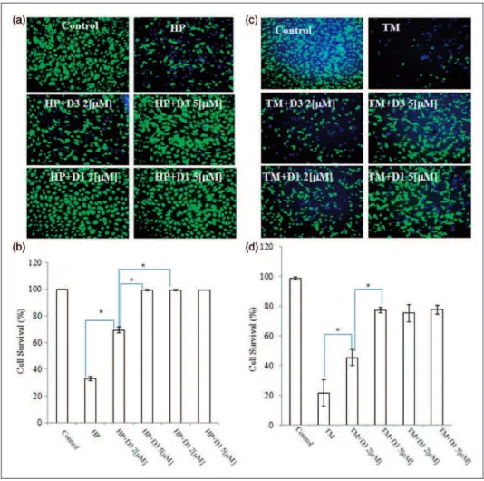
could protect neuronal cells against oxidative stress (Figure 5). As anin vitromodel of cell death related to AMD (Wada et al., 2001; Kim et al., 2003; Mandel et al., 2009), we exposed ARPE-19 cells to HP (1 mM) for 4 hr in serum-free medium (Figure 5(a) and (b)). In our stu-dies, we found that ortho-D3 was about twofold less potent thanpara-D1.
In addition to oxidative stress, we hypothesized that PEDs could also render cells resistant to ER stress by inducing HSPs, as shown previously (Satoh et al., 2011). Hence, we tested if the addition of D1 could pro-tect ARPE cells from ER stress elicited by 3mM TM. As
shown in Figure 5(c) and (d), exposure to TM (3mM)
induced cell death, but D1 (2-5mM) provided significant
protection. D3 was also effective against both oxidative
and ER stress, but proved to be a less potent proelectro-philic analog. In addition, although compound D3 afforded protection against TM, it did not activate the HSE. One possibility is that Nrf2 can induce HSPs in addition to phase 2 enzymes without affecting the HSE (Satoh et al., 2011).
Protective Effects in HT22 Cells
To confirm that para-D1 exhibited more potent neuro-protection than ortho-D3, we undertook an additional series of experiments examining neuronal cell death in the face of oxidative stress. We examined the protective effects of D1 and D3 against oxidative glutamate toxicity in mouse hippocampal HT22 cells. In these cells, high
Figure 5. Protective effects of various PEDs on ARPE-19 cells. (a and c) ARPE-19 cells were plated at 1105cells/cm2and incubated for 24 hr. Then, the medium was changed to serum-free medium containing vehicle (DMSO), D1 or D3, and the cells were incubated for an additional 24 hr. Thereafter, they were incubated with 1 mM HP, 3mM TM, or vehicle for 4 hr, and then stained with fluorescein diacetate
concentrations of glutamate induce cell death through depletion of glutathione, which is caused by inhibition of the glutamate-cystine antiporter (Satoh et al., 2008a, 2008b). We performed both the MTT assay for cell sur-vival and LDH release assay for cell death. D1 signifi-cantly protected cells against oxidative glutamate toxicity, whereas D3 was less potent, as assessed by MTT assay (Figure 6(a)). Again, ortho-D3 was about twofold less potent thanpara-D1. These protective effects were confirmed by the LDH release assay (Figure 6(b)).
Discussion
In the present study, we compared the chemical and bio-logical effects ofortho- andpara-PEDs in providing neu-roprotective activity. We performed a series of biochemical experiments, measuring oxidation potential, transcriptional activation, induction of phase 2 enzymes and HSPs, and neuroprotection, to compare the com-pounds D1 and D3, which share similar chemical struc-tures except that they arepara-andortho-hydroquinones, respectively. The para-electrophilic compound D1 acti-vated neuroprotective signaling pathways to a greater degree than the ortho-compound D3. These results are consistent with our CV experiments showing that the sequence of oxidation potential modulation for theses isomers is para (hydroquinone)>ortho (catechol)> >
meta (resorcinol) as shown in Table 1. We consistently found that the oxidation potential for the para-hydroquinone D1 was lower than that of the
ortho-hydroquinone D3 (Figure 2). Overall, the para -compound exhibited at least a twofold increase in the potency of neuroprotection over the ortho-compound. Whether this improvement will be reflected when para -versusortho- compounds are compared inin vivodisease models remains to be determined in future studies.
In terms of developing clinically tolerated drugs, we sought to learn principles from other recent ventures in successful central nervous system drug development. Along these lines, our development of the FDA-approved drug memantine, an N-methyl-D-aspartate receptor
antagonist, was in part based on the principle that drugs should interact with their target only during states of pathological hyperactivation and not during normal physiological function (Lipton, 2004, 2006, 2007). Drugs that have been developed using this strategy are designated PAT drugs. PEDs are candidate PAT drugs because conversion to the active quinone form is redox-controlled and thus enhanced by the very oxidative stress that these drugs then counteract (Satoh and Lipton, 2007; Satoh et al., 2008b, 2013). Moreover, in terms of their druggability, published work has shown that these PEDs can translocate into the retina and brain of mice and rats at levels sufficient to protect against significant
oxidative insults, including light-induced retinal degener-ation and middle cerebral artery occlusion (Satoh and Lipton, 2007; Satoh et al., 2008b, 2013).
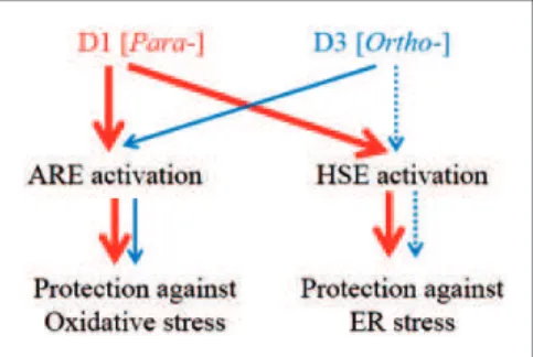
Overall, our results support the notion that compound D1 activates both Nrf2 and HSF-1, while compound D3 activates these pathways less potently. Moreover, the measured electrochemical oxidation potentials of these PEDs can be used to predict their activation of protective pathways. The differential ability of PEDs to activate the transcription factors in these pathways is inversely corre-lated to their potential for oxidation from the hydroquin-one to the quinhydroquin-one form (Figure 7). Importantly, the redox state of the cells also affects the ability to transform PEDs to their active quinone form. The Cu2þ/Cuþredox system regulates oxidative reaction from hydroquinone to quinone (Satoh et al., 2009; Wang et al., 2010), and this system is highly influenced by the availability of electron acceptors in the cell; under pathological conditions, such electron acceptors are represented by reactive oxygen spe-cies (Satoh et al., 2009; Wang et al., 2010). Redox state-dependent regulation of quinone formation dictates that PEDs, such as CA and D1, are PATs in the sense that they are converted from the hydroquinone to the quinone by the very reactive oxygen species that they then combat via transcriptional activation (Lipton, 2004, 2006, 2007). Accordingly, activated PEDs S-alkylate critical cysteine residues on KEAP1 and HSP90, leading to activation of the transcription factors Nrf2 and HSF-1, respectively (Satoh et al., 2011; Zhang et al., 2011). This approach thus represents a novel strategy against neurodegenera-tive disorders, providing disease-modifying electrophilic drugs in the context of pathological insult.
For many years, the herb rosemary has been reported to manifest antioxidant and anti-inflammatory activity.
We have previously shown that CA, present in rosemary extract, crosses the blood–brain barrier to exert neuro-protective effects by upregulating endogenous antioxi-dant enzymes via the Nrf2 transcriptional pathway (Rezaie et al., 2012; Satoh et al., 2013). The antioxidant and neuroprotective activities in retinal cell lines exposed to oxidative stress and in a rat in vivo model of light-induced retinal degeneration suggest that PEDs, such as D1 and CA, may potentially have clinical application to retinal diseases, including AMD and retinitis pigmentosa, in which oxidative stress is thought to contribute to dis-ease progression (Rezaie et al., 2012).
In conclusion, our findings suggest that para -hydro-quinones are both more potent and efficacious than their
ortho-hydroquinone homologues in terms of transcrip-tional activation, induction of phase 2 enzymes and HSPs, and neuroprotective effects against oxidative and ER stress. Nonetheless, other factors that determine druggability, such as pharmacokinetics, bioavailability, stability, metabolism, and translocation into the brain, will also be important in determining which structures are most effectivein vivo. Critically, however, this is the first report to our knowledge to demonstrate that pos-itional isomers of proelectrophilic hydroquinones are an important determinant in the activation of Nrf2- and HSF-1-mediated stress responses, and the disparate effect of these isomers is closely tied to their oxidation potentials.
Acknowledgments
The authors thank Dr. Larry D. Frye for editorial help with the article. The authors also thank Dr. Toby R. Long for the synthesis of compounds.
Author contributions
The experiments were conceived and designed by T. S., S. A. L., and G. P. R and performed by T. S., R. S., S. R. M., and R. E. W.
Funding
The authors disclosed receipt of the following financial support for the research, authorship, and/or publication of this article: This work was supported in part by a grant to T.S. from MEXT KAKENHI Grant Number 13382632 and in part by NIH grants to S. A. L. (P01 ES016738, P01 HD29587, R01 NS086890, R21 NS080799, and P30 NS076411, and funds from the Michael J. Fox Foundation).
Declaration of Conflicting Interests
The authors declared no potential conflicts of interest with respect to the research, authorship, and/or publication of this article.
References
Aruoma, O. I., Halliwell, B., Aeschbach, R., & Lo¨ligers, J. (1992). Antioxidant and pro-oxidant properties of active rosemary con-stituents: Carnosol and carnosic acid.Xenobiotica,22, 257–268. Figure 7. Proposed protective mechanisms of PEDs. Red and
Astudillo, P. D., Tiburcio, J., & Gonza´lez, F. J. (2007). The role of acids and bases on the electrochemical oxidation of hydroquin-one: Hydrogen bonding interactions in acetonitrile.Journal of Electroanalytical Chemistry,604, 57–64.
Bensasson, R. V., Zoete, V., Dinkova-Kostova, A. T., & Talalay, P. (2008). Two-step mechanism of induction of the gene expres-sion of a prototypic cancer-protective enzyme by diphenols. Chemical Research in Toxicology,21, 805–812.
Bredesen, D. E. (2008). Programmed cell death mechanism in neurological diseases.Current Molecular Medicine,8, 173–186. Calabrese, V., Cornelius, C., Dinkova-Kostova, A. T., Calabrese, E. J., & Mattson, M. P. (2010). Cellular stress responses, the horm-esis paradigm, and vitagenes: Novel targets for therapeutic intervention in neurodegenerative disorders. Antioxidants & Redox Signaling,13, 1763–1811.
Cano, M., Thimmalappula, R., Fujihara, M., Nagai, N., Sporn, M., Wang, A. L.,...Handa, J. T. (2010). Cigarette smoking, oxida-tive stress, the anti-oxidant response through Nrf2 signaling, and Age-related Macular Degeneration. Vision Research, 50, 652–664.
Groeger, A. L., & Freeman, B. A. (2010). Signaling actions of electrophiles: Anti-inflammatory therapeutic candidates. Molecular Interventions,10, 39–50.
Hara, M. R., & Snyder, S. H. (2007). Cell signaling and neuronal death. Annual Review of Pharmacology and Toxicology, 47, 117–141.
Itoh, K., Tong, K. I., & Yamamoto, M. (2004). Molecular mech-anism activating Nrf2-Keap1 pathway in regulation of adaptive response to electrophiles.Free Radical Biology & Medicine,36, 1208–1213.
Johansson, M. H. (2012). Reversible Michael additions: Covalent inhibitors and prodrugs.Mini Reviews in Medicinal Chemistry, 12, 1330–1344.
Kim, I., Xu, W., & Reed, J. C. (2008). Cell death and endoplasmic reticulum stress: Disease relevance and therapeutic opportu-nities.Nature Reviews Drug Discovery,7, 1013–1030. Kim, M. H., Chung, Y., Yang, J. W., Chung, S. M., Kwag, N. H.,
Yoo, J. S. (2003). Hydrogen peroxide-induced cell death in a human retinal pigment epithelial cell line, ARPE-19. Korean Journal of Ophthalmology,17, 19–28.
Kosaka, K., Mimura, J., Itoh, K., Satoh, T., Shimojo, Y., Kitajima, C,...Shirasawa, T. (2010). Role of Nrf2 and p62/ZIP in the neurite outgrowth by carnosic acid in PC12h cells.Journal of Biochemistry,147, 73–81.
Lipton, S. A. (2004). Concepts: Turning down but not off—Neuroprotection requires a paradigm shift in drug devel-opment.Nature,428, 473.
Lipton, S. A. (2006). Paradigm shift in neuroprotection by NMDA receptor blockade: Memantine and beyond. Nature Reviews Drug Discovery,5, 160–170.
Lipton, S. A. (2007). Pathologically-activated therapeutics.Nature Reviews Neuroscience,8, 803–808.
Mandel, M. N., Patlolla, J. M., Zheng, L., Agbaga, M. P., Tran, J. T., Wicker, L.,...Anderson, R. E. (2009). Curcumin protects retinal cells from light-and oxidant stress-induced cell death. Free Radical Biology & Medicine,46, 672–679.
Maruoka, H., Sasaya, H., Sugihara, K., Shimoke, K., & Ikeuchi, T. (2011). Low-molecular-weight compounds having neurotrophic activity in cultured PC12 cells and neurons. Journal of Biochemistry,150, 473–475.
Mimura, J., Kosaka, K., Maruyama, A., Satoh, T., Harada, N., Yoshida, H.,...Itoh, K. (2010). Nrf2 regulates NGF mRNA induction by carnosic acid in T98G glioblastoma cells and normal human astrocytes. Journal of Biochemistry, 150, 209–217.
Morimoto, R. I. (2008). Proteotoxic stress and inducible chaperone networks in neurodegenerative disease and aging. Genes & Development,22, 1427–1438.
Nakamura, T., & Lipton, S. A. (2009). Cell death: Protein misfold-ing and neurodegenerative diseases.Apoptosis,14, 455–468. Nasr, B., Abdellatif, G., Canizares, P., Saez, C., Lobato, J.,
Rodrigo, M. A. (2005). Electrochemical oxidation of hydroquin-one, resorcinol, and catechol on boron-doped diamond anodes. Environmental Science & Technology,39, 7234–7239. Nematollahi, D., & Mohammadi-Behzad, L. (2009).
Electrochemical oxidation of catechol in the presence of some azacrown ethers and transition metal ions in acetonitrile. International Journal of Electrochemical Science, 4, 1583–1592.
Prasad, A. S. (2009). Zinc: Role in immunity, oxidative stress and chronic inflammation. Current Opinion in Clinical Nutrition and Metabolic Care,12, 646–652.
Ramkumar, H. L., Zhang, J., & Chan, C. C. (2010). Retinal ultra-structure of murine models of dry age-related macular degener-ation (AMD). Progress in Retinal and Eye Research, 29, 169–190.
Rezaie, T., McKercher, S. R., Kosaka, K., Seki, M., Wheeler, L., Viswanath, V.,...Lipton, S. A. (2012). Protective effect of car-nosic acid, a pro-electrophilic compound, in models of oxidative stress and light-induced retinal degeneration. Investigative Ophthalmology & Visual Science,53, 7847–7854.
Sagara, Y., Ishige, K., Tsai, C., & Maher, P. (2002). Tyrphostins protect neuronal cells from oxidative stress. The Journal of Biological Chemistry,277, 36204–36215.
Sasaki, S., Tozawa, T., Sugamoto, K., Matsushita, Y., & Satoh, T. (2013). Diterpene para-hydroquinone compounds derived from cryptoquinone inhibit adipocyte differentiation of mouse 3T3-L1 cells and activate the Nrf2/ARE pathway. Bioscience, Biotechnology, and Biochemistry,77, 2131–2133.
Sasaki, S., Tozawa, T., Van Wagoner, R. M., Ireland, C. M., Harper, M. K., Satoh, T. (2011). Strongylophorine-8, a pro-electrophilic compound from the marine sponge Petrosia (Strongylophora) corticata, provides neuroprotection through Nrf2/ARE pathway. Biochemical and Biophysical Research Communications,415, 6–10.
Satoh, T., Baba, M., Nakatsuka, D., Ishikawa, Y., Aburatani, H., Furuta, K.,...Watanabe, Y. (2003). Role of heme oxygenase-1 protein in the neuroprotective effects by cyclopentenone pros-taglandin derivatives as a sustained phase of neuronal survival promoting mechanism under oxidative stress.European Journal of Neuroscience,17, 2249–2255.
Satoh, T., Furuta, K., Tomokiyo, K., Nakatsuka, D., Tanikawa, M., Nakanishi, M.,...Watanabe, Y. (2000). Facilitatory roles of novel compounds designed from cyclopentenone prostaglandins on neurite outgrowth-promoting activities of nerve growth factor.Journal of Neurochemistry,75, 1092–1102.
hydroxyl moieties-dependent manners. Neuroscience Letters, 434, 260–265.
Satoh, T., Kosaka, K., Itoh, K., Kobayashi, A., Yamamoto, M., Shimojo, Y.,...Lipton, S. A. (2008b). Carnosic acid, a cate-chol-type electrophilic compound, protects neurons both in vitro and in vivo through activation of the Keap1/Nrf2 path-way via S-alkylation of specific cysteines. Journal of Neurochemistry,104, 1116–1131.
Satoh, T., & Lipton, S. A. (2007). Redox regulation of neuronal survival by electrophilic compounds.Trends in Neurosciences, 30, 38–45.
Satoh, T., McKercher, S. R., & Lipton, S. A. (2013). Nrf2/ARE-mediated antioxidant actions of pro-electrophilic drugs. Free Radical Biology & Medicine,65, 645–657.
Satoh, T., Okamoto, S., Cui, J., Watanabe, Y., Furuta, K., Suzuki, M.,...Lipton, S. A. (2006). Activation of the Keap1/Nrf2 path-way for neuroprotection by electrophilic phase II inducers. Proceedings of the National Academy of Sciences of the United States of America,103, 768–773.
Satoh, T., Rezaie, T., Seki, M., Sunico, C. R., Tabuchi, T., Kitagawa, T.,...Lipton, S. A. (2011). Dual neuroprotective pathways of a pro-electrophilic compound via HSF-1-activated heat-shock proteins and Nrf2-activated phase 2 antioxidant response enzymes.Journal of Neurochemistry,119, 569–578. Satoh, T., Saitoh, S., Hosaka, H., & Kosaka, K. (2009). Simple
ortho- and para-hydroquinones as neuroprotective compounds against oxidative stress associated with a specific transcriptional activation. Biochemical and Biophysical Research Communications,379, 537–541.
Takahashi, T., Tabuchi, T., Tamaki, Y., Kosaka, K., Takikawa, Y., Satoh, T. (2009). Carnosic acid and carnosol inhibit adipocyte differentiation in mouse 3T3-L1 cells through induction of phase 2 enzymes and activation of glutathione metabolism. Biochemical and Biophysical Research Communications,382, 549–554.
Takii, R., Inouye, S., Fujimoto, M., Nakamura, T., Shinkawa, T., Prakasam, R.,...Nakai, A. (2010). Heat shock transcription factor 1 inhibits induction of IL-6 through inducing activation transcription factor 3.Journal of Immunology,184, 1041–1048. Talalay, P. (2000). Chemoprotection against cancer by induction of
phase 2 enzymes.Biofactors,12, 5–11.
Tamaki, Y., Tabuchi, T., Takahashi, T., Kosaka, K., & Satoh, T. (2010). Activated glutathione metabolism participates in pro-tective effects of carnosic acid against oxidative stress in neur-onal HT22 cells.Planta Medica,76, 683–688.
Tan, S., Sagara, Y., Liu, Y., Maher, P., & Schubert, D (1998). The regulation of reactive oxygen species during programmed cell death.The Journal of Cell Biology,15, 1423–1432.
Wada, A. M., Gelfman, C. M., Handa, J. T., & Hjelmeland, L. M. (2001). Downregulation of differentiation-specific gene expres-sion by oxidative stress in ARPE-19 cells. Investigative Ophthalmology & Visual Science,42, 2706–2713.
Wang, X. J., Hayes, J. D., Higgins, L. J., Wolf, C. R., & Dinkova-Kostova, A. T. (2010). Activation of the Nrf2 signaling pathway by copper-mediated redox cycling of para- and ortho-hydroqui-nones.Chemistry & Biology,17, 75–85.
Yanagitai, Y., Kitagwa, T., Itoh, S., Takenouchi, T., Kitani, H., Satoh, T. (2012). Carnosic acid, a pro-electrophilic compound, inhibits LPS-induced activation of microglia.Biochemical and Biophysical Research Communications,418, 22–26.
Zhang, Y., Ahn, Y. H., Benjamin, I. J., Honda, T., Hicks, R. J., Calabrese, V.,...Dinkova-Kostova, A. T. (2011). HSF1-dependent upregulation of Hsp70 by sulfhydryl-reactive inducers of the KEAP1/Nrf2/ARE pathway. Chemistry & Biology,18, 1355–1361.