Resumo
Óxidos de ferro (goethita e hematita) pulverizados foram irradiados com pul-sos laser objetivando verificar o resultado da interação desses pós com a radiação in-fravermelha concentrada em pulsos de curta duração e altamente energéticos. Esses experimentos se justificam diante da utilização desses pós, em obras de arte, como pigmentos, desde as épocas mais remotas. Na atualidade, tornou-se frequente recorrer a pulsos laser como ferramenta de limpeza de obras de arte, o que justifica a neces-sidade do desenvolvimento de conhecimentos relacionados à interação laser-pigmen-tos. Evitar danos às obras de arte, recentes ou não, é uma exigência inevitável. Os resultados mostraram que os pulsos laser Nd:YAG fornecem energia suficiente para promover, instantaneamente, mudanças químicas e estruturais nos pigmentos mine-rais estudados. Material vitrificado (amorfo) foi identificado nas amostras irradiadas e sua formação deveu-se à solidificação ultra-rápida ocorrida no locais onde a energia disponibilizada pelos pulsos laser foi suficiente para fundir as partículas do pó. Em consequência das mudanças químicas e estruturais induzidas, houve alteração da cor original do material irradiado. Essas alterações cromáticas foram quantificadas por intermédio de um método desenvolvido para tal finalidade e que é baseado na variação da intensidade das componentes RGB das cores digitais.
Palavras-chave: Laser Nd-YAG, óxido de ferro, material amorfo, goethita, hematita, difração de raios X, MEV – Microscopia Eletrônica de Varredura, Obras de Arte, Evolução das cores RGB.
Abstract
Laser irradiation of powdered iron oxides (goethite and hematite) was per-formed in order to obtain information about their interaction with short duration near-infrared pulses. Results have shown that under some conditions, Nd:YAG laser provides enough energy to induce fast chemical and structural transformations of the goethite and hematite. This kind of information is of great interest for professionals working with artwork conservation because this technique is used in the conservation of cultural heritage. Depending on the laser’s working conditions, glassy (amorphous) material was detected and its presence was related to areas of fast solidification where the energy delivered was enough to melt the powder particles. Color changes were
Metalurgia e materiais
Metallurgy and materials
Evolução das cores RGB:
uma ferramenta digital para
monitorar mudanças visuais em
pigmentos irradiados por pulsos de
laser infravermelho
RGB color evolution: a digital tool
for monitoring the visual changes of iron
oxide pigments irradiated by infrared laser pulses
Adilson Rodrigues da Costa
D. Sc., Prof. Associado III, DEMET – Departamento de Engenharia Metalúrgica e de Materiais – Escola de Minas – UFOP, REDEMAT (UFOP-CETEC-UEMG) adilson@em.ufop.br
Flávio Sandro Lays Cassino
D. Sc., Prof. Adjunto IV, DEMET – Departamento
de Engenharia Metalúrgica e de Materiais – Escola de Minas – UFOP, REDEMAT (UFOP-CETEC-UEMG)
fcassino@em.ufop.br
Rémy Chapoulie
D. Sc., Directeur de Recherches, Université de Bordeaux III, Maison d`Archéologie, Institut de Recherche sur les Archéomatériaux – Bordeaux – França
remy.chapoulie@u_bordeaux.fr
Florence Rud
M. Sc., Université de Bordeaux III,
observed and quantified by means of an RGB color measurement method developed to show the evolution of each color component.
Keywords: Nd:YAG laser, iron oxide, amorphous materials, goethite, hematite, X-ray diffraction, Scanning Electron Microscopy, artworks, RGB color evolution.
1. Introduction
Amongst the available surface cle-aning methodologies, Lasers play an im-portant role because they enable accurate control coupled with material selectivity and fast feedback. Notwithstanding, it is imperative to optimize the cleaning parameters to ensure that any damage to the remaining surfaces is minimal and well understood (Pouli et al., 2010) and (Zafiropoulos et al., 1998). Otherwise it is well established that Q-switched laser equipment can deliver pulses of high energy that can promote a great variety of structural and state changes (Castille-jo et al., 2003); (Cooper, 1998); (Costa, 2002); (Dickmann et al., 2005); (Fotakis et al., 1997); (Gaspar et al., 2000). These changes are related to the great amount of energy transferred to materials as thermal and mechanical energy (shock waves). The main purpose of this work is to analyze, quantitatively, the variations
in color due to laser-induced changes on previous structures, involving two iron oxides extensively used as pigments in modern and ancient artworks: goethite and hematite. Experiments were perfor-med with a Nd:YAG laser operating in its fundamental mode (1064 nm). In order to understand iron oxide interaction with short duration laser pulses (4 ns), we have studied the reaction products under specific laser treatment conditions (fluency, frequency, etc.). Dehydration and reduction processes were observed simultaneously for amorphous compound formation as reported elsewhere by Costa (2002), where samples had been analyzed by X-ray diffractometry, detecting struc-tural changes including amorphous phase formation. Visual and optical observa-tions revealed changes in color; specifically in the darkening of the original powder particles. These kinds of alterations have
been reported by Castillejo et al. (2003) and Rodriguez-Navarro et al. (2003). A method allowing fast characterization of these color changes was developed and ap-plied in this work. The method is based on the quantification of the three fundamen-tal digifundamen-tal colors Red, Green and Blue and its own evolution (variation) accordingly to the corresponding modification obser-ved after laser treatment. Color variations were measured accordingly to this specific method, being based on the R, G and B color evolution. These three fundamental colors were digitally treated according to this analytical procedure, taking into ac-count the light reflected under controlled conditions (Oshiro, 2008). SEM/EDS characterization was also realized and the results are presented and discussed here in order to better understand the nature and extent of laser-induced physicochemical alterations to the involved materials.
2. Materials and methods
During the last few years many research groups interested in surface cleaning have employed pulsed laser test-ing supports, different soiltest-ing layers, ar-cheological artifacts, ancient and modern paints, manufactured components deal-ing with several related challenges, and darkening of treated surfaces, which isthe worst (Asmus, 1994). Observed chromatic variations are associated with eventual changes due to laser interaction with pig-ments or chromophores. This discolor-ation phenomenon was well investigated by Sansonetti and Realini (2000), for six different pigments often employed in the art field. In our work, powders of goethite (HFeO2) and hematite (Fe2O3) were pre-pared by mechanical milling according to conventional laboratory procedures under irradiation with pulses of an Nd:YAG laser operating in its fundamental mode. Samples were then submitted to irradiation (Nd:YAG laser at 1064 nm, 10Hz and 4 ns pulse duration) in a way as to allow the continuous variable delivery of energy, so that a track of reaction products could be obtained. A moveable inclined plate adapted to the laser table was conceived,
allowing these experimental conditions. The amount of reaction products be-ing variable implies a continuous color modification line. (see Figure 1). The tool we developed, a software, is capable of measuring, in real time, the amount of each fundamental color component. This free software, named QuantiColor 1.0, is well adapted to converting digital color information captured in 24 bits bitmap format to numerical data that can be easily treated in a graphic manner where percentages of digital data are plotted. In other words, R, G and B percentages are calculated and displayed, providing quantified information from the examined area of the digital image (Oshiro, 2008 and Rud, 2007).
Chromatic studies were performed upon goethite and hematite powders adhered to supports made with inert material in order to avoid eventual unde-sirable interactions. These specimens had been irradiated in a way so as to allow a continuous energy variation thanks to a movable inclined plate placed in front of the laser beam. Thus, a track of reaction products is obtained showing a
continu-ous variation in color and morphology. Afterwards, irradiation specimens were analyzed to evaluate the color differences between the irradiated and non-irradiated zones of the specimens. These differences, representing discrete intensities of each component, enabled us to visualize the evolution of the three R, G and B com-ponents when plotted against the length of the treated track. (Oshiro, 2008 and Rud, 2007).
In the beginning of the track, the spot size touching the sample was 1.5 ± 0.3 mm in diameter, implying that there is an energy density of 36 ± 0.7 mJ/mm2 that diminishes as the diameter spot increases. Along this track, the intensity, or laser fluency, falls to 14.1mJ/mm2 in the end of
and published elsewhere by Costa (2002).
Concerning the morphological stud- ies, the SEM/EDS characterization was performed at the Microanalysis Labora- tory of Ouro Preto’s School of Mines (Universidade Federal de Ouro Preto).
3. Results and discussions
As mentioned, the software to measure R, G and B evolution, named QuantiColor, is able to quantify, in terms of RGB component percentages, the variations in color level with a pixel resolution.
The developed method allows us to represent the evolution of the three fun-damental colors, R, G and B, measured in a defined area as small as a pixel or greater. Photographs were taken under controlled conditions in order to
guar-antee illumination reproducibility and reflectability of selected areas. Digital color reconstitution is possible when taking the matrix information and as-sociating it to the color measurements (Oshiro, 2008).
R
G B
E v o l u t i o n
40
30
20
10
0
-10
1 2 3
cm
Goethite
4 5 5
R
G B
E v o l u t i o n
40
30
20
10
0
-10
-1 0 1 2
cm
Hematite
4
3 5 6
a b
a b
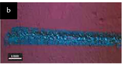
Figure 1
Typical features of an irradiated track in goethite (a) and hematite (b) powders. Damage areas are due to ejected material during high irradiation occurring at the beginning of the track.
Figure 2 Evolution of fundamental colors,
R, G and B, along the laser treated track upon goethite (a) and hematite (b) powders. The corresponding digital reconstitution of color evolution is placed below the graphics. (Rud, 2007)
As reported in a previous work by Costa (2002), the browning of treated goethite is due to dehydration, while the darkening of hematite is related to its re-duction to magnetite. Under such intense heating levels, and no oxygen availability
Fe2O3 is converted to Fe3O4: iron – oxygen bonds being broken by photolysis. Other-wise, goethite dehydration involves the re-lease of a water molecule as a consequence of chemical bond breaking. Instantaneous browning of goethite particles is evidence
of its transformation into hematite. Related diagrams showing R, G and B evolutions are presented in Figure 2. They represent, quantitatively, the color evolution along the track. So, one can verify how chemical modifications may
1200
1000
800
600
400
200
0
0 2 4 6 8 10
Full scale counts: 1078 florence_C_2_pt1
keV
Figure 3 (a) SEM micrographs showing irradiated area with glassy phase upon non altered hematite powder. (b) Faceted crystals (detail) emerging from glassy phase resemble octahedral magnetite crystals. (c) Typical EDS spectrum of distinct points signaled by arrows.
a b
induce color changes in these experi-ments. The continuous variable energy delivery idealized system (Rud, 2007) al-lowed us to infer the threshold value of the energy capable to induce changes. These values correspond to the non detectable color modification point along the track: approximately 4.5 cm for the goethite sample and 5.0 cm for the hematite one. Information like this is of great interest to professionals in charge of conserving
surfaces (Shekede, 1992). The RGB color evolution diagram for goethite powder begins at 1.5cm, due to the impossibility of taking measurements at the beginning of the track because it was damaged by an excess of absorbed energy in that area.
Concerning the micro structural studies, SEM characterization, coupled with EDS analysis, shows interesting morphological features. In Figures 3 and 4, vitreous matter can be observed that
results from rapid cooling of melted oxide. The presence of holes in the vitreous phase infers that gas expansion and release oc-curred, probably originating from the dehydration and desorption of powder particles on the surface. In some cases, cracks are related to contraction during rapid solidification of melted material; eventually they are due to the preparation of the samples.
400
300
200
100
0
0 2 4 6 8 10
Full scale counts: 332 florence_C_6_pt2
keV
a b
Figure 4
(a) SEM micrograph showing irradiated area with glassy phase upon non altered goethite powder. (b) Typical EDS spectrum of distinct points signaled by arrows.
4. Conclusions
The darkening and browning of iron oxides treated by an Nd:YAG laser can be quantified by RGB color evolu-tion according to a method based on digital color intensity measurements, treated as a percentage of each color component.
Color change studies in goethite and magnetite powders allow us to show that there exists a threshold of
energy necessary to promote chemical changes and glassy phase formation. It can be visually detected by observing the graphics where color evolution (the RGB components) is displayed related to the spot size or energy density delivered. Pigments of goethite and hematite are stable beyond this level. The accuracy of this method is much better than the conventional visual control.
Digital control of color change may help professionals to avoid damage to the surfaces being treated with lasers.
The suggested methodology is proven to reliably and accurately detect changes influencing the visible reflec-tance of the surface, and thus, it can serve as a monitoring tool to ensure good results when surfaces are treated by lasers pulses.
5. References
ASMUS J. Spectral control in laser restoration of archaeological treasure. SPIE Proceedings 2273, p. 207-213, 1994.
CASTILLEJO M., MARTIN M., OUJJA M., REBOLLAR E., DOMINGO C., GARCIA-RAMOS J. V., SANCHEZ-CORTES S. Effect of wavelength on the laser cleaning of polychromes on wood, J. Cult. Herit., v.4, p. 243–249, 2003.
COOPER M. Laser cleaning in conservation: an introduction. Oxford: Butterworth Heine-man, 1998.
COSTA A. R. da. Ultra-fast dehydration and reduction of iron oxides by infrared pulsed radia-tion. Scripta Materialia, v. 47, p. 327- 330, 2002.
DICKMANN K., FOTAKIS C., ASMUS, J.F. (Eds.). Lasers in the conservation of artworks, LACONA V Proceedings..., Osnabrueck, Springer Proceedings in Physics 100, Germany, 2005.
FOTAKIS, C., ANGLOS, D., BALAS, C., GEORGIOU S., VAINOS N.A., ZERGIOTI I., ZA-FIROPOULOS V. Laser technology in art conservation. In: TAM, A.C. (Ed.). OSA TOPS on Lasers and Optics for Manufacturing. Optical Society of America, v. 9, 1997.
GASPAR P., KEARNS A., VILAR R., WATKINS K., MALHOA GOMES M. M. A study of the effect of Wavelength on Q-Switched Nd:YAG Laser Cleaning of Eighteenth-Century Portuguese Tiles. Studies in Conservation, v. 45, n. 3, p.189-200, 2000.
OSHIRO, P. S. Desenvolvimento de um software para análise cromática quantitativa de ima-gens digitais. Ouro Preto: Universidade Federal de Ouro Preto, REDEMAT, 2008. 87 p. (Dissertação de Mestrado em Engenharia de Materiais).
RODRIGUEZ-NAVARRO, C., ELERT, K., SEBASTIAN, E., ESBERT, R. M., MARIA-GROSSI, C., ROJO, A., ALONSO, F. J., MONTOTO, M., ORDAZ, J. Laser cleaning of stone materials: an overview of current research. Reviews in Conservation, v. 4, p. 65-82, 2003.
RUD, F. Altérations chromatiques des pigments minéraux irradiés par laser Nd:YAG. Univer-sité de Bordeaux I. 2007. 94 p. (Dissertação de Mestrado em Química).
SANSONETTI, A., REALINI, M., J. Cult Heritage. v.1, p. 189-198, 2000.
SHEKEDE L. The effects of laser radiation on polychromed surfaces. The United Kingdom Institute for Conservation of Historic and Artistic Works, p. 23-25, 1992.
ZAFIROPOULOS, V., FOTAKIS, C., COOPER, M. (Ed.). Laser cleaning in conservation: an introduction. Oxford: Butterworth-Heinemann, 1998. Chapter 6.