87 87 87 87 87 Mem Inst Oswaldo Cruz, Rio de Janeiro, Vol. 94(1): 87-93, Jan./Feb. 1999
Myofibroblasts in Schistosomal Portal Fibrosis of Man
Zilton A Andrade/
+, Sylviane Guerret*, André LM Fernandes
Centro de Pesquisas Gonçalo Moniz-Fiocruz , Rua Valdemar Falcão 121, 40295-001 Salvador, BA, Brasil *Laboratoire Marcel Merieux, Avenue Tony Garnier, Lyon, France
Myofibroblasts, cells with intermediate features between smooth muscle cells and fibroblasts, have been described as an important cellular component of schistosomal portal fibrosis. The origin, distribu-tion and fate of myofibroblasts were investigated by means of light, fluorescent, immunoenzymatic and ultrastructural techniques in wedge liver biopsies from 68 patients with the hepatosplenic form of schistosomiasis. Results demonstrated that the presence of myofibroblasts varied considerably from case to case and was always related to smooth muscle cell dispersion, which occurred around medium-sized damaged portal vein branches. By sequential observation of several cases, it was evident that myofibroblasts derived by differentiation of vascular smooth muscle and gradually tended to disappear, some of them further differentiating into fibroblasts. Thus, in schistosomal pipestem fibrosis myofibroblasts appear as transient cells, focally accumulated around damaged portal vein branches, and do not seem to have by themselves any important participation in the pathogenesis of hepatosplenic schistoso-miasis.
Key words: myofibroblast - schistosomiasis - pipestem fibrosis - liver
The characteristic hepatic lesion seen in patients with hepatosplenic schistosomiasis is represented by systematized portal fibrosis which appears at the cut surface of the liver as whitish plaques against a background of normal looking paren-chyma, the so-called Symmers’ clay pipestem fi-brosis (Symmers 1904). The microscopic coun-terpart is also characteristic, being represented by expansion of the portal spaces by fibrosis, with total or partial destruction of the portal vein branch contrasting with the preservation of the biliary and hepatic arterial structures (Andrade et al. 1992). Ultrastructural findings have revealed another im-portant feature: the outstanding presence of myofibroblasts, a cell-type with intermediate fea-tures of both fibroblasts and smooth muscle cells (Gabbiani 1996), that in schistosomal portal fibro-sis was noted to be a prominent cell type (Grimaud & Borojevic 1977, 1986).
Besides the capacity to synthesize extracel-lular matrix, myofibroblasts could play a role in the pathogenesis of portal hypertension due to their contractile properties. The myofibroblast is a prominent cell-type in abnormal scar tissue, such as occurs in Peronye’s disease, Dupuytren’s
dis-Supported by Pronex.
Corresponding author. Fax: +55-71-356.8787. E-mail: zilton@server01.cpqgm.fiocruz.br
Received 26 June 1998 Accepted 26 October 1998
ease, keloids and scar contractures, especially those due to burns (Gabbiani & Majno 1972, Eddy et al. 1988, Kuhn & McDonald 1991, Berndt et al. 1994). These implications contribute to give the par-ticipation of myofibroblasts in hepatic schistoso-miasis a special interest. However, the true signicance of myofibroblasts in schistosomal portal fi-brosis has not been sufficiently established. The present investigation concerns a morphological study on the presence, distribution, origin and fate of myofibroblasts in portal fibrosis due to advanced schistosomiasis in human patients.
MATERIALS AND METHODS
Wedge liver biopsies were performed upon a total of 68 patients, 53 males and 15 females, dur-ing splenectomy, accompanied or not by vascular shunts, and done as a surgical treatment to amelio-rate the manifestations due to portal hypertension. The ages of the patients varied from 16 to 61 years (average 30.5). The diagnosis of hepatosplenic schistosomiasis was made on clinico-parasitologi-cal grounds in all patients. The presence of schis-tosomal portal fibrosis (Symmers’ pipestem fibro-sis), was confirmed histologically. No other ma-jor hepatic disease was present.
Histology - Liver tissue was either fixed in pH
88 88 88 88
88 Myofibroblasts and Schistosomiasis Zilton A Andrade et al.
Immunohistochemistry - Formalin-fixed
par-affin-embedded sections were also submitted to immunohistochemical investigation with previous thermal micro-wave treatment and application of a stretptavidin-biotin-peroxidase method, for the identification of actin, desmin, vimentin, and elas-tin. The following primary antibodies were used: mouse monoclonal anti-actin (Sigma, A2547, 1:200), rabbit polyclonal anti-desmin (Sigma, D8281, 1:20), mouse monoclonal anti-vimentin (Boehringer, 814318, 1:5) and rabbit polyclonal anti-elastin (Institut Pasteur, Lyon, 15011AC1, 1:50). For negative control sections, the primary antibody was replaced by normal mouse serum.
Immunofluorescence - Fresh fragments taken
separately from eight samples were immersed in Tissue-Tek (Miles Inc., Elkhart, IN, USA) and immediately frozen in liquid nitrogen. Blocks were kept in air-tight plastic boxes at -70oC until the moment they were sectioned in a cryostat at -20oC. Sections were fixed in dehydrated acetone and treated for immunofluorescence study for the iden-tification of smooth muscle-related structures (a
-actin, desmin) and elastic tissue (elastin). Dilu-tion for the primary antibodies were the same as mentioned above. Secondary fluoresceinated an-tibodies (Diagnostic Pasteur, rabbit anti-IgG, 74.561 and mouse anti-IgG, 74.641) were diluted at 1:40.
Electron microscopy - From six cases with
outstanding portal fibrosis, tiny pieces of liver tis-sue were taken for electron microscope study. Fixa-tion was made in 0.2% glutaraldehyde in 0.1 mol/ l, sodium cacodylate buffer, pH 7.4, for 2 hr. After thorough washing in buffer, the fragments were post-fixed in 1% osmium tetroxide for 1 hr and embedded in Epon resin (Polybed 812TM). Areas selected after examination of semi-thin sections, were submitted to ultrathin sectioning in a Reichert Ultra Cut automatic ultramicrotome with diamond knife. Sections were contrasted with uranyl acetate and lead citrate, and examined with a Zeiss EM-109 electron microscope at 50 kV.
RESULTS
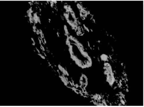
The characteristic fibrous expansion of the por-tal tracts associated with signs of porpor-tal vein dam-age and preservation of arterial and biliary struc-tures was documented in all cases. Feastruc-tures of chronic hepatitis of variable degree were present in 65% of the cases. Variable degree of venous muscular coat dispersion was histologically ob-served in all the cases of this series. It varied from the conspicuous presence of many smooth muscle fibers and cells radiating from the main portal vein and “buried” within the dense portal fibrous tis-sue (Fig. 1) to a few remnants or the complete
disappearance of the vein, with its replacement by fibrosis. Immunohistochemical and immunofluo-rescent data confirmed the histological findings. Dispersed smooth muscle cells were strongly stained by specific anti-a-smooth muscle actin
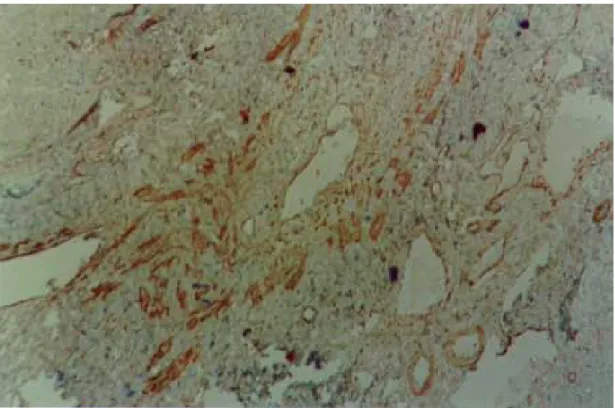
an-tibodies (Fig. 2). Other fusiform connective cells, faintly expressing actin, probably myofibroblasts, also appeared in variable number, dispersed among the smooth muscle cells and shortly beyond (Fig. 3). Actin-positive cells were observed around dam-aged portal veins, but not anywhere else in the dense fibrous tissue of the portal space.
Many cell-types in the vicinity of muscular coat dispersion were positive for desmin and vimentin. In relation to the peri-vascular muscular disper-sion these elongated and fusiform desmin and vimentin-positive cells were proportionally more evident while scattered actin-positive cells were becoming less and less numerous. Elastin was expressed in the same structures histologically stained by orcein. Elastic fibers appeared as con-tinuous or fragmented membranes delimiting por-tal vein structures, and laying free in variable amount, in diffuse or focal distribution, within the portal fibrous tissue. A more dense concentration of orcein-stained fibers were seen associated with areas of smooth muscle cell dispersion.
Myofibroblasts were positively identified by means of the electron microscope only in area where the smooth muscle cells were dispersed, which oc-curred in the vicinity of damaged small or medium-sized portal vein branches. Several attempts were made to identify myofibroblasts within the dense fi-brous tissue of the portal spaces away from the areas of venous changes, but without success. Collagen fibers with their typical periodicity were usually ar-ranged in compact parallel bundles, sometimes ac-companied by dark amorphous materials (elastin). Cellular elements were represented by typical fibro-blasts and slender cytoplasmic prolongations, prob-ably belonging to fibroblasts, and occasional leu-kocytes. Foci of collagen degradation showing rar-efied areas with fragmented collagen fibers of dif-ferent diameters and presence of granular electron dense material were sometimes seen.
89 89 89 89 89 Mem Inst Oswaldo Cruz, Rio de Janeiro, Vol. 94(1), Jan./Feb. 1999
Fig 1: large branch of the portal vein suggestive of old thrombosis with organization and re-canalization, showing dispersion of smooth muscle fibers around it. The presence of such vascular lesion in a fibrotic portal space with preservation of biliary and arterial structures is almost pathognomonic of schistosomal “pipestem” fibrosis. Hematoxylin-Eosin, 200 X.
90 90 90 90
90 Myofibroblasts and Schistosomiasis Zilton A Andrade et al.
Fig 3: dispersion of actin-positive cells around a damaged branch of the portal vein. There are elongated cells of different sizes, exhibiting different degrees of staining, suggestive of transformation and also disappearance of some dispersed smooth muscle cells. Streptavidin-biotin peroxidase method, 200 X.
91 91 91 91 91 Mem Inst Oswaldo Cruz, Rio de Janeiro, Vol. 94(1), Jan./Feb. 1999
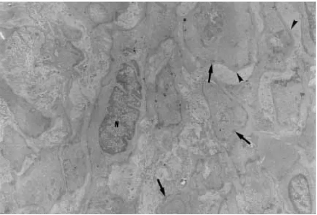
a clear perinuclear area. They appeared dispersed in several directions and isolated in the middle of collagen fibers, but when in contact with other similar cells, they frequently exhibited points of intimate cellular adhesion (Fig. 5). At some dis-tance from the vessel wall the dispersed smooth muscle cells appeared more or less modified and sometimes assumed the features of typical myofibroblasts. Further out the myofibroblasts gradually diminished in numbers while typical fi-broblasts were more frequently seen. There fre-quently appeared to be a transition between myofibroblasts and fibroblasts within these areas (Fig. 6).
DISCUSSION
Results from several studies have indicated that myofibroblasts are transient cells in which stress filaments (a-actin) are expressed in
connective-tis-sue cells in response to mechanical traction or con-traction (Gabbiani et al. 1972, Gabbiani 1996). In wound healing myofibroblasts usually disappear after some time through apoptosis (Gabbiani 1996), but in other circumstances they can probably re-vert to smooth muscle cells and/or fibroblasts.
However, as far as human schistosomal hepatic fibrosis is concerned, the only previous studies made on this subject indicate that myofibroblasts may have a more lasting and decisive presence (Grimaud & Borojevic 1977, 1986). Based on ul-trastructural findings, these authors suggested that severe hepatic lesions in schistosomiasis do not depend on the parasite eggs, but rather on a series of factors, culminating in vascular damage, myofibroblast proliferation and the production of excessive amount of extracellular matrix by these hyperplastic cellular elements. Present light, ultra-structural, immunoenzymatic and immunofluores-cent microscopic findings reveal that the main por-tal vein branch within the expanded porpor-tal space is often completely replaced by fibrosis, leaving a few or no traces of the muscular coat “buried” into the fibrous tissue. When remnants of the vascular muscular coat are present, their amount varies considerably from case to case or even in a same case. Numerous bundles of smooth muscle may appear in a section and only of few traces of actin-positive fibrils are detected in another. Therefore, observation of multiple sections are in keeping with the interpretation that myofibroblasts are
92 92 92 92
92 Myofibroblasts and Schistosomiasis Zilton A Andrade et al.
sient cells, most of them rapidly disappearing, some exhibiting signs of differentiation into fibroblasts. No ultrastructural evidence of apoptosis in dis-persed smooth muscle cells or in myofibroblasts was seen, at least as an outstanding finding, but disappearance through apoptosis cannot be ruled out. Probably this sequence of events seen in por-tal tracts is far from being peculiar to schistoso-miasis. As a matter of fact, such a sequence can be even experimentally demonstrated in vascular le-sions during the evolution of venous grafts (Shi et al. 1996, O’Brien et al. 1997).
Experimental findings in mice have shown that myofibroblasts derived from fat-storing cells par-ticipate in the formation of periovular granulomas in portal spaces (Barbosa Jr et al. 1993, Boloukhere et al. 1993). If the same holds true for human pa-thology, has not been determined, but during the present study the presence of myofibroblasts (left by healed granulomas?) in the dense portal fibrous tissue, away from peri-vascular muscular coat dis-persion, was not observed. Therefore, schistoso-mal portal fibrosis does not have the peculiar mor-phology of abnormal scars and myofibroblasts,
transiently appearing around damaged portal vein branches, do not exhibit evidence indicating a peculiar and important participation in the patho-genesis of pipestem fibrosis.
REFERENCES
Andrade ZA, Peixoto E, Guerret S, Grimaud JA 1992. Hepatic connective tissue changes in hepatosplenic schistosomiasis. Hum Pathol23: 566-573. Barbosa Jr AA, Pfeifer U, Andrade ZA 1993. Role of
fat-storing cells in schistosomal hepatic fibrosis of mice. Virchows Archiv B Cell Pathology64: 91-96. Berndt A, Kosmehl H, Katenkamp D, Tauchmann V 1994. Appearance of the myofibroblast phenotype in Dupuytren’s disease is associated with a fibronectin, laminin, collagen type IV and tenascin extracellular matrix. Pathobiology62: 55-58. Boloukhere M, Baldo-Correa E, Borojevic R 1993.
Ex-perimental schistosomiasis mansoni: characterization of connective tissue cells in hepatic periovular granu-lomas. J Submicrosc Cytol 25: 505-517. Darby I, Skalli O, Gabbiani G. 1990. a-smooth muscle
ac-tin is transiently expressed in myofibroblasts during experimental wound healing. Lab Invest63: 21-29. Eddy RJ, Petro JÁ, Tomasek JJ 1988. Evidence for the
non-muscle nature of the myofibroblasts of
93 93 93 93 93 Mem Inst Oswaldo Cruz, Rio de Janeiro, Vol. 94(1), Jan./Feb. 1999
lation tissue and hypertrophic scar. An immunofluo-rescence stydy. Am J Pathol130: 252-260. Gabbiani G 1996. The cellular derivation and the life
span of the myofibroblast. Pathol Res Pract192: 708-711.
Gabbiani G, Majno G 1972. Dupuytren’ s contracture: fibroblast contraction? An ultrastructural study. Am J Pathol 66: 131-146.
Gabbiani G, Hirschel BJ, Ryan GB, Statkov PR, Majno G 1972. Granulation tissue as a contratile organ. J Exp Med135: 719-734.
Grimaud J-A, Borojevic R 1977. Myofibroblasts in he-patic schistosomal fibrosis. Experiencia33: 890-892. Grimaud J-A, Borojevic R 1986. Portal fibrosis: intrahe-patic portal vein pathology in chronic human schisto-somiasis mansoni. J Submicrosc Cytol18: 783-793.
Kuhn C, McDonald JA 1991. The roles of the myofibroblasts in idiopathic pulmonary fibrosis. Ultrastrucctural and immunohistochemical features of sites of active extracellular matrix synthesis. Am J Pathol138: 1257-1265.
O’ Brien JE, Shi Y, Fard A, Buer T, Zalewski A, Mannion JD 1997. Wound healing around and within saphe-nous vein bypass grafts. J Thorac Cardiovasc Surg 114: 38-45.
Shi Y, O’Brien JE, Fard A, Mannion JD, Wang D, Zalewski A 1996. Adventitial myofibroblasts con-tribute to neointimal formation in injured porcine coronary arteries. Circulation 94: 1655-1664. Symmers WStC 1904. Note on a new form of liver
94 94 94 94