699 699 699 699 699 Mem Inst Oswaldo Cruz, Rio de Janeiro, Vol. 92(5): 699-706, Sep./Oct. 1997
An Experimental Approach to the Pathogenesis of
“Pipestem” Fibrosis (Symmers’ Fibrosis of the Liver)
Zilton A Andrade
+, Luciana M Silva, Marcia Maria de Souza
Laboratório de Patologia Experimental, Centro de Pesquisas Gonçalo Moniz -FIOCRUZ, Rua Valdemar Falcão 121, 40295-001 Salvador, BA, Brasil
Pathogenesis of schistosomal hepatic fibrosis (“pipestem” fibrosis of the liver) was investigated by means of the murine model. Although worm load appears as the main pathogenetic factor, alone it is not sufficient to produce that characteristic lesion. By comparing the findings in animals with heavy and prolonged Schistosoma mansoni infection, which developed or not “pipestem” fibrosis, it was observed that the lesion was more frequent in intact animals than in the splenectomized one. However, the size of the spleen, the number of recovered worms, the number of eggs per gram of liver tissue, the level of serum idiotype and anti-idiotype antibodies, the size and volume of periovular granulomas formed in the liver, all that failed to show statistically significant differences between the two groups. After analysing all these data, other factors, that apparently have been hitherto negleted, rested to explain the findings. Among them, the timing and sequence of the egg-induced intrahepatic vascular changes seemed cru-cial. The sequential development of intrahepatic portal vein obstruction, followed by the opening of periportal collateral veins and the continous arrival of schistosome eggs going to be lodged into the latter, appeared as essential steps in the pathogenesis of “pipestem” fibrosis.
Key words: Schistosoma mansoni - “pipestem” fibrosis - pathogenesis
One of the most characteristic, if not pathog-nomonic, lesion of human pathology is that first described by Symmers (1904), and since then known as “pipestem” fibrosis of the liver. It repre-sents the morphological counterpart of hepatosplenic schistosomiasis, which is clinically characterized by hepato-splenomegaly, portal hy-pertension (esophageal varices) and variable de-grees of pancytopenia (hypersplenism), usually in the absence of signs of hepato-cellular failure. The lesion involves every branch of the portal tree, the fibrous tissue that enlarges the portal spaces appearing at the cut surface of the liver as whitish plaques against a background of normal-looking parenchyma (Fig. 1).
Lichtenberg and Sadun (1968) listed the fol-lowing anatomical characteristics for the lesion: (a) grossly evident, diffuse, stellate fibrosis and en-largement of the large and medium-sized portal fields; (b) variable portal inflammatory infiltration and crowding of the portal areas with schistosome eggs and granulomas (active stage) or scar tissue containing egg shells and pigment (late stage); (c) conservation of the lobular architecture, except for porto-central fibrous banding or post-necrotic
scar-Financial support was provided by PAPES-1 (FIOCRUZ).
+Corresponding author. Fax: +55-71-356.4292
Received 16 April 1997 Accepted 30 June 1997
ring usually confined to the subcapsular paren-chyma; (d) relatively normal hepatocytic structure and function; (e) destructive lesions of the intra-hepatic portal radicles.
Pathogenesis of “pipestem” fibrosis is appar-ently simple. Heavily infected patients develop periportal fibrosis with vascular obstruction due to the deposition of numerous schistosome eggs along the trajectory of the portal vein. This expla-nation is in keeping with quantitative findings ob-tained by Cheever (1965) who counted worms by doing perfusion of the portal system of cadavers. It also agrees with the fact that severe schistoso-miasis with “pipestem” fibrosis is not to be seen in subjects with mild schistosome infections. As a matter of fact, a good rule of the general pathol-ogy of helminthic infections indicates that severe clinical manifestations are related to heavy worm burden. On the other hand, it is common experi-ence that people with heavy Schistosoma mansoni
700 700 700 700
700 Pipestem Fibrosis in Mice • ZA Andrade et al.
Attempts to investigate the problem of addi-tional factors in the pathogenesis of hepatosplenic schistosomiasis in humans have not been very suc-cessful. This subject has been reviewed by Prata (1991). Considerable attention has been paid to genetic background, especially to race and HLA determinants of hepatosplenic patients, but results have so far been inconclusive. The age of the pa-tients when first infected, the frequency of expo-sure to contaminated waters, the failure to gener-ate anti-idiotypic antibodies having a modulatory influence on periovular granulomatous formation represent the main lines of research. Results have been so variable, that the need for a good experi-mental model is well recognized.
EXPERIMENTAL APPROACH
Warren (1966) observed that mice, infected with 1-2 worm pairs of S. mansoni and left for prolonged periods of time (more than 16 weeks), developed systematized periportal fibrosis due to a concentration of periovular granulomas and fi-brosis along the portal spaces. Although the lesion was only observed microscopically, its similarity to human “pipestem” fibrosis was unquestionable. At first it was stated that chronic infection in mice
did not lead to periportal fibrosis (Cheever 1965) and this was probably a common opinion, since this interesting model was not further explored for some time after Warren’s paper.
While performing studies on S. mansoni infec-tion in chimpanzees, Sadun et al. (1970) observed the development of typical, gross and microscipical “pipestem” fibrosis in some heavily infected ani-mals. Difficulties of several types, especially con-cerning the obtaining, maintenance and manage-ment of these primates, turned the model of little practical usefulness.
In the last 10 years we became interested in the mouse model of “pipestem” fibrosis. Our attention to the subject came by sheer chance. While inves-tigating whether fibrosis in late periovular granu-lomas would also be reversible after treatment, we submitted mice to light infection with only 15-20 cercariae in order to decrease mortality, and waited up to 20 weeks before treating them. As a result we observed that some mice with prolonged in-fection not only developed periportal fibrosis as Warren (1966) had described, but that periportal fibrosis was also susceptible to degradation fol-lowing specific chemotherapy (Andrade & Grimaud 1986). Our interest specially arouse
701 701701 701701 Mem Inst Oswaldo Cruz, Rio de Janeiro, Vol. 92(5), Sep./Oct. 1997
cause of earlier findings with the technique of plas-tic vascular injection followed by corrosion in strong acid for the obtaining of portal vein casts.
S. mansoni infected mice revealed the development of a periportal cuffing of collateral vessels (Fig. 2, Andrade & Brito 1981). This finding appeared then as the missing link necessary to explain the some-what curious distribution of eggs along periportal spaces (Andrade 1987). The questions now were: how frequently the periportal lesion developed in mice with chronic infection and how representa-tive of human schistosomisis it was? The material then studied included a group of outbred mice and several inbred strains. This study was comple-mented with material taken from the files of the Laboratory of Parasitic Diseases, NIH, Bethesda, MD, USA. The development of schistosomal peri-portal fibrosis occurred in both outbred and in-bred strains of mice, indicating that genetic back-ground did not seem to be a crucial factor. The percentages of “pipestem” fibrosis was quite vari-able in the several groups of mice, in general around 50%. Table shows the distribution of the lesions according to mouse strains. Mice that failed to de-velop the “pipestem” lesion showed scattered iso-lated granulomas instead. As to the significance of the model, it was considered valid as an experi-mental model of human “pipestem” fibrosis. Ac-tually, the lesion was represented by a concentra-tion or periovular granulomas along periportal ar-eas, rather than a diffuse periportal fibrosis. But, there occurred portal vein obstructive lesions and a certain amount of extra-granulomatous peripor-tal fibrosis (Andrade & Cheever 1993).
A mouse model with marked and dense peri-portal fibrosis was described by Henderson et al. (1993) in about 20% of male CBA/J inbred mouse chronically infected with approximately 45 S. mansoni cercariae. With this model “pipestem” fi-brosis was accompanied by massive splenomegaly,
a feature not so impressive in previous studies. Furthermore, the animals with “pipestem” fibrosis exhibited a peculiar anti-idiotypic profile against anti-egg antigen, which was distinct from those mice that did not develop the lesion. Similar sero-logical findings have been obtained by the same group in hepatosplenic patients (Colley et al. 1986). This led to the concept that immunological modu-latory failure during periovular granuloma forma-tion is an important pathogenic factor for the de-velopment of “pipestem” fibrosis.
RECENT EXPERIMENTS
Generalities - Experiments in our laboratory have been performed in outbred albino young (15-18 g) Swiss mice of both sexes. Animals were sub-mitted to infection by the transcutaneous route with about 20-30 recently shed S. mansoni cercariae. Only those animals which were eliminating viable eggs in the stools were included in the study. Sac-rifice took place about 20-22 weeks from cercarial exposure. Paraffin embedded histological sections stained with sirius red (Junqueira et al. 1979) were then examined with the low power of the
micro-Fig. 2: vascular plastic cast from the portal system of a mouse with chronic schistosomiasis. Numerous fine collaterals give a hairy appearance to the portal vein branches.
TABLE
General data on the murine model of schistosomal “pipestem” fibrosis of the liver
Strain Duration of “Pipestem” fibrosis Eggs/liver Spleen wt.
infection (wks) Yes (%) No (%) (x1,000) (mg)
Outbred 23 27 (54) 23 (46) 30 No data
C57BL/8 20 6 (42.8) 8 (57.2) 24 249
C57B1/6 52 21 (70) 9 (30) 62 217
BALB/c 52 8 (80) 2 (20) 50 225
BALB/c 69 10 (79.9) 3 (23.1) 74 265
C3H/HeN 22 6 (66.6) 3 (33.3) 25 336
C3H/HeN 41 7 (63.6) 4 (36.4) 27 407
C3H/HeN 52 5 (100) 0 46 420
702 702 702 702
702 Pipestem Fibrosis in Mice • ZA Andrade et al.
scope. Infected mice, harboring 1-2 pairs of worms presented with either one of the following lesions: isolated periovular granulomas (Fig. 3) or perio-vular granulomas and fibrosis preferentially dis-tributed along the periportal spaces (Fig. 4). Usu-ally, the portal spaces in this last group appeared amplified by fibrosis and stood out as dense stel-late areas on a background of unstained paren-chyma, forming a very characteristic lesion (Fig. 5). Discrimination between the two types of lesions was usually clear-cut. Only rarely did a mixture of the two lesions occur, in such a way as to make the decision difficult.
Role of the spleen and other factors - With this very simple basic methodology, experiments were performed to compare “pipestem” positive and negative animals according to the following pa-rameters: the influence of the spleen (splenecto-mized versus sham-operated animals), spleen weight and splenic microscopic changes, worm load (number of eggs per gram of liver tissue), morphometric measurements of periovular granu-lomas and general immunological abnormalities (idiotype and idiotype schistosome anti-bodies).
In these recent sets of experiments (Andrade et al. manuscript in preparation), “pipestem” fibro-sis of the liver appeared in 38.3% of 94 mice. In 60 splenectomized animals (61.8%) the lesion was present in 15 (25%) and absent in 45 (75%). Among 34 (36.2%) sham-operated animals the lesion
de-veloped in 21 (61.8%) and was absent in 13 (36.2%). These results pointed out, in a statisti-cally significant way, that the presence of the spleen does favour the development of “pipestem” fibro-sis. However, no clear-cut differences appeared be-tween splenectomized and sham-operated animals regarding the levels of serum antibodies against S. mansoni or the levels of anti-idiotypic antibodies tested against whole adult worm antigens, the size of the spleen (measured as splenic index), and the number of eggs per gram of liver tissue or the number of worms recovered from perfusion of the portal system.
Experiments with undernourished mice - An attempt to investigate the influence of nutrition on the development of “pipestem” fibrosis was per-formed in two groups of mice (Coutinho et al. manuscript in preparation): one was maintained on a basically low-protein diet that has been intended to represent the basic regional diet of poor people living in endemic areas for schistosomiasis in northeastern Brazil (Coutinho 1980); the other was a control group of mice maintained on a normal commercial balanced chow. The same basic meth-odology described above was used. Undernour-ished animals reached only half the weight of the controls at the end of experiments. They had fewer worms and fewer eggs per worm pairs, smaller liver and spleen, both in absolute terms and in re-lation to body weight. Hepatic periovular granulo-mas were significantly smaller than those in
703 703703 703703 Mem Inst Oswaldo Cruz, Rio de Janeiro, Vol. 92(5), Sep./Oct. 1997
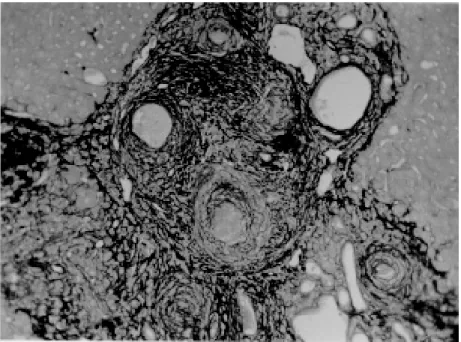
Fig. 4: periovular granulomas are numerous and show a tendency to accumulate along the periportal space. Note that the portal space is amplified by fibrosis and the main portal vein branch presents sub-intimal thickening. Picro-sirius-red method for col-lagen, X 150.
704 704 704 704
704 Pipestem Fibrosis in Mice • ZA Andrade et al.
trol mice. None of the 16 mice maintained on defi-cient diet developed “pipestem” fibrosis at the end of 16 weeks of infection, while 7 (43.7%) out of 16 on a normal diet did. The total amount of fibro-sis (collagen) in the liver was decreased in under-nourished mice as compared to controls when evaluated both by morphometry and biochemistry (hydroxiproline).
DISCUSSION
The task of evaluating the results obtained in our laboratory with the murine model is not a simple one. First of all we have to question whether the model really represents the lesion seen in ad-vanced schistosomiasis of man. As a matter of fact, this contention appeared even before (Cheever 1965) the model was first described by Warren (1966). A critical appraisal of the murine model of “pipestem” fibrosis has already been attempted (Andrade & Cheever 1993). Weighing similarities and differences, it has been concluded that the model stands up as a miniature of the human “pipestem” fibrosis and, being so, the model is worth of experimental investigation. Not only the eggs and the granulomas they elicited were con-centrated within the portal spaces, but there were also extra-granulomatous fibrosis and vascular le-sions (Fig. 6). As in the human lesion, portal
vas-cular lesions include angiogenesis, phlebosclero-sis, narrowing and obstruction (Fig. 7).
Considering the worm load of the animals, one important point was raised by Cheever (1968). He estimated that the heaviest infection reported in man at autopsy have seldom been grater that 5 worm-pairs per kg of body weight, whereas in a 20 g mouse the lightest infection corresponds to about 50 worm pairs per kilogram of body weight. Therefore, animals in the experiments reported above were all heavily infected. They harbored 1-2 worm pairs and their livers contained from 6,400 to 10,300 schistosome eggs. This means that in animals developing periportal fibrosis other factor(s) might have influenced the distribution of eggs all along the periportal spaces.
The murine “pipestem” fibrosis is neither seen in early (8-12 weeks), nor in late, massive (50 cer-cariae up) infections. In these latter infections eggs are not orderly distributed and concentrated in portal spaces, but appear throughout the liver, pro-ducing fibrosis and distorting the hepatic vascula-ture without a definite pattern. The importance of the “right” number of eggs and the timing of their deposition within the liver, probably in concert with a sequence of vascular changes are therefore ap-parent. In view of the negative results obtained when such factors as genetical, immunological and
705 705705 705705 Mem Inst Oswaldo Cruz, Rio de Janeiro, Vol. 92(5), Sep./Oct. 1997
parasitological features were investigated, the im-portance of the factor sequential vascular changes
assumes a prime importance for explaining the pathogenesis of “pipestem” fibrosis in mice.
Studies with injection of plastic material into the portal vein, followed by corrosion in strong acid, have yielded casts which showed numerous collateral vessels sprouting out from the branches of the intrahepatic portal vein in mice chronically infected with S. mansoni, a finding not seen in non-infected controls (Andrade & Brito 1981). These vascular changes allow for the schistosome eggs to be lodged in small vessels distributed along the periportal tissues.
For the development of this crucial vascular alteration, it is presumably necessary a series of sequential events: a somewhat disseminated and sudden obstruction of portal radicles by periovu-lar granulomas, generation of intrahepatic portal hypertension, opening of periportal collaterals, ar-rival of schistosome eggs into these new vascular canals. Factors influencing this chain of events are probably multifactorial. Of course, the produc-tion of numerous parasite eggs is essential. A proper (normal) immunological response is also important, as the data from undernourished mice indicated. But the timing of the vascular changes and the arrival of new eggs seem essential for the end-result of “pipestem” fibrosis.
Formation of peri-portal vascular cuffing was a prominent finding seen in human “pipestem”
fi-brosis when plastic casts were obtained with the injection/corrosion methodology (Bogliolo 1957, Andrade & Cheever 1971).
It seems still premature to say that the patho-genesis of “pipestem” fibrosis of the liver is the same for mouse and man.
In some patients with hepatosplenic schistoso-miasis pulmonary arteritis develops as a compli-cation. It has been shown that when a few eggs reach the lungs they are lodged in alveolar pre-capillaries, forming discrete granulomas. Severe lesion does not result from gradual accumulation of eggs, but from a sudden and massive emboliza-tion to the lungs. Then, intra-pulmonary hyperten-sion causes dilatation of fine collaterals that come out at right angles from large and medium-sized pulmonary arteries. If the arrival of eggs contin-ues, many of them are stopped at the entrance of those collaterals causing severe arterial and arteri-olar damage, with vascular obstruction that results in chronic “cor pulmonale”. This is a good example of vascular changes occurring in concert with para-site stimuli to produce a sequence of changes to-ward a characteristic schistosomal picture (Andrade & Andrade 1970). No particular racial or immunological features were apparent in those 14 out of 78 hepatosplenic patients (17.9%) who concomitantly developed chronic schistosomal
“cor pulmonale”.
The pathogenesis now postulated for the patic lesion is basically the same for both the
706 706 706 706
706 Pipestem Fibrosis in Mice • ZA Andrade et al.
patic and the pulmonary lesions of advanced schistosomiais.
REFERENCES
Andrade ZA 1987. Pathogenesis of pipe stem fibrosis of the liver (experimental observation on murine schistosomiasis). Mem Inst Oswaldo Cruz82: 325-334.
Andrade ZA, Andrade SG 1970. Pathogenesis of pul-monary schistosomiasis. Am J Trop Med Hyg19: 305-310.
Andrade ZA, Brito PA 1981. Evolution of schistosomal hepatic vascular lesions after specific chemotherapy.
Am J Trop Med Hyg30: 1223-227.
Andrade ZA, Cheever AW 1971. Alterations of the in-trahepatic vasculature in hepatosplenic schistosomia-sis mansoni. Am J Trop Med Hyg20: 425-432. Andrade ZA, Cheever AW 1993. The characterization
of the murine model of schistosomal periportal fi-brosis (“pipestem” fifi-brosis). Int J Exp Med74: 195-202.
Andrade ZA, Grimaud J-A 1986. Evolution of the schis-tosomal hepatic lesions in mice after curative che-motherapy. Am J Pathol124: 59-65.
Bogliolo L 1957. The anatomical picture of the liver in hepatosplenic schistosomiasis mansoni. Ann Trop Med Parasitol51: 1-14.
Cheever AW 1965. A comparative study of Schistosoma mansoni infections in mice, gerbils, multimammate rats and hamsters. II - Qualitative pathological dif-ferences. Am J Trop Med Hyg14: 227-238. Cheever AW 1968. A quantitative post-mortem study of
schistosomiasis mansoni in man. Am J Trop Med Hyg 17: 38-64.
Colley DG, Garcia AA, Lambertucci JR, Parra JC, Katz N, Rocha RS, Gazinelli G 1986. Immune responses during human schistosomiasis. XII. Differential
re-sponsiveness in patients with hepatosplenic disease.
Am J Trop Med Hyg 35: 793-802.
Coura JR, Conceição MJ 1981. Correlação entre carga parasitária de S. mansoni e gravidade das formas clínicas em uma comunidade rural de Minas Gerais.
Rev Soc Bras Med Trop19: 69-73.
Coutinho EM 1980. Patobiologia da desnutrição nas doenças parasitárias. Mem Inst Oswaldo Cruz75: 63-76.
Henderson GS, Nix NA, Montesano MA, Gold D, Free-man Jr GL, McCurley TL, Colley DG 1993. Two distinct pathological syndromes in male CBA/J in-bred mice with chronic Schistosoma mansoni infec-tions. Am J Pathol142: 703-714.
Junqueira LCU, Bignolas G, Brentani R 1979. Picrosirius staining plus polarization microscopy, a specific method for collagen detection in tissue sec-tions. Histochem J11: 447-455.
Lichtenberg F, Sadun EH 1968. Experimental produc-tion of bilharzial pipe-stem fibrosis in the chimpan-zee. Exp Parasitol22: 264-278.
Prata A 1991. Fatores determinantes das formas anátomo-clínicas e evolução da esquistossomose, p. 3-12. In FP Castro, PRS Rocha, AS Cunha (eds),
Tópicos em Gastroenterologia, MEDSI-Editora Médica e Científica Ltda, Rio de Janeiro.
Sadun EH, Lichtenberg F, Cheever AW, Erickson DG 1970. Schistosomiasis mansoni in the chimpanzee. The natural history of chronic infections after single and multiple exposures. Am J Trop Med Hyg19: 258-277.
Symmers WStC 1904. Note on a new form of liver cir-rhosis due to the presence of the ova of Bilharzia haematobia. J Pathol Bacteriol9: 237-239. Warren KS 1966. The pathogenesis of “clay-pipe stem