Effect of strenuous maternal exercise
before and during pregnancy on rat
progeny renal function
Departamento de Biologia, Faculdade de Filosofia Ciências e Letras de Ribeirão Preto, Universidade de São Paulo, Ribeirão Preto, SP, Brasil
A.O. Oliveira, C. Fileto and M.S. Melis
Abstract
The effects of strenuous exercise before and during pregnancy on the renal function and morphological alterations of the progeny were determined in a study on female Wistar rats. This research was done based on a previous study carried out in our laboratory, which showed morphological alterations in rats submitted to this kind of exercise. As the form is related to the function, the physiological relevance of submitting a pregnant female to a high-intensity exercise training regimen could be explained by the fact that morphological alterations can influence kidney function. The animals were assigned to one of two groups: control animals that did not exercise during pregnancy and trained animals that swam for 120 min 5 days a week for 8 weeks before pregnancy and daily for 60 min over a period of 8 weeks starting on the second day of pregnancy. Seven rats of each group were analyzed for morphological alterations and for renal function. The progeny of the rats used for morphological evaluation were born by cesarean section and the progeny of the animals used to evaluate renal function were born normally. The progeny were two months old when renal function was evaluated. Fertility and morbidity were the same for both groups. Strenuous maternal exercise had no significant influence on glomerular filtration rate (GFR) but renal plasma flow was lower in the progeny of the trained group (mean ± SD, 16.65 ± 3.77 ml min-1 kg-1) compared to the progeny of the control group (33.42 ± 2.56 ml min-1 kg-1). Antidiuretic and antinatriuretic effects on the progeny of the trained group were observed, since urine flow as percentage of GFR and the fraction of urinary sodium excretion were lower in this group (1.38 ± 0.10 and 0.60 ± 0.04%, respectively) compared to the progeny of the control group (2.36 ± 0.11 and 1.55 ± 0.20%, respec-tively). Moreover, in this exercise program, fetuses from trained animals were small-sized (2.45 ± 0.19 vs 4.66 ± 2.45 g for control animals) and showed lower differentiation compared to fetuses from the control group. These effects were probably caused by caloric restriction, hypoxia and reduction of umbilical cord length.
Correspondence
M.S. Melis
Laboratório de Fisiologia Departamento de Biologia FFCLRP, USP
Av. Bandeirantes, 3900 14040-901 Ribeirão Preto, SP Brasil
E-mail: [email protected]
C. Fileto is the recipient of a CNPq fellowship (No. 522151-94.9).
Publication supported by FAPESP.
Received December 2, 2002 Accepted December 1, 2003
Key words
•Renal function
•Exercise during pregnancy •Fetus
Conflicting results have been reported concerningthe effect of endurance exercise before or during pregnancy on fetal develop-ment. Studies on rats and mice have indi-cated that endurance exercise during preg-nancy causes a decrease in fetal size (1,2). However, other investigators did not find a correlation between maternal endurance ex-ercise and fetal birth weight (3). Recent stud-ies have shown that moderate physical exer-cise practiced before and during pregnancy by rats (4) and by women (5) did not affect fetal development, while strenuous antena-tal exercise caused reduced birth weight, umbilical cord length and placental weight (1). These conflicting results about the ef-fects of maternal exercise on the fetus are probably due to the different exercise inten-sities to which the mothers were submitted, to animal familiarity with the physical exer-cise or to different species of animals used (6).
Little information is available about the modifications of renal parameters induced by physical exercise in the progeny, but re-nal function during exercise has been stud-ied extensively in man and animals. Pioneer-ing experiments carried out on humans by Wesson Jr. (7) and later by Castenfors (8) showed that physical exercise reduces renal blood flow and glomerular filtration rate (GFR). A decrease in urinary flow and in renal sodium excretion was also observed, probably caused by a higher antidiuretic hor-mone and aldosterone release during exer-cise (8). These results were later confirmed in humans and rats (9) and in horses (10).
Renal responses to physical exercise are related to exercise intensity. At low exercise intensity, urinary flow and sodium excretion tend to increase while at high exercise inten-sity both parameters are decreased consider-ably (11). Similar results were observed in a study of different renal responses to exercise in humans that ran various distances (12). Thus, the kidney, whose primary function is to regulate the volume and the extracellular
liquid composition, can suffer changes in hemodynamics and in sodium and water ex-cretion during exercise, and the magnitude of these alterations is related to exercise intensity.
The effects of endurance exercise train-ing before and durtrain-ing pregnancy on the renal function of the progeny have not been fully clarified. Considering the importance re-cently attributed to physical activities, in-cluding those practiced by pregnant women in physical exercise programs, the purpose of the present study was to investigate the effects of forced training of female rats
be-fore and during pregnancy on a)
morpho-logical alterations of the fetus, placenta and umbilical cord, and b) renal function of prog-eny (renal plasma flow, GFR, urine flow, and urinary sodium excretion).
The study was conducted on female Wistar rats weighing 110 to 130 g. The ani-mals were fed a standard pellet diet (Purina Nutrimentos Ltda., Campinas, SP, Brazil) and had free access to water until the morn-ing of the first day of forced swimmmorn-ing.
of 130 g, when renal function was analyzed. The animals that were not exercised received the same treatment as the trained ones, ex-cept for the swimming sessions.
Rats born to mothers submitted or not to exercise were anesthetized with sodium pen-tobarbital (30 mg/kg), placed on a tempera-ture-regulated table and submitted to trache-otomy. Their body temperature was 37º to 37.5ºC. The carotid artery was catheterized for blood collection and polyethylene cath-eters were also inserted into the jugular vein for infusion of Ringer solution. The bladder was catheterized through an abdominal inci-sion for urine collection.
Isotonic Ringer solution containing 10% inulin and 2% p-aminohippuric acid (PAH) was infused iv at the rate of 0.03 ml/min
throughout the course of the experiment. After infusion was started, the animals were allowed to equilibrate for 40 to 60 min, and urine was then collected from the bladder at 30-min intervals. Blood was withdrawn at the midpoint of each clearance period.
Clearance studies were performed on two groups of animals: 1) control progeny, born to mothers which did not exercise, and 2) trained progeny, born to mothers that had exercised. The group of exercised mothers swam 5 days a week for 8 weeks, for 120 min before and 60 min during pregnancy. Each group contained seven animals.
Inulin concentrations in plasma and urine were determined by the anthrone method (13). Plasma and urinary PAH concentra-tions were measured by the method of Smith et al. (14). Sodium and potassium concentra-tion in urine and plasma were determined with a Klina flame photometer (Beckman Instruments, Fullerton, CA, USA).
The Student t-test was used for statistical
analysis of the data, and the results are re-ported as the mean ± SD, with the critical level of significance set at P < 0.05.
The final body weights of animals of the control and trained groups were 234.60 and 219.00 g, respectively (Table 1). A
signifi-cant difference was found only during week 8 of training. Animals of the control group gained approximately 17.9 g more weight than those of the trained group.
Mean fetal body weight (on pregnancy day 21) was 4.66 g for the control group and 2.45 g for the trained group (P < 0.01; Figure 1).
Mean placental and umbilical cord weights were significantly higher in fetuses of the control group compared to those of the trained group (P < 0.01; Table 1). The mean length of the umbilical cord of the fetuses of the trained group was shorter compared to the control group (P < 0.01). Kidney weight was similar for the animals of both groups.
Endurance exercise did not affect the GFR of the progeny, which was the same for both groups, but a significant decrease in urinary flow was observed for the trained group (Table 1). Renal plasma flow decreased and filtration fraction increased in the trained group. Fractional sodium excretion de-creased, suggesting a potential antinatriuretic effect of endurance exercise on the progeny, and the plasma sodium concentration was similar for the control and trained groups (Table 1).
Figure 1. Pups born to mothers of the control (C) and trained (T) groups. Fetal (g) and placental (mg) weights are reported as means ± SD for 7 pups in each group.
C T
4.66 ± 2.45 2.45 ± 0.19
The mean body weight of trained fetuses was lower than that of control fetuses. Fetal growth seems to be influenced by maternal activity, as shown in some investigations which reported significantly larger babies born to moderately trained women compared to non-trained or heavily trained women (2). In the latter group, the reduction could be explained by a lower neonatal fat mass. Therefore, it has been suggested that intense maternal exercise may compromise fetal de-velopment (1,4,5,15).
In our experiments, the placental weight of rats submitted to exercise was lower than the controls, in agreement with some studies (4) that showed a decrease of placental weight parallel to the decrease of fetal body weight. A smaller placenta probably has a lower blood flow, resulting in significant fetal hy-poxia that may lead to retarded intrauterine growth (4). On the other hand, moderate maternal exercise enhances fetal and placen-tal growth (3).
In the present study, the umbilical cords of fetuses whose mothers were submitted to endurance exercise were shorter, indicating limitation of fetal movement (1).
It is important to emphasize that there
have been no previous reports of renal func-tion in pups born to mothers who practiced moderate or endurance exercise. Studies on humans using PAH clearance have demon-strated a decrease in renal plasma flow after physical exercise which was related to exer-cise intensity (16,17). An explanation for this reduced flow could be the intrarenal blood flow distribution, leading to a drop in the rate of renal PAH extraction during exer-cise (17).
In the present study, we observed a marked decrease in renal plasma flow in the progeny of trained mothers compared to con-trol, which agrees with the results cited above. During exercise, blood flow can be deviated from the renal circulation to active muscle
(18), but it has been shown that GFR is
maintained constant until exercise intensity is increased (9,16,19). In horses, GFR de-creases by about 40% at any exercise inten-sity (10); in man, GFR seems to be more resistant to the effects of exercise, but it is hard to compare exercise intensity between such different species. Despite the signifi-cant decrease in renal plasma flow induced by strenuous exercise, GFR did not change in our experiments. Probably, the balance of water and salt was maintained in the animal body and/or the modification of the tonus of afferent and efferent arterioles kept the GFR constant.
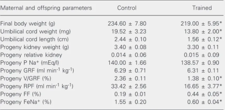
We also observed a decrease in urinary flow in animals born to mothers that exer-cised compared to the control mothers, even when this parameter was calculated in terms of GFR percentage. It is commonly found in the literature that increased antidiuretic hor-mone and aldosterone release due to endur-ance exercise plays a role in the process of urinary flow reduction observed in animals exercised during pregnancy (8,9). However, no experiments were done concerning this point in the present study. A reduction of sodium excretion in animals whose mothers were submitted to swimming during preg-nancy was also observed. The literature Table 1. Effect of strenuous maternal exercise before and during pregnancy on rat
progeny renal function.
Maternal and offspring parameters Control Trained
Final body weight (g) 234.60 ± 7.80 219.00 ± 5.95*
Umbilical cord weight (mg) 19.52 ± 3.23 13.80 ± 2.00*
Umbilical cord length (cm) 2.44 ± 0.10 1.56 ± 0.12*
Progeny kidney weight (g) 3.40 ± 0.08 3.30 ± 0.11
Progeny relative kidney 0.014 ± 0.06 0.015 ± 0.09
Progeny P Na+ (mEq/l) 140.00 ± 1.66 138.57 ± 0.90
Progeny GRF (ml min-1 kg-1) 6.29 ± 0.71 6.31 ± 0.11
Progeny V/GRF (%) 2.36 ± 0.11 1.38 ± 0.10*
Progeny RPF (ml min-1 kg-1) 33.42 ± 2.56 16.65 ± 3.77*
Progeny FF (%) 0.19 ± 0.01 0.44 ± 0.05*
Progeny FeNa+ (%) 1.55 ± 0.20 0.60 ± 0.04*
Data are reported as means ± SD for 7 animals per group. FeNa+ = fraction of urinary
sodium excretion; FF = filtration fraction; GRF = glomerular filtration rate; P Na+ =
shows that the decrease of sodium and water excretion during strenuous exercise in spite of an unchanged GFR may be explained by high production of aldosterone and by the deviation of blood from the kidneys to other organs during exercise (11,17). Therefore, the model used in the present study does not discriminate between the direct and indirect effects of endurance training on renal so-dium reabsorption.
In summary, in Wistar rats strenuous physical exercise during pregnancy can re-sult in smaller and less differentiated fe-tuses, effects probably related to caloric re-striction, hypoxia and reduction of umbilical cord length. Thus, maternal physical exer-cise decreases renal plasma flow and has antidiuretic and antinatriuretic effects on the progeny.
References
1. Ribeiro CAL, Maia Campos G, Melis MS, Sala MA & Lopes RA (1991). Alteraciones morfológicas y estereológicas del epitelio lin-gual de fetos de rata provocadas por el ejercicio materno. Archivos de la Facultad de Medicina de Zaragoza, 30: 84-90.
2. Riemann MK & Hansen ILK (2000). Effects on the fetus of exercise in pregnancy. Scandinavian Journal of Medicine and Science in Sports, 10: 12-19.
3. Bell R & Palma S (2000). Antenatal exercise and birthweight. Aus-tralian and New Zealand Journal of Obstetrics and Gynaecology, 40: 70-73.
4. Houghton PE, Mottola MF, Plust JH & Schachter CL (2000). Effect of maternal exercise on fetal and placental glycogen storage in the mature rat. Canadian Journal of Applied Physiology, 25: 443-452. 5. Clapp 3rd JF, Kim H, Burciu B, Schmidt S, Petry K & Lopez B
(2002). Continuing regular exercise during pregnancy: Effect of exercise volume on fetoplacental growth. American Journal of Obstetrics and Gynecology, 186: 142-147.
6. Pivarnik JM (1996). Cardiovascular responses to aerobic exercise during pregnancy and postpartum. Seminars in Perinatology, 20: 242-249.
7. Wesson Jr LG (1974). Kidney Function in Exercise. 2nd edn. How & Harper, New York.
8. Castenfors J (1967). Renal function during exercise. Acta Physi-ologica Scandinavica, 70: 1-44.
9. Poortmans JR & Vanderstareten J (1994). Kidney function during exercise in healthy and diseased humans. An update. Sports Medi-cine, 18: 419-437.
10. Gleadhill A, Marlin D, Harris PA & Michell AR (2000). Reduction of renal function in exercising horses. Equine Veterinary Journal, 32: 509-514.
11. Shizuru EM, Freud BJ, Hashiru GM & Clay-Baugh JR (1991). Hormo-nal, electrolyte, and renal responses to exercise are intensity de-pendent. Journal of Applied Physiology, 70: 900-906.
12. Poortmans JR (1995). Renal response to exercise in healthy and diseased patients. Nephrologie, 16: 317-324.
13. Fuehr J, Kaczmarzik J & Kruttgen CD (1955). Eine einfache kolorimetrische Methode zur Inulin Bestimmung für Nierenclearance Untersuchungen bei Stoffwechsel Gesunden und Diabetikern. Klini-sche Wochenschrift, 33: 729-730.
14. Smith HW, Einkelstein N, Aliminosa L, Crawford B & Graber M (1945). The renal clearances of substituted hippuric acid derivatives and other aromatic acids in dog and man. Journal of Clinical Investi-gation, 24: 388-404.
15. Terada M (1974). Effects of physical activity before pregnancy on fetuses of mice exercised forcibly during pregnancy. Teratology, 10: 141-144.
16. Clapp 3rd JF, Kim H, Burciu B & Lopez B (2000). Beginning regular exercise in early pregnancy: Effect on fetoplacental growth. Ameri-can Journal of Obstetrics and Gynecology, 183: 1484-1488. 17. Poortmans JR (1990). Postexercise proteinuria in normal and
dis-eased humans. Japanese Journal of Constitutional Medicine, 54: 8-18.
18. Hammond RL, Augustyniak RA, Rossi NF, Churchill PC, Lapanowski K & O’Leary DS (2000). Heart failure alters strength and mechan-isms of the muscle metaboreflex. American Journal of Physiology, 278: H818-H828.