Evaluation by blue native
polyacrylamide electrophoresis
colorimetric staining of the effects
of physical exercise on the activities of
mitochondrial complexes in rat muscle
1Departamento de Bioquímica, Instituto de Biologia,
Universidade Estadual de Campinas, Campinas, SP, Brasil
2Department of Pharmacology, University of Padova, Padova, Italy
A.M. Molnar1,
A.A. Alves1,
L. Pereira-da-Silva1,
D.V. Macedo1 and
F. Dabbeni-Sala2
Abstract
Blue native polyacrylamide electrophoresis (BN-PAGE) is a tech-nique developed for the analysis of membrane complexes. Combined with histochemical staining, it permits the analysis and quantification of the activities of mitochondrial oxidative phosphorylation enzymes using whole muscle homogenates, without the need to isolate muscle mitochondria. Mitochondrial complex activities were measured by emerging gels in a solution containing all specific substrates for NADH dehydrogenase and cytochrome c oxidase enzymes (com-plexes I and IV, respectively) and the colored bands obtained were measured by optique densitometry. The objective of the present study was the application of BN-PAGE colorimetric staining for enzymatic characterization of mitochondrial complexes I and IV in rat muscles with different morphological and biochemical properties. We also investigated these activities at different times after acute exercise of rat soleus muscle. Although having fewer mitochondria than oxidative muscles, white gastrocnemius muscle presented a significantly higher activity (26.7 ± 9.5) in terms of complex I/V ratio compared to the red gastrocnemius (3.8 ± 0.65, P < 0.05) and soleus (9.8 ± 0.9, P < 0.001) muscles. Furthermore, the complex IV/V ratio of white gastrocnemius muscle was always significantly higher when compared to the other muscles. Ninety-five minutes of exhaustive physical exercise induced a decrease in complex I/V and complex IV/V ratios after all resting times (0, 3 and 6 h) compared to control (P < 0.05), probably reflecting the oxidative damage due to increasing free radical production in mitochondria. These results demonstrate the possible and useful ap-plication of BN-PAGE-histochemical staining to physical exercise studies.
Correspondence
D.V. Macedo
Departamento de Bioquímica Instituto de Biologia, UNICAMP Caixa Postal 6109
13083-970 Campinas, SP Brasil
Fax: +55-19-3788-6129 E-mail: [email protected]
Research supported by FAPESP and CNPq (Nos. 00/07962-2 and 523383-96-7). A.M. Molnar is the recipient of a PhD grant from CAPES.
Received August 28, 2003 Accepted March 18, 2004
Key words •BN-PAGE
Introduction
Skeletal muscles have a highly variable requirement for energy, depending on the demand placed on them. Skeletal muscle has four major fiber types, which can be bio-chemically and physiologically distinguished and are classified according to the velocity of ATP hydrolysis by the myosin heavy chain as follows: fibers I, IIa, IIb and IIx/d. The myosin heavy chain composition of a muscle fiber is strongly correlated with the maxi-mum velocity of the fiber contraction (1-3). In general, the fibers are identified by tech-niques such as histochemical staining and SDS-electrophoresis. These techniques have been well established for classifying fiber differences although there are still discrep-ancies among the different classifications and the metabolic properties of the fibers, which must have a consistently specialized machinery for their specific action (4).
Skeletal muscles possess quite a remark-able capacity of adaptation to changes in metabolic demands and physical exercise induces changes in many structural and bio-chemical components of this tissue (5-7). Different muscle fiber types respond differ-ently depending on the intensity, type and duration of the exercise. Sprint training seems to be associated with an increase of fast-twitch fibers (8). Other studies report adap-tations induced by endurance training in vari-ables responsible for the improvement of oxidative metabolism such as a coordinated increase in the capacity for fatty acid, carbo-hydrate, ketone and amino acid oxidation and in the enzymatic pathways required for handling the reducing equivalents (9-11). Despite the beneficial adaptations induced by physical exercise, which leads to qualita-tive changes in muscle metabolism, an im-portant side effect should be noted, such as the increased generation of reactive oxygen species (ROS), especially during exhaustive and unaccustomed exercise (12-15).
Mitochondrial electron transport has long
been recognized as a major intracellular source of oxygen radicals and hydrogen per-oxide during exercise (16-18). Elevated ROS production exceeding the cellular antioxi-dant defense system might result in an in-creased oxidative stress level. Several inves-tigators have shown greater amounts of lipid peroxidation and protein breakdown prod-ucts in rodent and human muscles after an acute bout of exhaustive exercise than at rest (13,16,17,19). It is also known that antioxi-dant enzymes have fiber-type dependent ac-tivities, and that oxidative damage is one of the determinants of mitochondrial turnover (16,20-22). Finally, recent reports have as-sociated ROS production with apoptosis af-ter physical effort, a situation in which the apoptotic mitochondrial pathways may play a major role by releasing cytochrome c and activating initiators such as caspases (23-25).
Within this context, with mitochondria playing a major role in several mechanisms of physiological and biochemical muscle adaptations, our objective was to adapt the recently developed blue native polyacryl-amide gel electrophoresis (BN-PAGE) method to the analysis of mitochondrial oxi-dative phosphorylation (OXPHOS) enzymes in different muscles. For this purpose we used the quantification of skeletal muscle OXPHOS enzymes by colorimetric enzy-matic staining of BN-PAGE. This is a method recently developed for analyzing membrane complexes (26-28), which has the important advantage of using small quantities of muscle for analysis and which does not require the isolation of mitochondria. Moreover, it spe-cifically measures the OXPHOS enzymes whereas most spectrophotometric assays also detect other cellular activities (27,28).
gastrocnemius and semitendinosus muscles. We also analyzed the activities of mitochon-drial complexes I and IV at different times after an acute bout of exhaustive exercise to determine if this technique could be applied to physical exercise studies.
Material and Methods
Animals and muscle sampling
The experiments were performed using 2-month-old male Wistar rats maintained on an inverted 12-h dark-light cycle, at 22ºC, with food and water ad libitum. The animals were anesthetized with an intraperitoneal injection of 10% chloral hydrate (w/v), 0.3 ml/100 g body weight. The red gastrocne-mius, white gastrocnegastrocne-mius, soleus, and semi-tendinosus muscles were carefully dissected, immediately frozen in liquid nitrogen and subsequently stored at -70ºC until analysis. The experimental procedures were approved by the Committee for Animal Use in Re-search of the Biological Institute, UNICAMP, Brazil.
Sample preparation and electrophoresis technique
The method of sample preparation for BN-PAGE was that originally described by Zerbetto et al. (27), with the following modi-fications: muscle samples (30 mg) were minced and homogenized in 1 ml of a cooled solution containing 20 mM MOPS, 440 mM sucrose, 1 mM EDTA and 5 mM PMSF, pH 7.2, in a Kinematica Polytron Homogenizer (Westbury, NY, USA) with a generator di-ameter of 7 mm at the highest speed for 10 s. After centrifugation at 20,000 g for 20 min the supernatants were submitted to the en-zyme assay (citrate synthase), as described by Srere (29). The pellet was re-suspended in 80 µl of 1 M 6-aminocaproic acid and 50 mM Bis-Tris, pH 7.0, containing 5 mM PMSF. The membranes were solubilized by
the addition of 30 µl of freshly prepared 10% n-dodecylmaltoside. After 25 min of cen-trifugation at 100,000 g, the supernatants were collected and stored in small aliquots of 20 µl each at -70ºC until analysis. These aliquots were charged with 1 µl of 5% serva blue G diluted in 1 M 6-aminocaproic acid. Then, 5 µl of each sample was loaded per slot of a 5-11% polyacrylamide gradient gel. BN-PAGE was performed as described else-where (26). Protein concentration was deter-mined by the method of Bradford (30). These procedures were performed at 4ºC.
All chemicals were obtained from Sigma, except for 6-aminocaproic acid and n-dodecylmaltoside, which were obtained from Fluka and Boehringer Mannheim, respec-tively.
Colorimetric enzymatic staining of BN-PAGE gels
the gel image and the area was quantified by the Image Master 1D-program (Amersham Pharmacia). The areas of the bands are re-ported either in arbitrary units or relative to the area of Coomassie blue-stained complex V, thus permitting the BN-PAGE results to be reported quantitatively.
Exercise protocol
Twenty rats that showed willingness to
run on a motorized treadmill were randomly assigned to a sedentary control group (5 animals) or to an exhaustion exercise group (15 animals). All the animals were habitu-ated to a low intensity treadmill running daily session of 5 min at a speed of 5 m/min for one week. This regimen was used to ensure that the rats could run as outlined in Table 1, but would be minimally influenced by a training effect. The results showed that 95 min was the maximum time the sedentary rats could run before exhaustion, when they refused to continue running. Soleus muscles were removed either 0, 3 or 6 h after the end of the exercise protocol (5 animals for each time) and the same procedure was per-formed with the control group.
Statistical analysis
Before applying the statistical tests, a pre-test was performed to determine whether a parametric or nonparametric test should be used. Citrate synthase activity, protein deter-mination, evaluation of colorimetric enzy-matic staining, and kinetics experiments af-ter exercise are reported as means ± SEM and statistical comparison was performed by one-way ANOVA, with the level of signifi-cance set at P ≤ 0.05. The graphs for the correlation between complex V area and citrate synthase activity had a 95% confi-dence interval, assuming Gaussian distribu-tions, and P ≤ 0.05 was considered to be significant.
Results
Mitochondrial complexes I and IV from different types of skeletal muscle fibers
Figure 1A shows the Coomassie blue staining of the gels for all OXPHOS en-zymes and the colorimetric enzymatic stain-ing of NADH and COX complexes of four rat muscles (soleus, red gastrocnemius, white gastrocnemius, and semitendinosus) after
Table 1. Protocol of exhaustive physical exercise.
Time (min) Speed (m/min)
0-15 5
15-30 10
30-50 15
50-70 20
70-80 25
80-90 30
90-95 35
Animals (N = 15) were submitted to this protocol until they refused to run, and were then divided into three groups, resting for 0, 3 and 6 h after the end of exhaustive physical exercise.
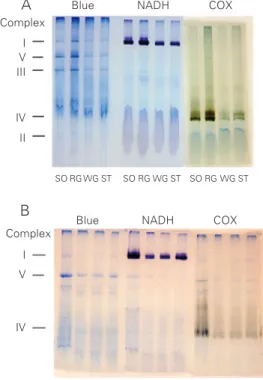
Figure 1. A, Colorimetric enzy-matic staining on BN-PAGE gels of different rat muscle samples. Coomassie blue staining of all complexes (Blue), complex I (NADH dehydrogenase) and complex IV (cytochrome c oxi-dase, COX). SO = soleus, RG = red gastrocnemius, WG = white gastrocnemius, ST = semiten-dinosus. B, Colorimetric enzy-matic staining on BN-PAGE gels of soleus muscles from exhaus-tively exercised rats. Coomassie blue staining of all complexes (Blue), complex I (NADH) and complex IV (COX). Control group (CO) and experimental samples were collected imme-diately (0), 3 and 6 h after ex-haustive exercise. The Figure is representative of several experi-ments performed.
Complex
I V III
IV
II
I
V
IV
Blue NADH COX
SO RG WG ST SO RG WG ST SO RG WG ST
Blue NADH COX
CO 0 3 6 CO 0 3 6 CO 0 3 6
Complex
A
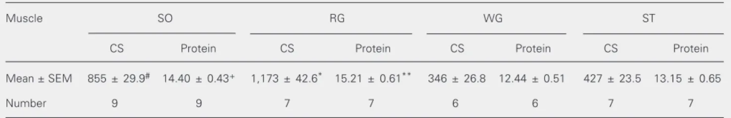
detergent extraction and BN-PAGE. The five major bands represent the oxidative phos-phorylation enzymes although the best reso-lutions were consistently achieved for com-plexes I, III and V. Comcom-plexes IV and I were well identified using the enzymatic colori-metric reactions set up for histochemical analysis. The colorimetrically stained enzy-matic activities of the complexes were local-ized specifically in a single or double band, while after Coomassie blue staining the com-plexes were generally identified as a single band, less strong than that obtained by color-imetric staining. Only the COX activity ap-peared in a single or double staining band due to its monomeric and dimeric forms. There were apparent differences in the width of the gel bands representing OXPHOS en-zymes from different muscles, mainly by histochemical staining. Broader bands were observed for the more oxidative muscles (soleus and red gastrocnemius) and narrower bands were observed for more glycolytic muscles (white gastrocnemius and semiten-dinosus). This result may reflect the higher and lower oxidative potentials of these muscles, respectively. This observation is confirmed and quantified in Table 2 which shows the activity of citrate synthase, used as a mitochondrial content marker. Table 2 also shows the total protein content of the same samples. It is evident that this measure-ment could not be used as a good marker of mitochondrial content because it does not
reflect the oxidative capacity of different muscles.
As shown in Figure 2, a linear correlation between complex V area and citrate syn-thase activity was found for all four muscles. This indicates that the Coomassie blue-stained complex V area is a better indicator of mitochondrial protein content than the total protein content shown in Table 2, and that the complex V area is a useful internal standard of the OXPHOS proteins loaded onto the gel, as shown by Zerbetto et al. (27). Table 3 shows the stained area of complexes I and IV reported as a function of the stained area of the Coomassie blue-stained complex V (internal standard). The ratio between the colorimetric and Coomassie blue staining of oxidative muscles, soleus and red gastrocnemius, fell within a narrow range of values for both complexes and also showed a linear correlation with the internal standard, complex V (Table 3). The same ratios were more variable in glycolytic muscles, white gastrocnemius and semiten-dinosus, and presented higher values when compared to those for the oxidative muscles. There were significant differences in the complex I/V ratio only when white nemius muscle was compared to red gastroc-nemius and semitendinosus muscles (Table 3) while there was a significant difference in the complex IV/V ratio between white gas-trocnemius and all the other muscles ana-lyzed (Table 3).
Table 2. Mitochondrial citrate synthase activity and protein content of different rat skeletal muscles.
Muscle SO RG WG ST
CS Protein CS Protein CS Protein CS Protein
Mean ± SEM 855 ± 29.9# 14.40 ± 0.43+ 1,173 ± 42.6* 15.21 ± 0.61** 346 ± 26.8 12.44 ± 0.51 427 ± 23.5 13.15 ± 0.65
Number 9 9 7 7 6 6 7 7
Citrate synthase (nmol min-1 mg tissue-1) was determined in the 20,000 g supernatant of a 30-mg homogenate. Protein content (mg/ml) was
determined in the 20,000 g supernatant by the method of Bradford. CS = citrate synthase; RG = red gastrocnemius; SO = soleus; ST = semitendinosus; WG = white gastrocnemius.
*P < 0.001 compared to all muscles; #P < 0.001 compared to WG and ST (one-way ANOVA). **P < 0.01 compared to WG and ST; +P = 0.01
1050 1000 950
Citrate synthase activity (nmol min -1 mg tissue -1)
900 850 800 750 700
0
1350
1300
1250
Citrate synthase activity (nmol min -1 mg tissue -1)
1200
1150
1100
1050 200
550
500
450
Citrate synthase activity (nmol min -1 mg tissue -1)
400
350
300
0 450
400
350
Citrate synthase activity (nmol min -1 mg tissue -1)
300
100 0
0
0 40 60 80 100 120 140 Complex V area
0 20 40 60 80 100
Complex V area
0 20 40 60 80 100 Complex V area
120 140 160 180 0 100 200 300 400 500
Complex V area
Soleus muscle Red gastrocnemius muscle
White gastrocnemius muscle Semitendinosus muscle Figure 2. Correlation between
the complex V area and citrate synthase activity of different rat muscles. The area of complex V is reported in arbitrary units. So-leus muscle, N = 8, P = 0.006, r = 0.94; red gastrocnemius muscle, N = 7, P = 0.003, r = 0.92; white gastrocnemius muscle, N = 6, P = 0.003, r = 0.95; semitendinosus muscle, N = 7, P = 0.015, r = 0.85.
Table 3. Complex I (NADH dehydrogenase) and IV (cytochrome c oxidase) activities in rat muscles determined by colorimetric enzymatic staining of BN-PAGE gel assays.
Muscle SO RG WG ST
I/V IV/V I/V IV/V I/V IV/V I/V IV/V
Mean ± SEM 9.8 ± 0.90 2.7 ± 0.25 3.8 ± 0.65 2.7 ± 0.38 26.7 ± 9.52*# 31.8 ± 12.07*# 12.1 ± 6.89 3.3 ± 1.36
Number 6 9 10 10 7 7 8 10
Quantitative data were obtained from the ratio of the activity and the internal standard of each muscle. RG = red gastrocnemius; SO = soleus; ST = semitendinosus; WG = white gastrocnemius.
I/V: *P < 0.001 compared to RG; #P < 0.05 compared to SO. IV/V: *P < 0.01 compared to RG and ST; #P < 0.05 compared to SO (one-way
ANOVA).
Soleus muscle mitochondrial complex I and IV activities after exhaustive exercise
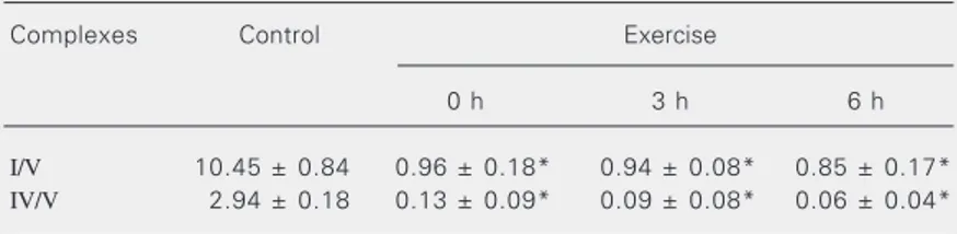
The colorimetric enzymatic staining of OXPHOS enzymes was applied to the anal-ysis of the effect of exhaustive physical exer-cise on the mitochondrial complexes IV and I of rat soleus muscle. Samples were col-lected at different resting periods after exer-cise and the results are presented in Figure 1B and Table 4. There was a significant reduction of the complex I/V and complex IV/V ratios, detected immediately after the end of exhaustive exercise (0 h). These
de-creased ratios persisted for 3 and 6 h after the end of exercise (P < 0.01 and P < 0.05) for both complexes.
Discussion
complex III runs on the gel as a dimer with a molecular mass of 500 kDa. Complex IV, 200 kDa, runs in its monomeric form, but occasionally a second band for this complex is detected, possibly representing the dimeric form of the same complex. Complex II runs at the bottom of the gel, with a molecular mass of 130 kDa. Studies combining BN-PAGE with colorimetric enzymatic staining have been recently performed in an attempt to simultaneously identify and quantify the activity of the OXPHOS complexes on the gel. These methods have been successfully applied to samples of skeletal muscles from humans, cows and rats (27). However, the results obtained in the analysis of rat skeletal muscles were not totally satisfactory. In an attempt to improve this characterization, the morphological (myosin heavy chain compo-sition of a muscle fiber) and functional (oxi-dative and glycolytic metabolism) differences between fiber types were considered. The present results showed that BN-PAGE in combination with the colorimetric enzymatic staining permits the identification and quan-tification of the activities of mitochondrial complexes I and IV from rat skeletal muscles with different oxidative capacities, as al-ready shown for human skeletal muscles.
According to Delp and Duan (31), the white gastrocnemius and semitendinosus muscles showed the higher percentage of type IIb fibers, 95 and 74% respectively, whereas the soleus and red gastrocnemius muscles presented a higher percentage of type I fibers, 86 and 55%, and type IIa fibers, 6 and 36%, respectively. Citrate synthase activity, used as a marker of mitochondrial protein content (Table 2), apparently reflect-ed the same pattern of fast-twitch glycolytic/ fast-twitch oxidative and slow-twitch oxida-tive classification of muscles by percentage of type fibers although we used a different animal breed. However, the metabolic char-acteristics and fiber composition of both animals and humans and how they correlate with the activity of a specific muscle group
have been poorly explored in the literature. Furthermore, the mitochondrial content of the working muscle cells is thought to act as a key regulatory mechanism in substrate se-lection (32).
An interesting observation made here was that the white gastrocnemius muscle, which has fewer mitochondria, showed high cata-lytic activities of complexes I and IV in comparison with the other muscles, prob-ably to compensate for its low oxidative potential. In agreement with our data, Howlett and Willis (32) observed that the isocitrate dehydrogenase (NAD-IDH) activity is higher in mitochondria from type IIb muscle, sug-gesting that type IIb mitochondria may rely on this enzyme as a regulatory site. This enzyme catalyzes the first dehydrogenase reaction in the TCA cycle and produces NADH + H+, which is the substrate of NADH
dehydrogenase. This may indicate a higher activity of NADH dehydrogenase, demon-strated in the present study.
Conflicting results regarding the effect and the role of exhaustive physical exercise on mitochondria and their production of ROS have been reported in the literature (13,16, 22,33,34). It seems that ROS play a primor-dial role in altering mitochondrial function in a variety of patho-physiological conditions (35), and in physical exercise situations mi-tochondrial alterations seem to depend on exercise schedule, previous training and
Table 4. Activity ratios of complexes I and IV in soleus muscle after exhaustive physical exercise.
Complexes Control Exercise
0 h 3 h 6 h
I/V 10.45 ± 0.84 0.96 ± 0.18* 0.94 ± 0.08* 0.85 ± 0.17* IV/V 2.94 ± 0.18 0.13 ± 0.09* 0.09 ± 0.08* 0.06 ± 0.04*
The values were obtained by densitometric analysis of the colorimetric enzymatic stained areas of complexes I and IV and the Coomassie blue-stained area of complex V of the control group (internal standard) run on the same gel. Quantitative data were determined by the ratio between the activity and the internal standard (N = 5). Results are reported as means ± SEM.
muscle fiber type. Zhang et al. (36) showed mitochondrial enzyme inactivation as the first step of oxidative damage, which was further related to exercise-induced oxidative stress. Leeuwenburgh et al. (37) and Radák et al. (38) found increased levels of oxidized amino acids in muscle mitochondria but not in cytosolic proteins, suggesting that exer-cise is a physiological source of oxidative stress. It has also been suggested that oxida-tive injury might cause irreversible cell dam-age through the loss of homeostasis and the loss of mitochondrial function (39). Given the high oxygen consumption during exer-cise, mitochondria would be requested for
energy supplying and might simultaneously produce ROS. These considerations agree with our results, which show decreasing activities of mitochondrial complexes I and IV no matter the time of sampling, and this may indicate that mitochondria are the main target of exercise-induced ROS production and attack. In addition, COX appears to have an indirect antioxidant action because of its affinity for O2,reducing O2•••••- formation (40).
Thus, the decreased complex IV activity shown in our experiment supports the prob-ability of ROS attack in mitochondrial mem-brane complexes.
References
1. Pette D & Staron RS (1990). Cellular and molecular diversities of mammalian skeletal muscle fibers. Review of Physiology,
Biochem-istry, and Pharmacology, 116: 1-76.
2. Gunning O & Hardeman E (1991). Multiple mechanisms regulate muscle fiber diversity. FASEB Journal, 5: 3064-3070.
3. Schiaffino S & Reggiani C (1994). Myosin isoforms in mammalian skeletal muscle. Journal of Applied Physiology, 77: 493-501. 4. Rivero JL, Talmadge RJ & Edgerton VR (1999). Interrelationships of
myofibrillar ATPase activity and metabolic properties of myosin heavy chain-based fibre types in rat skeletal muscle.
Histochemis-try and Cell Biology, 111: 277-287.
5. Holloszy JO (1967). Effects of exercise on mitochondrial oxygen uptake and respiratory enzyme activity in skeletal muscle. Journal
of Biological Chemistry, 242: 2278-2282.
6. Hoppeler H, Howald H, Conley K, Lindstedt SL, Claassen H, Vock P & Weibel ER (1985). Endurance training in humans: aerobic capacity and structure of skeletal muscle. Journal of Applied Physiology, 59: 320-327.
7. Henriksson J (1995). Effect of training and nutrition on the develop-ment of skeletal muscle. Journal of Sports Sciences, 13: S25-S30. 8. Linossier MT, Dormois D, Geyssant A & Denis C (1997).
Perfor-mance and fibre characteristics of human skeletal muscle during short sprint training and detraining on a cycle ergometer. European
Journal of Applied Physiology, 75: 491-498.
9. Green HJ, Jones S, Ball-Burnett M, Farrance B & Ranney D (1995). Adaptations in muscle metabolism to prolonged voluntary exercise and training. Journal of Applied Physiology, 78: 138-145.
10. McArdle WD, Katch FI & Katch VL (1996). Exercise Physiology:
Energy, Nutrition, and Human Performance. 4th edn. Williams &
Wilkins, Baltimore, MD, USA.
11. Chilibeck PD, Bell RP & Martin TP (1998). Higher mitochondrial fatty acid oxidation following intermittent versus continuous endurance exercise training. Canadian Journal of Physiology and Pharmacolo-gy, 76: 891-894.
12. Alessio HM & Goldfarb AH (1988). Lipid peroxidation and scavenger enzymes during exercise: adaptive response to training. Journal of
Applied Physiology, 64: 1333-1336.
13. Alessio HM, Hagerman AE, Fulkerson BK, Ambrose J, Rice RE & Wiley RL (2000). Generation of reactive oxygen species after exhaustive aerobic and isometric exercise. Medicine and Science
in Sports and Exercise, 32: 1576-1581.
14. Packer L (1997). Oxidants, antioxidant nutrients and the athlete.
Journal of Sports Sciences, 15: 353-363.
15. Sastre J, Asensi M, Gasco E, Pallardo FV, Ferrero JA, Furukawa T & Vina J (1992). Exhaustive physical exercise causes oxidation of glutathione status in blood: prevention by antioxidant administra-tion. American Journal of Physiology, 263: R992-R995.
16. Davies KJ, Quintanilha AT, Brooks GA & Packer L (1982). Free radicals and tissue produced by exercise. Biochemical and
Bio-physical Research Communications, 107: 1198-1205.
17. Sjödin B, Westing HY & Apple FS (1990). Biochemical mechanisms for oxygen free radical formation during exercise. Sports Medicine, 10: 236-254.
18. Moyes CD & Hood DA (2003). Origins and consequences of mito-chondrial variation in vertebrate muscle. Annual Review of Physiolo-gy, 65: 177-201.
19. Smolka MB, Zoppi CC, Alves AA, Silveira LR, Marangoni S, Pereira-da-Silva L, Novello JC & Macedo DV (2000). HSP72 as a comple-mentary protection against exercise-induced oxidative stress in the soleus muscle of rats. American Journal of Physiology, 279: R1539-R1545.
20. Criswell D, Powers S, Dodd S, Lawler J, Edwards W, Renshler K & Grinton S (1993). High intensity training-induced changes in skeletal muscle antioxidant enzyme activity. Medicine and Science in Sports
and Exercise, 25: 1135-1140.
21. Powers SK, Criswell D, Lawler J, Ji LL, Martin D, Herb RA & Dudley G (1994). Influence of exercise and fiber type on antioxidant en-zyme activity in rat skeletal muscle. American Journal of Physiolo-gy, 266: R375-R380.
22. Ji LL & Fu R (1992). Responses of glutathione system and antioxi-dant enzymes to exhaustive exercise and hydroperoxide. Journal of
Applied Physiology, 72: 549-554.
Oxford University Press, Oxford, UK, 90-106.
24. Podhorska-Okolow M, Sandri M, Zampieri S, Brun B, Rossini K & Carraro U (1998). Apoptosis of myofibres and satellite cells: exer-cise induced damage in skeletal muscle of the mouse.
Neuropa-thology and Applied Neurobiology, 24: 518-531.
25. Augusti GNA, Sauleda J, Miralles C, Gomez C, Togores B, Sala E, Batle S & Busquets X (2002). Skeletal muscle apoptosis and weight loss in chronic obstructive pulmonary disease. American Journal of
Respiratory and Critical Care Medicine, 166: 485-489.
26. Schagger H & Jagow G (1991). Blue native electrophoresis for isolation of membrane protein complexes in enzymatically active form. Analytical Biochemistry, 199: 223-231.
27. Zerbetto E, Vergani L & Dabbeni-Sala F (1997). Quantification of muscle mitochondrial oxidative phosphorylation enzymes via his-tochemical staining of blue native polyacrylamide gels.
Electropho-resis, 18: 2059-2064.
28. Nijtmans LGJ, Henderson NS & Holt IJ (2002). Blue native electro-phoresis to study mitochondrial and other protein complexes. Meth-ods, 26: 327-334.
29. Srere PA (1969). Citrate synthase. Methods in Enzymology, 13: 3-5. 30. Bradford MM (1976). A rapid and sensitive method for the quantifi-cation of microgram quantities of protein utilizing the principle of protein-dye binding. Analytical Biochemistry, 72: 255-260. 31. Delp MD & Duan C (1996). Composition and size of type I, IIA, IID/X,
and IIB fibers and citrate synthase activity of rat muscle. Journal of
Applied Physiology, 80: 261-270.
32. Howlett RA & Willis WT (1998). Fiber-type-related differences in the enzymes of a proposed substrate cycle. Biochimica et Biophysica
Acta, 1363: 224-230.
33. Vina J, Gomez-Cabrera MC, Lloret A, Marquez R, Minana JB, Pallardo FV & Sastre J (2000). Free radicals in exhaustive physical exercise: mechanism of production, and protection by antioxidants IUBMB. Life, 50: 271-277.
34. Di Meo S & Venditti P (2001). Mitochondria in exercise-induced oxidative stress. Biological Signals and Receptors, 10: 125-140. 35. Supinski G (1998). Free radical induced respiratory muscle
dysfunc-tion. Molecular and Cellular Biochemistry, 179: 99-110.
36. Zhang Y, Marcillat O, Giulivi C, Ernster L & Davies KJA (1990). The oxidative inactivation of mitochondrial electron transport chain com-ponents and ATPase. Journal of Biological Chemistry, 265: 16330-16336.
37. Leeuwenburgh C, Hansen PA, Holloszy JO & Heinecke JW (1999). Hydroxyl radical generation during exercise increases mitochondrial protein oxidation and levels of urinary dityrosine. Free Radical
Biol-ogy and Medicine, 27: 186-192.
38. Radák Z, Sasvári M, Nyakas C, Taylor AW, Ohn H, Nakamoto H & Goto S (2000). Regular training modulates the accumulation of reactive carbonyl derivatives in mitochondrial and cytosolic frac-tions of rat skeletal muscle. Archives of Biochemistry and Biophys-ics, 383: 114-118.
39. Liu J, Yeo HC, Overvik-Douki E, Hagen T, Doniger SJ, Chu DW, Brookes GA & Ames BN (2000). Chronically and acutely exercised rats: biomarkers of oxidative stress and endogenous antioxidants.
Journal of Applied Physiology, 89: 21-28.