Effect of salbutamol on innervated
and denervated rat soleus muscle
Department of Anatomy, Medical Faculty, University of Rijeka, Rijeka, Croatia T. Šoiƒ-Vraniƒ, D. Bobinac,
S. Bajek, R. Jerkoviƒ, D. Malnar-Dragojeviƒ
and M. Nikoliƒ
Abstract
The objective of the present investigation was to perform a 14-day time-course study of treatment with salbutamol, a ß2 adrenoceptor
agonist, on rat soleus muscle in order to assess fiber type selectivity in the hypertrophic response and fiber type composition. Male Wistar rats were divided into four groups: control (N = 10), treated with salbutamol (N = 30), denervated (N = 30), and treated with salbutamol after denervation (N = 30). Salbutamol was injected intraperitoneally in the rats of the 2nd and 4th groups at a concentration of 0.3 mg/kg twice a day for 2 weeks. The muscles were denervated using the crush method with pean. The animals were sacrificed 3, 6, 9, 12, and 14 days after treatment. Frozen cross-sections of soleus muscle were stained for myosin ATPase, pH 9.4. Cross-sectional area and percent of muscle fibers were analyzed morphometrically by computerized im-age analysis. Treatment with salbutamol induced hypertrophy of all fiber types and a higher percentage of type II fibers (21%) in the healthy rat soleus muscle. Denervation caused marked atrophy of all fibers and conversion from type I to type II muscle fibers. Denervated muscles treated with salbutamol showed a significantly larger cross-sectional area of type I muscle fibers, 28.2% compared to the dener-vated untreated muscle. Moreover, the number of type I fibers was increased. These results indicate that administration of salbutamol is able to induce changes in cross-sectional area and fiber type distribu-tion in the early phase of treatment. Since denervadistribu-tion-induced atro-phy and conversion from type I to type II fibers were improved by salbutamol treatment we propose that salbutamol, like other ß2
adreno-ceptor agonists, may have a therapeutic potential in improving the condition of skeletal muscle after denervation.
Correspondence
T. Šoiƒ-Vraniƒ
Department of Anatomy Medical Faculty University of Rijeka Braƒe Branchetta 20, 51000 Rijeka Croatia
E-mail: [email protected]
Received September 29, 2004 Accepted June 1, 2005
Key words •Salbutamol •Rat skeletal muscle •Soleus muscle •Denervation •Histochemistry •Muscle fibers
Introduction
Salbutamol is a ß2 adrenoceptor agonist (BAA) known to induce bronchiolar relax-ation, higher vascularization of skeletal muscle, as well as muscle hypertrophy (1). Martineau et al. (2) demonstrated that short-term administration of salbutamol increases
also demonstrated in skeletal muscles with altered physiological conditions or functional demands. Zeman et al. (5) showed that clenbuterol, another BAA, is able to retard muscle atrophy in denervated muscles. Clenbuterol treatment reduced the loss of wet weight and of protein content and also increased the cross-sectional area of dener-vated solei. Data reported by Maltin et al. (6) indicate that clenbuterol not only inhibits but also partially reverses denervation-in-duced atrophy of rat soleus muscle. This was evident from measurements of both muscle protein content and fiber cross-sectional ar-eas. Furthermore, the hypertrophic effect of BAA seems to be selective for certain fiber types. Most studies have demonstrated that BAA increase the cross-sectional area of fast muscle fibers (7-9) while the results of studies of the effects of BAA on slow muscle fibers have not been consistent. Some stud-ies have demonstrated that the cross-sec-tional area of slow twitch oxidative fibers is not changed by chronic administration of BAA (8,10,11) while others have shown that all fiber types are affected equally (12,13). Recent reports have established that clen-buterol is able to induce changes in fiber type distribution. After 2 weeks of clenbuterol treatment there was a pronounced change in fiber type distribution, i.e., slow-to-fast tran-sitions (7). Lynch et al. (14) have demon-strated slow-to-fast transformation in rat so-leus muscle after 15 weeks of clenbuterol administration. Zeman et al. (8) have shown that a treatment period of more than 2 weeks was required to increase fast-twitch fiber composition. On the other hand, after four weeks of clenbuterol administration there were no significant fiber type alterations in tibialis anterior and intercostal muscles which are fast-twitch contracting muscles (15).
The aim of the present investigation was to perform a 14-day time-course study of salbutamol administration with two main objectives: 1) to elucidate the fiber type selectivity in the hypertrophic response and
2) to explore the changes in fiber type distri-bution in slow rat skeletal muscle, both in the presence and in the absence of the nerve.
Material and Methods
Animals
We used 3-month-old male Wistar rats weighing approximately 200-250 g (N = 100), with free access to standard laboratory food and water. The animals were divided into four groups as follows: 1) control (N = 10), 2) treated with salbutamol (N = 30), 3) denervated (N = 30), and 4) treated with salbutamol after denervation (N = 30).
Surgical procedure and treatment with salbutamol
The animals from the 3rd and 4th groups were anesthetized with an intraperitoneal injection of ketamine hydroxychloride (25 mg/kg, 1 mg/ml ketamine; Parke-Davis, Milan, Italy). The right sciatic nerve of the thigh was exposed and crushed proximal to its branching for denervation, with pean last-ing 2 min. The overlylast-ing muscles and skin were then sutured. Salbutamol (Pliva, Zagreb, Croatia), diluted in 0.3 mg/kg 0.9% NaCl, was injected intraperitoneally in the rats of the 2nd and 4th groups twice a day for 2 weeks. The first salbutamol dose was in-jected 1 h after the surgical procedure.
Animals from the control group were analyzed as a zero time control. The animals from the other groups (salbutamol, dener-vated and denerdener-vated plus salbutamol) were then sacrificed by cervical hyperextension 3, 6, 9, 12, and 14 days after denervation. The soleus muscles were dissected from ten-don to tenten-don from the right hindlimb.
Muscle analysis
upright cylinder in embedding medium (Tis-sue-Tek, Sakura, Netherlands) while still frozen. Serial 8-µm thick transverse sections were cut with a cryostat at -20ºC, attached to glass slides and stainedfor myosin ATPase, pH 9.4 (16,17). Sections were then fixed at 40ºC for 5 min in 20% (v/v) paraformalde-hyde, 0.1 M sodium cacodylic acid contain-ing 0.4 M saccharose and rinsed well with running tap water. Rinsed sections were in-cubated at 37ºC for 30 min in the following solution: 5 mg ATP was dissolved in a few drops of distilled water, 10 ml 0.2 M sodium barbiturate buffer containing 0.1 M CaCl2 in distilled water was added and the pH was adjusted to 9.4. After washing with distilled water, sections were immersed in 2% CaCl2 for three rinses of 1 min each. Specimens were rinsed again in distilled water and im-mersed in diluted (1:10) ammonium sulfide solution for 30 s. After a final rinsing in running tap water the sections were dehy-drated, cleared and mounted in glycerine.
In treated and untreated soleus muscles, fibers showing the densest reaction product were identified as fast-twitch oxidative glyco-lytic fibers, fibers showing no reaction prod-uct were identified as slow-twitch fibers, and fibers showing an intermediate reaction prod-uct were identified as fast-twitch glycolytic fibers. However, since fast-twitch glycolytic fibers represented less than 1.5% of all the fibers detected in the three experimental groups, i.e., treatment with salbutamol, denervation and denervation plus treatment with salbuta-mol, fast-twitch glycolytic fibers were ex-cluded from the measurements and calcula-tions as a separate group and were included in population of type II fibers. The cross-sec-tional areas of 200-300 fibers were measured with the SFORM image analyzer (VAMS, Zagreb, Croatia) in random fields of muscle sections obtained from each group. Counts of type I or type II fibers were used to calculate fiber type percentages. Data are reported as means ± SD. The control group was used for comparison with the healthy group treated
with salbutamol and the denervated group was used for comparison with the denervated plus salbutamol group. Statistical analyses were performed by the Student t-test, with the level
of significance set at P < 0.05.
Results
Changes in the cross-sectional area of muscle fibers
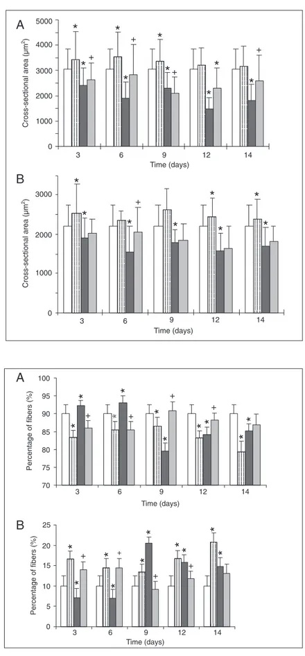
After 14 days of salbutamol administra-tion, the cross-sectional areas of type I and type II muscle fibers were larger in healthy soleus muscle than in muscles of the un-treated control group (Figures 1A,B and 2A,B). A statistically significant effect of salbutamol on fiber size was observed as
Figure 2. Changes in cross-sectional area of soleus muscle type I (A) and type II (B) fibers from the four treated groups. Open columns = control group; vertical lined columns = group treated with salbutamol; dark gray columns = denervated group; light gray columns = group treated with salbutamol following denervation. Data are reported as means ± SD. *P < 0.05 compared to control; +P < 0.05 compared to the denervated group
(Student t-test).
Figure 3. Distribution of type I and II fibers in soleus muscle. A, Type I fibers from the four treated groups; B, type II fibers from the four treated groups. Open col-umns = control group; vertical lined colcol-umns = group treated with salbutamol; dark gray columns = dener-vated group; light gray columns = group treated with salbutamol following denervation. *P < 0.05 compared to control; +P < 0.05 compared to the denervated group
(Student t-test).
A
B
A
early as 3 days after treatment (P < 0.05). After denervation, the cross-sectional areas of both types of muscle fibers decreased significantly (P < 0.05; Figures 1C and 2A,B). By the 14th day of denervation the cross-sectional area of type I fibers was reduced to 59.4% and that of type II fibers was reduced to 6.7% compared to healthy muscle (Figure 2A,B). The cross-sectional area of type I fibers of denervated muscles treated with salbutamol increased from 1815.3 ± 650.6 to 2529.7 ± 1018.1 µm2 (P < 0.05), correspond-ing to a 28.24% increase compared to dener-vated untreated muscle (1815.3 ± 650.6 µm2) at the end of experiment (Figures 1C,D and 2A). Type II fibers of denervated muscles treated with salbutamol were not signifi-cantly affected although a transient increase of 24.03% in fiber size was observed on the 6th day (P < 0.05).
Changes in the proportions of muscle fiber types I and II
Soleus, as a slow-twitch muscle, is com-posed of 90% type I fibers and 10% type II fibers. The influence of salbutamol adminis-tration was observed in the early phase of the experiment starting on the 3rd day. In con-trol muscle, salbutamol increased the num-ber of type II finum-bers, from 10 to 21%, a fact that was most evident by the 14th day of treatment (21%; P < 0.05; Figure 3B). The number of type II fibers increased from the 9th day of denervation (P < 0.01; Figure 3B) and was clearly demonstrable until the end of the experiment. Furthermore, a higher percentage of type II fibers was observed in denervated soleus during the first 6 days of salbutamol administration (Figure 3B). Sal-butamol increased the percentage of type I fibers until the end of the experiment (Figure 3A).
Discussion
The aim of this investigation was to
After 2 weeks of salbutamol administra-tion, we observed a more pronounced effect on fiber composition than on the anabolic response, in agreement with data reported by Ricart-Firinga et al. (7). Over 14 days the number of type II fibers increased signifi-cantly from the initial 10% observed in healthy soleus up to 21%, in agreement with data reported by Zeman et al. (8). This oc-curs also during mechanical unweighting (10,19) and during the period of denerva-tion. Actually, during muscle maturation the number of type I fibers in soleus increases at the expense of the number of type II fibers. Administration of clenbuterol prevents the conversion of type II fibers to type I, nor-mally occurring in the process of matura-tion, due to the effect of the drug on in-creased synthesis and accumulation of fast myosin light chains in soleus (8). Stevens et al. (20) demonstrated that, during 2 weeks of administration, clenbuterol produced changes in the group of type II fibers. Treat-ment with clenbuterol causes the upregula-tion of IIA fibers (23%) and, in addiupregula-tion, the induction, of considerable amounts of the faster IIX (3%) and fastest IIB (2%) fiber
types. These data demonstrated the high plas-ticity of muscle spanning the spectrum of fiber types.
The number of type I fibers decreased during the first 6 days in denervated soleus treated with salbutamol compared to dener-vated untreated muscle. However, from the 6th day to the end of the experiment the number of type I fibers was higher in the former than in the latter. On day 9 the activ-ity of salbutamol resulted in such a signifi-cant increase in the number of these fibers that it almost reached the corresponding value in control muscle. Incidentally, the same day marked the lowest value for type I fibers. This leads us to conclude that the most sig-nificant changes in fiber distribution occurred on day 9. Indeed, on day 9 there was the most profound conversion of type I to type II fibers in denervated muscles and of type II to type I in denervated muscles under the influ-ence of salbutamol. Thus, we may postulate that salbutamol reestablishes the process of growth and development in denervated muscles with the occurrence of the process of conversion of type II to type I fibers (21).
References
1. Apperley GH, Daly MJ & Levy GP (1976). Selectivity of ß-adreno-ceptor agonists and antagonists on bronchial, skeletal, vascular and cardiac muscle in the anesthetized cat. British Journal of Pharma-cology, 57: 235-246.
2. Martineau L, Horan MA, Rothwell NJ et al. (1992). Salbutamol, a ß2
-adrenoceptor agonist, increases skeletal muscle strength in young men. Clinical Science, 83: 615-621.
3. Emery PV, Rothwell NJ, Stock MJ et al. (1984). Chronic effects of ß-adrenergic agonists on body composition and protein synthesis in the rat. Bioscience Reports, 4: 83-91.
4. Li JB & Jefferson LS (1977). Effect of isoproterenol on amino acid levels and protein turnover in skeletal muscle. American Journal of Physiology, 232: E243-E249.
5. Zeman RJ, Ludemann R & Etlinger JD (1987). Clenbuterol, a beta-2-agonist, retards atrophy in denervated muscles. American Journal of Physiology, 252: E152-E155.
6. Maltin CA, Reeds PJ, Delday MI et al. (1986). Inhibition and reversal of denervation induced atrophy by the beta-agonist growth pro-moter, clenbuterol. Bioscience Reports, 6: 811-818.
7. Ricart-Firinga C, Stevens L, Canu MH et al. (2000). Effects of ß2
-agonist clenbuterol on biochemical and contractile properties of unloaded soleus fibers of rat. American Journal of Physiology, 278: C582-C588.
8. Zeman RJ, Ludemann R, Easton TG et al. (1988). Slow to fast alterations in skeletal muscle fibers caused by clenbuterol, a beta2
-receptor agonist. American Journal of Physiology, 254: E726-E732. 9. Criswell DS, Powers SK & Herb RA (1996). Clenbuterol-induced fiber type transition in the soleus of adult rats. European Journal of Applied Physiology, 74: 391-396.
10. Maltin CA, Delday MI, Hay SM et al. (1990). Effects of bovine pituitary growth hormone alone or in combination with the beta-agonist clenbuterol on muscle growth and composition in veal calves. British Journal of Nutrition, 63: 535-545.
11. Miller MF, Garcia DK, Coleman ME et al. (1988). Adipose tissue, longissimus muscle and anterior pituitary growth and function in clenbuterol-fed heifers. Journal of Applied Physiology, 66: 12-20. 12. Maltin CA, Delday MI, Hay SM et al. (1992). Denervation increases
14: 188-192.
13. Maltin CA, Delday MI & Reeds PJ (1986). The effect of a growth promoting drug, clenbuterol, on fiber frequency and area in hind limb muscles from young male rats. Bioscience Reports, 6: 293-299. 14. Lynch GS, Hayes A, Campbell SP et al. (1996). Effects of ß2-agonist
administration and exercise on contractile activation of skeletal muscle fibers. Journal of Applied Physiology, 81: 1610-1618. 15. Polla B, Cappelli V, Morello F et al. (2001). Effects of the beta
2-agonist clenbuterol on respiratory and limb muscles of weaning rats. American Journal of Physiology, 280: 862-869.
16. Round JM, Matthews Y & Jones DA (1980). A quick simple and reliable method for ATPase in human muscle preparations. His-tochemical Journal, 12: 707-709.
17. Brooke MH & Kaiser KK (1970). Muscle fiber types: how many and
what kind. Archives of Neurology, 23: 369-379.
18. Maltin CA, Hay SM, Delday MI et al. (1987). Clenbuterol, a beta agonist, induces growth in innervated and denervated rat soleus muscle via apparently different mechanisms. Bioscience Reports, 7: 525-532.
19. Stevens L, Gohlsch B, Mounier Y et al. (2000). Upregulation of myosin heavy chain MHClα in rat muscles after unweighting and clenbuterol treatment. Biochemical and Biophysical Research Com-munications, 275: 418-421.