Physical Therapy Department, Universidade Federal de Minas Gerais (UFMG), Belo Horizonte, MG, Brazil Received: 01/05/2013 Revised: 04/01/2013 Accepted: 04/08/2013
a r t i c l e
Strength of the respiratory and lower limb muscles and
functional capacity in chronic stroke survivors with
different physical activity levels
Janaine C. Polese, Marina B. Pinheiro, Christina D. C. M. Faria, Raquel R. Britto, Verônica F. Parreira, Luci F. Teixeira-Salmela
ABSTRACT | Background: The assessment of strength and its relationships with functional capacity could contribute to
more speciic and effective disability management of stroke survivors. Objective: To compare and investigate associations
between measures of strength and functional capacity of 98 chronic stroke survivors, stratiied into three groups, according to their physical activity levels. Method: The physical activity levels were classiied as impaired, moderately active, and
active, based on their Human Activity Proile (HAP) scores. Strength was assessed by the maximal inspiratory (MIP) and expiratory (MEP) pressures and by the residual deicits (RDs) of work of the lower limb and trunk muscles, whereas functional capacity was evaluated by the distance covered during the six-minute walking test (6MWT). Results: One-way
analyses of variance revealed signiicant differences between the groups, except between the active and moderately active groups regarding the RDS of the hip and knee lexors/extensors and ankle dorsilexors (2.91<F<8.62; 0.001<p<0.01). Differences between the groups were found for the 6MWT (F=10.75; p<0.001), but no differences were found for the MIP and MEP measures (0.92<F<2.13; 0.13<p<0.40). Signiicant, negative, and fair correlations were observed between the RDS of the hip and knee muscles and the 6MWT (0.30<r<–0.43; p<0.01) and the HAP (–0.28<r<–0.41; p<0.01). Moderate to good correlations were found between the 6MWT and the HAP (r=0.50; p<0.0001). There were no signiicant correlations between measures of respiratory strength and any of the investigated variables (–0.11<r<0.12; 0.26<p<0.56). Conclusions: Lower strength deicits and higher functional capacity were associated with higher physical
activity levels. However, the moderately active and active groups demonstrated similar strength deicits.
Keywords: stroke; respiratory muscles; muscular weakness; exercise capacity; physical activity levels; rehabilitation.
HOW TO CITE THIS ARTICLE
Polese JC, Pinheiro MB, Faria CDCM, Britto RR, Parreira VF, Teixeira-Salmela LF. Strength of the respiratory and lower limb muscles and functional capacity in chronic stroke survivors with different physical activity levels. Braz J Phys Ther. 2013 Sept-Oct; 17(5):487-493. http://dx.doi.org/10.1590/S1413-35552012005000114
Introduction
The physical consequences of stroke are variable and may include impairments in the motor, emotional, and sensory systems, language, perception, and cognitive functions1. Hemiparesis, the most frequently observed motor deicit, affects more than 80% of these individuals2 and is related to decreased exercise
capacities and physical function3. Clinically, muscular weakness has been recognized as a limiting factor in the motor rehabilitation of stroke patients and as the most contributing factor to post-stroke disabilities4. Therefore, strengthening exercises have been
widely employed to increase functional capacity in
individuals with hemiparesis5-8. However, further studies are required to investigate other variables,
which could interfere with functional capacities with
this population.
Previous studies have reported decreased respiratory strength in individuals during both the
acute and chronic post-stroke stages9-12. Within this context, weaknesses of the respiratory muscles and changes in the thoraco-abdominal motion could be related to decreased tidal volumes, and consequently, lower exercise tolerance, as observed in healthy individuals13. Additionally, in accordance with Patterson et al.14, the ability to generate force with
the paretic lower limb is the major determinant of the
ability to perform short or long walks.
The preventive and rehabilitative approaches of individuals with hemiparesis are of utmost relevance due to the increased survival rates of this population15. Thus, to assess muscular parameters of individuals
with chronic hemiparesis and its relationships with functional capacities could contribute to a more
speciic management of this condition. Therefore, the
aim of this study was to compare motor performance
muscles) and functional capacity, as assessed by the distance walked during the six-minute walk test (6MWT), between individuals with hemiparesis with various levels of physical activity. In addition, it investigated possible associations between motor parameters and functional capacities in individuals with chronic hemiparesis. It was hypothesized that subjects with chronic stroke who had higher levels of physical activity would demonstrate higher levels of functional capacity and lower strength deicits of the respiratory and lower limb muscles.
Method
Participants
Ninety-eight subjects with unilateral stroke, who had residual weakness and/or spasticity of the affected lower extremity, were recruited on a volunteer basis from the general community of Belo Horizonte, MG, Brazil. All participants were
screened to ensure that their mean time since the onset
of the stroke was at least nine months; were above 20 years of age; were able to walk with or without assistive devices; had no facial palsy which could prevent proper labial occlusion during tests; and had no cognitive impairments, as assessed by the Mini Mental State Examination16, according to the cut-off scores recommended by Brucki et al.17. They should
also be able to understand and perform all of the
test procedures. Those individuals with
non-stroke-related disabilities and who had respiratory diseases
were excluded. All participants provided consent based upon approval by the Ethical Review Board of Universidade Federal de Minas Gerais (UFMG), Belo Horizonte, MG, Brazil (protocol 172/02).
Instruments and procedures
Initially, the participants underwent a physical examination and an interview for the collection of anthropometric, demographic, and clinical data, such as age, gender, body mass, height, time since the onset of stroke, paretic side, and the use of medications. The participants were stratiied into three groups based upon their physical activity levels, which were determined by the self-reported activity scores on the Brazilian version of the Human Activity Proile (HAP), which is considered a valid and reliable
tool18. The participants’ physical activity levels were classiied according to their adjusted activity scores (AAS) either as impaired (<53), moderately active (53-74) or active (>74)19.
Lower limb muscular strength
Bilateral measures of strength of the major muscular groups of the ankle, knee, and hip joints were obtained with the Biodex System 3Pro®
isokinetic dynamometer (Biodex Medical System Inc., Shirley, NY) in the concentric-concentric mode at angular speeds of 60°/s. Isokinetic measures have shown to be reliable for evaluating the strength of individuals with hemiparesis20,21 and all procedures were carried out following previously described
protocols8,21. The residual deficits (RDs) of the normalized work measures, in Joules, were used
for analyses and were calculated according to the
following formula: 100 – (paretic/non-paretic lower limb * 100).
Six-minute walk test
The distances covered during the 6MWT, which appropriately relect the physical capacity to perform daily tasks, were registered for analyses22-24. The test was carried out, following the procedures and recommendations of the American Thoracic Society25, which were adapted to the Brazilian Portuguese language26.
Respiratory muscular strength
Respiratory strength was evaluated by measures of maximal inspiratory (MIP) and expiratory (MEP) pressures, which were obtained by a previously calibrated manovacuometer with operating intervals of ±300 cmH2O (GeRar®, São Paulo, Brazil). All of the test procedures were conducted according to
previous recommendations27,28. These measures have shown adequate psychometric properties in previous
studies29,30.
Statistical analysis
Descriptive statistics, tests for normality (Kolmogorov-Smirnov), and equality of variances (Levene) were carried out for all outcome variables. One-way analyses of variance (ANOVAs) followed by LSD post-hoc tests were employed to investigate
differences between the groups (impaired, moderately active, and active) regarding the RDs of the paretic lower limb muscles, the distances covered during the 6MWT, and the respiratory pressures. Pearson correlation coeficients were calculated to explore the relationships between these selected variables and were classiied as low or none (0.00<r<0.25),
fair (0.25<r<0.50), moderate to good (0.50<r<0.75),
and good to excellent (r>0.75)31. All analyses were
Results
Participants’ characteristics
Ninety-eight individuals, 54 men and 44 women, participated in this study. They had a mean age of 56.4±12.5 years and a time since the onset of stroke ranging from six to 240 months. No significant differences between the three physical activity level groups were observed regarding the participants’ ages and the time since the onset of stroke (p=0.84 and p=0.68, respectively). Their clinical, demographic,
and anthropometric data are described in Table 1. Six individuals were unable to perform the respiratory pressure procedures (four of the impaired and two of the moderately active group) and the isokinetic work data of eight individuals were
excluded due to technical problems (two of the impaired, six of the moderately active, and one from the active group). Strength data for the ankle plantar and dorsal lexor muscles were obtained for only 40 individuals (four from the impaired, 25 from the moderately active, and 11 from the active group), since 58 participants were unable to generate enough torque to perform the tests.
Comparisons between the groups
Table 2 provides the results of the between-group comparisons for all of the variables. No signiicant differences between the groups were observed for the RDs of the ankle plantar lexor muscles [F(2, 37)=0.67; p=0.52]. However, for all the other
muscular groups, signiicant differences between the groups were found, except between the moderately
Table 1. Demographic and anthropometric characteristics of the participants.
Characteristic n=98
Age (years), mean±SD, (range: min-max) 56.4±12.5 (24-86)
Gender, male, n (%) 54 (55)
Paretic side, right, n (%) 58 (59)
Time since the onset of stroke (months), mean±SD, (range: min-max) 64.8±53.6 (6-240)
Use of medications (number), mean±SD 3±2
BMI (Kg/m2), mean±SD, (range: min-max) 25.9±4 (15.24-36.51)
SD=Standard deviation; BMI= Body Mass Index.
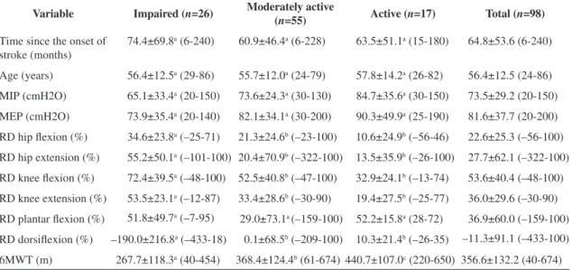
Table 2. Demographic and clinical data (means±standard deviations and range [minimum-maximum] of the participants, according to their physical activity levels and comparisons between the groups.
Variable Impaired (n=26) Moderately active
(n=55) Active (
n=17) Total (n=98)
Time since the onset of
stroke (months) 74.4±69.8
a (6-240) 60.9±46.4a (6-228) 63.5±51.1a (15-180) 64.8±53.6 (6-240)
Age (years) 56.4±12.5a (29-86) 55.7±12.0a (24-79) 57.8±14.2a (26-82) 56.4±12.5 (24-86)
MIP (cmH2O) 65.1±33.4a (20-150) 73.6±24.3a (30-130) 84.7±35.6a (30-150) 73.5±29.2 (20-150)
MEP (cmH2O) 73.9±35.4a (20-140) 82.1±34.1a (30-200) 90.3±49.9a (25-190) 81.6±37.7 (20-200)
RD hip lexion (%) 34.6±23.8a (–25-71) 21.3±24.6b (–23-100) 10.6±24.9b (–56-46) 22.6±25.3 (–56-100)
RD hip extension (%) 55.2±50.1a (–101-100) 20.4±70.9b (–322-100) 13.5±35.9b (–26-100) 27.7±62.1 (–322-100)
RD knee lexion (%) 72.4±39.5a (–48-100) 52.5±40.8b (–47-100) 32.9±24.1b (–13-74) 53.6±40.4 (–48-100)
RD knee extension (%) 53.5±23.1a (–12-87) 33.4±28.6b (–30-90) 19.4±27.5b (–25-77) 36.0±29.6 (–30-90)
RD plantar lexion (%) 51.8±49.7a (–7-95) 29.0±73.1a (–159-100) 52.2±15.8a (28-72) 36.9±60.0 (–159-100)
RD dorsilexion (%) –190.0±216.8a (–433-18) 0.1±68.5b (–209-100) 10.3±21.4b (–26-35) –11.3±91.1 (–433-100)
6MWT (m) 267.7±118.3a (40-454) 368.4±124.4b (61-674) 440.7±107.0c (220-650) 356.6±132.2 (40-674)
active and active groups (4.91<F(2, 89)<8.62;
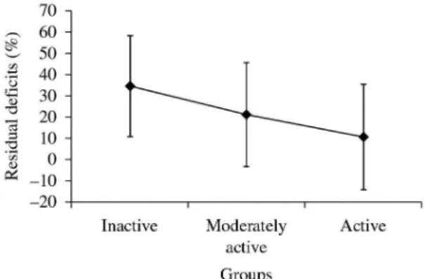
0.01<p<0.001). As illustrated in Figure 1, the RDS
of the hip lexor muscles demonstrated a tendency for lower values with higher physical activity levels, although no signiicant differences between the moderately active and the active groups were observed. Similar patterns were found for the RD values for all of the other muscular groups.
Significant between-group differences were observed for the distance covered during the 6MWT [F (2, 95)=10.75; p<0.001]. However, no signiicant
differences between the groups were found for
the MIP [F (2, 91) =2.13; p=0.13] and MEP [F(2, 91)=0.92; p=0.40] measures.
Associations between the variables
Signiicant, negative, and fair correlations were found between the RDs of all the lower limb muscles, except for the ankle plantar and dorsal lexors, with the distance covered during the 6MWT (–0.30<r<–0.43; p<0.01) and the AAS scores (–0.28<r<–0.41; p<0.01). These associations indicated that the higher
the work differences between the paretic and non-paretic lower limbs, as expressed by the RDs, the lower were the AAS score and the distances covered during the 6MWT. In addition, moderate to good correlations between the distances covered during the 6MWT and the AAS scores were also found (r=0.51; p<0.0001). It is important to note that no signiicant
correlations were found between the respiratory
strength measures and any of the other investigated variables (–0.11<r<0.12; 0.26<p<0.56).
Discussion
The present study found signiicant differences
between the groups regarding the functional
capacities and the RDs of the paretic lower limb
muscles. However, regarding the RD, no signiicant differences were found between the moderately active and active groups. No signiicant between-group
differences were found for the respiratory strength
measures. Significant correlations were found between the RDs of the hip and knee muscles, the functional capacity, and the physical activity levels. Interestingly, no signiicant correlations were found
between the measures of respiratory strength and any
of the investigated variables.
To the best of our knowledge, this was the irst study to examine the relationships between the
strength of the respiratory and the lower limb muscles
and the distances covered during the 6MWT in individuals with chronic hemiparesis based upon their physical activity levels. It is reported that little attention is given to the pulmonary system during the evaluation and treatment of individuals with hemiparesis, although it is known that these individuals demonstrate deicits in this system32. Interestingly, there were no signiicant correlations
between the respiratory strength measures and
any of the other studied variables, nor differences in respiratory strength between the impaired, moderately active, and active groups.
Differences between the impaired, moderately active, and active groups were found for all work measures, except for the ankle plantarlexor muscles, demonstrating that individuals with higher levels of physical activity showed fewer strength discrepancies between their paretic and non-paretic lower limbs. Interestingly, the work measures were not different between the moderately active and active groups. These indings could be partially explained by the high levels of inter-subject variability, as evidenced by the high standard deviation values found for all groups. The RDs of the work measures were used for analyses, since their calculations are based upon the principle that the performance of the non-paretic lower limb of each individual was the reference for the performance of the paretic one. Thus, the use of this variable allows objective comparisons between groups and reduces the typical variability of the results in this population, which facilitates the interpretation of the indings33. However, the variability of the results still remained high, but this inding seems common with this population, when motor and functional parameters are investigated8,11,34,35.
Although previous studies have reported that the variable which best explained physical activity levels was walking speed36, and that plantar lexor strength has been identiied as an important component of
speed37, the work produced by the ankle plantar lexor Figure 1. Residual deicits of the hip lexor muscles (means and
muscles was the only variable which did not show differences between the groups. However, only 40 individuals were able to perform the ankle tests and this may have inluenced the results, in addition to the high inter-subject variability.
Most of the RD values did not show regular distribution patterns since some individuals showed positive values and others negative ones, which meant that in these cases, the paretic side was able to produce higher levels of work than the non-paretic one. Stabilization has been reported to be an important factor inluencing the amount
of muscular force produced during testing38-40. Although stabilization was performed following the manufacturer’s recommendations, perhaps the paretic limbs received greater stabilization from the non-paretic side, which could have contributed to the higher values found in the same muscles on the paretic side, compared to the non-paretic one, and could partially explain the negative values.
The RD levels of the isokinetic work generated by
the lower limb muscles were selected to demonstrate
muscular performance. Despite the fact that peak torque is considered an excellent indicator of maximum levels of forces that an individual is able to generate, this variable alone does not take into account the ability of maintaining this force over a certain range of motion. Work, on the other hand, demonstrates the ability of individuals to generate and sustain forces over a certain range of motion41. This fact might explain the fair correlations found between the isokinetic work and the distance covered during the 6MWT.
Despite reports that individuals with hemiparesis
show significant respiratory dysfunctions and
reduced exercise tolerance42, the present indings do not support the idea that individuals with various exercise capacities show differences in strength in their respiratory muscles. These indings could be partially explained by the fact that the included individuals had chronic hemiparesis and were
probably already functionally adapted and did not
demonstrate major respiratory strength deicits11. However, Sutbeyaz et al.32 showed that a six-week
strengthening program of the inspiratory muscles was
able to increase exercise capacity, decrease dyspnea, and improve the quality of life in individuals with
hemiparesis who demonstrated greater respiratory
strength deicits.
The absence of signiicant relationships between
measures of respiratory strength and the distance
covered during the 6MWT, could be related to the
distinct demands required for the measurement of
these variables. The manovacuometry, for instance, requires maximum effort over a short period of time, whose demands are not equivalent to those required for the execution of the 6MWT, in which the individual should maintain efforts throughout the test.
The signiicant differences between the groups regarding the distance covered during the 6MWT suggested that active individuals had higher physical capacities, corroborating previous reported indings
with the elderly26. From the functional point of view, the 6MWT is widely used for the evaluation of physical itness and exercise capacity with various
populations43 and consists of a simple and practical test, that can be widely used within clinical contexts. Moderate correlations were observed between the distances covered during the 6MWT and the AAS,
which demonstrated that they assessed similar
aspects.
It was observed that individuals with higher activity levels, as determined by their AAS scores, also had higher levels of functional capacity, as demonstrated by the greater distances covered during the 6MWT. Previous studies reported that individuals with hemiparesis have reduced abilities to exercise and showed a 40% reduction in their exercise capacities, compared to sedentary healthy individuals, matched by age and sex32. These indings
show the importance of maintaining high physical
activity levels for this population.
It should be noted that due to the design of the study, causal relationships cannot be determined. In addition, the fact that the sample size was also
not uniformly distributed between the groups may
have inluenced the results. It is important to point out that only individuals with chronic hemiparesis who demonstrated the ability to independently walk were included. Thus, the present indings cannot be generalized to subjects with different characteristics, such as being in the acute or sub-acute stages or being unable to walk. Furthermore, parameters related to the cardiovascular system were not taken into consideration and they may have inluenced the assessments, especially the 6MWT. This aspect should be investigated in future studies.
Conclusions
The findings of this study demonstrated that
individuals with hemiparesis with different physical activity levels showed differences in the distance walked during the 6MWT and work deicits of the paretic lower limb muscles. However, differences in
active and active groups. Measures of respiratory strength were similar between the groups, and these variables were not related to any of the other functional variables. Lower strength deicits and
higher functional capacities were associated with
higher physical activity levels.
Acknowledgements
To the Brazilian national funding agencies, Coordenação de Aperfeiçoamento de Pessoal de Nível Superior (CAPES), Conselho Nacional de Desenvolvimento Cientíico e Tecnológico (CNPQ), and Fundação de Amparo a Pesquisa do Estado de Minas Gerais (FAPEMIG).
References
1. Kelly-Hayes PM, Robertson JT, Broderick JP, Duncan PW, Hershey LA, Roth EJ, et al. The American Heart Association Stroke outcome classification. Stroke. 1998;29(6):1274-1280. PMid:9626308. http:// dx.doi.org/10.1161/01.STR.29.6.1274
2. LeBrasseur NK, Sayers SP, Ouellette MM, Fielding RA. Muscle impairments and behavioral factors mediate functional limitations and disability following stroke. Phys Ther. 2006;86(10):1342-50. PMid:17012638. http:// dx.doi.org/10.2522/ptj.20050162
3. Billinger SA, Coughenour E, MacKay-Lyons MJ, Ivey FM. Reduced Cardiorespiratory fitness after stroke: Biological consequences and exercise-induced adaptations. Stroke Res Treat. 2012;2012:1-11. PMid:21876848 PMCid:PMC3159380. http://dx.doi. org/10.1155/2012/959120
4. Canning CG, Ada L, Adams R, O’Dwyer NJ. Loss of strength contributes more to physical disability after stroke than loss of dexterity. Clin Rehabil. 2004;18(3):300-8. PMid:15137561. http://dx.doi. org/10.1191/0269215504cr715oa
5. Hill TR, Gjellesvik TI, Moen PM, Torhaug T, Fimland MS, Helgerud J, et al. Maximal strength training enhances strength and functional performance in chronic stroke survivors. Am J Phys Med Rehabil. 2012;91(5):393-400. PMid:22357133. http://dx.doi.org/10.1097/ PHM.0b013e31824ad5b8
6. Ouellette MM, LeBrasseur NK, Bean JF, Phillips E, Stein J, Frontera WR, et al. High-intensity resistance training improves muscle strength, self-reported function, and disability in long-term stroke survivors. Stroke. 2004;35(6):1404-9. PMid:15105515. http://dx.doi. org/10.1161/01.STR.0000127785.73065.34
7. Olney SJ, Nymark J, Brouwer B, Culham E, Day A, Heard J, et al. A randomized controlled trial of supervised versus unsupervised exercise programs for ambulatory stroke survivors. Stroke. 2006;37(2):476-81. PMid:16410482. http://dx.doi.org/10.1161/01.STR.0000199061.85897.b7
8. Teixeira-Salmela LF, Olney SJ, Nadeau S, Brouwer B. Muscle strengthening and physical conditioning to reduce impairment and disability in chronic stroke survivors. Arch Phys Med Rehabil. 1999;80:1211-1218. http://dx.doi. org/10.1016/S0003-9993(99)90018-7
9. Harraf F, Ward K, Man W, Rafferty G, Mills K, Polkey M, et al. Transcranial magnetic stimulation study of expiratory muscle weakness in acute ischemic stroke. Neurology. 2008;71(24):2000-7 PMid:19064882. http:// dx.doi.org/10.1212/01.wnl.0000336927.30977.56 10. Lanini B, Bianchi R, Romagnoli I, Coli C, Binazzi
B, Gigliotti F, et al. Chest wall kinematics in patients with hemiplegia. Am J Respir Crit Care Med. 2003;168(1):109 13. PMid:12714347. http://dx.doi. org/10.1164/rccm.200207-745OC
11. Teixeira-Salmela LF, Parreira VF, Britto RR, Brant TC, Inacio EP, Alcantara TO, et al. Respiratory pressures and thoracoabdominal motion in community-dwelling chronic stroke survivors. Arch Phys Med Rehabil. 2005;86(10):1974-8. PMid:16213241. http:// dx.doi.org/10.1016/j.apmr.2005.03.035
12. Ward K, Seymour J, Steier J, Jolley CJ, Polkey MI, Kalra L, et al. Acute ischaemic hemispheric stroke is associated with impairment of relex in addition to voluntary cough. Eur Respir J. 2010;36(6):1383-90. PMid:20413536. http:// dx.doi.org/10.1183/09031936.00010510
13. Harik-Khan RI, Wise RA, Fozard JL. Determinants of maximal inspiratory pressure. The Baltimore Longitudinal Study of Aging. Am J Respir Crit Care Med. 1998;158(5 Pt 1):1459-64. PMid:9817693. http://dx.doi.org/10.1164/ ajrccm.158.5.9712006
14. Patterson SL, Forrester LVV, Rodgers MM, Ryan AS, Ivey FM, Sorkin JD, et al. Determinants of walking function after stroke: Differences by deicit severity. Arch Phys Med Rehabil. 2007;88:115-119. PMid:17207686. http://dx.doi.org/10.1016/j.apmr.2006.10.025
15. Mukherjee D, Patil CG. Epidemiology and the global burden of stroke. World Neurosurg. 2011;76(6 Suppl):S85-90. PMid:22182277. http://dx.doi. org/10.1016/j.wneu.2011.07.023
16. Folstein MF, Folstein SE, McHugh PR. “Mini-mental state”: A practical method for grading the cognitive state of patients for the clinician. J Psychiatr Res. 1975;12:189-98. http://dx.doi.org/10.1016/0022-3956(75)90026-6 17. Brucki SMD, Nitrini R, Caramelli P, Bertolucci PHF,
Okamoto IH. Sugestões para o uso do mini-exame do estado mental no Brasil. Arq Neuropsiquiatr. 2003;61(3B):777-81. PMid:14595482. http://dx.doi.org/10.1590/ S0004-282X2003000500014
18. Souza AC, Magalhaes LC, Teixeira-Salmela LF. Adaptação transcultural e análise das propriedades psicométricas da versão brasileira do Peril de Atividade Humana. Cad Saude Publica. 2006; 22(12):2623-2636. PMid:17096041. http://dx.doi.org/10.1590/S0102-311X2006001200012 19. Davidson M, De MN. A systematic review of the
20. Dempster WJ. Space requirements of the seated operator. Wright Patterson Air Force Base; 1985. (Rep. No. WADC-TR-tt-159).
21. Sharp SA, Brouwer BJ. Isokinetic strength training of the hemiparetic knee: Effects of function and spasticity. Arch Phys Med Rehabil. 1997;78:1231-36. http://dx.doi. org/10.1016/S0003-9993(97)90337-3
22. Guyatt GH, Thompson PJ, Berman LB, Sullivan MJ, Townsend M, Jones NL, et al. How should we measure function in patients with chronic heart and lung disease? J Chronic Dis. 1985;38(6):517-24. http://dx.doi. org/10.1016/0021-9681(85)90035-9
23. Iwama AM, Andrade GN, Shima P, Tanni SE, Godoy I, Dourado VZ. The six-minute walk test and body weight- walk distance product in healthy Brazilian subjects. Braz J Med Biol Res. 2009;42(11):1080-5. PMid:19802464. http://dx.doi.org/10.1590/S0100-879X2009005000032 24. Soares MR, Pereira CAC. Six-minute walk test:
Reference values for healthy adults in Brazil. J Bras Pneumol. 2011;37(5):576-583.
25. ATS Committee on Proiciency Standards for Clinical Pulmonary Function Laboratories. ATS Statement: Guideline for the six-minute walk test. Am J Respir Crit Care Med. 2002;166:111-7. PMid:12091180. http://dx.doi. org/10.1164/ajrccm.166.1.at1102
26. Britto RR, Sousa LAP. Six minute walk test: A Brazilian Standardization. Fisioter Mov. 2006; 18(4):49-54. 27. Neder JA, Andreoni S, Lerario MC, Nery LE. Reference
values for lung function tests. II: Maximal respiratory pressures and voluntary ventilation. Braz J Med Biol Res. 1999;32:719-27. PMid:10412550. http://dx.doi. org/10.1590/S0100-879X1999000600007
28. Souza RB. Pressões respiratórias estáticas máximas. J Pneumol. 2002;28 Suppl 3:S155-65.
29. Hamnegard CH, Wragg S, Kyroussis D, Aquilina R, Moxham J, Green M. Portable measurement of maximum mouth pressures. Eur Respir J. 1994;7:398-401. PMid:8162993. http://dx.doi.org/10.1183/0903193 6.94.07020398
30. McConnell AK, Copestake AJ. Maximum static respiratory pressures in healthy men and women: Issues of reproducibility and interpretation. Respiration. 1999;66:251-8. http:// dx.doi.org/10.1159/000029386
31. Portney LG, Watkins MP. Foundations of Clinical Research: Applications to practice. 3rd ed. Upper Saddle River: Prentice-Hall; 2008.
32. Sutbeyaz ST, Koseoglu F, Inan L, Coskun O. Respiratory muscle training improves cardiopulmonary function and exercise tolerance in subjects with subacute stroke: A randomized controlled trial. Clin Rehabil. 2010;24(3):240-50. PMid:20156979. http:// dx.doi.org/10.1177/0269215509358932
33. Alon G. Deining and measuring residual deicits of the upper extremity following stroke: A new perspective.
Top Stroke Rehabil. 2009;16(3):167-76. PMid:19632961. http://dx.doi.org/10.1310/tsr1603-167
34. Adams RW, Gandevia SC, Skuse NF. The distribution of muscle weakness in upper motoneuron lesions affecting the lower limb. Brain. 1990;113(5):1459-76. PMid:2245306. http://dx.doi.org/10.1093/brain/113.5.1459
35. Dombovy M. Stroke: Clinical course and neurophysiologic mechanism of recovery. Phys Rehabil Med. 1991;2:171-88. 36. Teixeira-Salmela LF, Devaraj R, Olney SJ. Validation
of the human activity proile in stroke: A comparison of observed, proxy and self-reported scores. Disabil Rehabil. 2007;29(19):1518-24. PMid:17852225. http:// dx.doi.org/10.1080/09638280601055733
37. Brincks J, Nielsen JF. Increased power generation in impaired lower extremities correlated with changes in walking speeds in sub-acute stroke patients. Clin Biomech. 2012;27(2):138-44. PMid:21899933. http:// dx.doi.org/10.1016/j.clinbiomech.2011.08.007
38. Bohannon RW, Bubela DJ, Wang Y, Magasi SR, Gershon RC. Adequacy of belt-stabilized testing of knee extension strength. J Strength Cond Res. 2011;25(7):1963-67. PMid:21399535 PMCid:PMC3116990. http://dx.doi. org/10.1519/JSC.0b013e3181e4f5ce
39. Hanten WP, Ramberg CL. Effect of stabilization on maximal isokinetic torque of the quadriceps femoris muscle during concentric and eccentric contractions. Phys Ther. 1988;68(2):219-22. PMid:3340660.
40. Hart DL, Stobbe TJ, Till CW, Plummer RW. Effect of trunk stabilization on quadriceps femoris muscle torque. Phys Ther. 1984;64:1375-80. PMid:6473518.
41. Brown LE, Weir JP. ASEP procedures recommendation I: Accurate assessment of muscular strength and power. JEP. 2001;4(3):1-21.
42. Sezer N, Ordu NK, Sutbeyaz ST, Koseoglu BF. Cardiopulmonary and metabolic responses to maximum exercise and aerobic capacity in hemiplegic patients. Funct Neurol. 2004;19(4):233-8. PMid:15776791.
43. Pires SR, Oliveira AC, Parreira VF, Britto RR. Teste de caminhada de seis minutos em diferentes faixas etárias e índices de massa corporal. Rev Bras Fisioter 2007;11(2):147-51. http://dx.doi.org/10.1590/ S1413-35552007000200010
Correspondence
Luci Fuscaldi Teixeira-Salmela
Universidade Federal de Minas Gerais Departamento de Fisioterapia