http://dx.doi.org/10.1590/bjpt-rbf.2014.0117 Braz J Phys Ther. 2015 Sept-Oct; 19(5):429-432 429
clinical commentary
Clinical commentary of the evolution of the treatment for
chronic painful mid-portion Achilles tendinopathy
Håkan Alfredson1,2,3
ABSTRACT | Thechronic painful Achilles tendon mid-portion was for many years, and still is in many countries, treated with intratendinous revision surgery. However, by coincidence, painful eccentric calf muscle training was tried, and it
showed very good clinical results. This inding was unexpected and led to research into the pain mechanisms involved in
this condition. Today we know that there are very few nerves inside, but multiple nerves outside, the ventral side of the
chronic painful Achilles tendon mid-portion. These research indings have resulted in new treatment methods targeting
the regions with nerves outside the tendon, methods that allow for a rapid rehabilitation and fast return to sports.
Keywords: rehabilitation; tendinosis; eccentric training.
HOW TO CITE THIS ARTICLE
Alfredson H. Clinical commentary of the evolution of the treatment for chronic painful mid-portion Achilles tendinopathy. Braz J Phys Ther. 2015 Sept-Oct; 19(5):429-432. http://dx.doi.org/10.1590/bjpt-rbf.2014.0117
1 Department of Community Medicine and Rehabilitation, Sports Medicine Unit, Umeå University (UMU), Umeå, Sweden 2 Pure Sports Medicine Clinic, London, UK
3 The Institute of Sport Exercise & Health (ISEH), University College London Hospitals (UCLH), London, UK
Received: Feb. 26, 2015 Revised: May. 18, 2015 Accepted: June. 22, 2015
Background
Chronic painful mid-portion Achilles tendinopathy is a relatively common condition among recreational and elite athletes, but it is also seen in non-active individuals. It is most common between the age of 36 and 60 and very rare among individuals younger than 25 years. The etiology is unknown, but an altered
lipid proile with high cholesterol levels has been found
in 1/3 of the patients1. Excessive dorsilexion in the
ankle joint2 and low calf muscle strength have also been
suggested as possible etiological factors. Conservative
treatment with different loading regimens is the irst
line of treatment, and if that fails, surgical treatment is instituted. For surgical treatment, intratendinous revision via tenotomy followed by 4-6 months of rehabilitation has been the most commonly used procedure worldwide.
The purpose of this clinical commentary is to show how the results of research on the basic science for this condition has resulted in a completely new treatment
strategy with major advantages for the patients.
In the 1990s, the Sports Medicine Unit in Umeå, Sweden, as in most other countries, used intratendinous revision surgery to treat patients with chronic painful mid-portion Achilles tendinopathy. Patients not responding to conservative management
were treated with open surgery, including excision of
macroscopically abnormal tendon tissue via a central longitudinal tenotomy, followed by immobilization in a cast for 2-6 weeks, with a total 4-6 months rehabilitation period.
By coincidence, our group at the Sports Medicine Unit
in Umeå tried a modiied version of the Stanish et al.3
model for eccentric calf muscle training. We used a level of loading that was causing pain in the tendon
during the exercise and the exercises were done at a slow pace, in contrast to pain-free exercises and
gradually increased speed. We got surprisingly good clinical results. To achieve good clinical results after applying painful heavy loading on a chronic painful Achilles tendon was completely opposite to previous thinking around treatments of chronic painful tendons, and the good clinical results4 led to research into
the pain mechanisms involved in chronic painful mid-portion Achilles tendinopathy.
Painful eccentric calf muscle training
Our group designed an eccentric training regimen
modiied from the Stanish et al.3 model to be tried on
Alfredson H
430 Braz J Phys Ther. 2015 Sept-Oct; 19(5):429-432
Achilles tendinosis. The training program included
eccentric training over a step – 3x15 reps with straight and lexed knee performed 2 times/day, 7 days/week,
for 3 months4. The method was tested in scientiic
studies4-6, and the overall results were very good,
with around 80% satisied and pain-free patients.
After a while, we found out that high-level athletes,
especially runners and jumpers who wear spiked shoes,
did not have such good results with this treatment.
Also, we found it to be of signiicant importance to
establish that the patients had a correct diagnosis before the start of treatment. A partial rupture has
to be excluded, because using eccentric training on
a partially ruptured Achilles can further damage the tendon, possibly causing a lengthening of the tendon,
that is known to be very dificult to treat.
Ultrasound follow-ups were performed on patients with chronic Achilles tendinopathy and very interestingly showed that in the successfully treated patients the Achilles tendon thickness had decreased over time, and the structure looked more normal sonographically7. Consequently, it appeared that painful eccentric calf muscle training had the potential to remodel the
tendinosis tendon. From these research projects,
where high painful loads were applied to the thick and painful Achilles tendons, we also learned that tolerating these high eccentric loads clearly show that the Achilles tendinosis tendon is not what had previously been thought: a so-called degenerative and weak tendon. Instead, it might very well be a strong tendon!
New research on tendon histology
and imaging
We could not explain the background to the good
clinical results achieved with painful eccentric training,
and this led to extensive research together with Professor
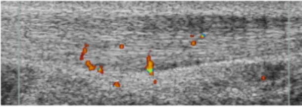
Sture Forsgren’s group at the Anatomy Department and Dr Lars Öhberg at the Department of Radiology at Umeå University. Using ultrasound+Doppler, we found
high blood low inside and outside the ventral side of
the Achilles tendon mid-portion in patients with chronic painful mid-portion Achilles tendinopathy, but not in normal Achilles tendons8 (Figure 1). In a following
study, ultrasound+Doppler-guided biopsies were taken
from the region with high blood low inside and outside
the Achilles mid-portion in patients with chronic painful tendinosis. Immune-histochemical analyses showed nerves in close relation to blood vessels outside the tendon, but very few nerves inside the tendon9.
An interesting observation was that these were mainly sympathetic nerves, but also a few sensory nerves9.
To try to trace the pain, ultrasound+Doppler-guided
injections of small volumes of the local anesthetic xylocain+Adrenaline were administered, targeting the regions with high blood low outside the tendon.
This temporarily cured the tendon pain10. These indings
clearly indicated that the pain in mid-portion Achilles tendinopathy comes from the nerves located on the ventral side of the Achilles, and that the nerves can indirectly be found by using ultrasound+Doppler to
ind the regions with high blood low (blood vessels
with accompanying nerves).
Ultrasound+Doppler-guided
sclerosing polidocanol injections
The new research findings related to the reduction of pain at the regions of highest blood
low led to the invention of a new treatment method: ultrasound+Doppler-guided injections of the sclerosing
substance polidocanol, targeting the regions with high
blood low and nerves outside the tendon. This type of treatment showed good clinical results with signiicantly
lowered pain scores (VAS) during Achilles tendon loading activity in pilot studies and in a randomized placebo-controlled study11,12. Ultrasound+Doppler
2-year follow-ups of patients treated with sclerosing
polidocanol injections showed decreased tendon
thickness and improved structure (less irregular structure with less hypo-echoic regions) over time13,
indicating a high potential in the soft tissues outside the ventral side of the Achilles tendon. The limitations
with ultrasound+Doppler-guided polidocanol injections
are that it is technically demanding, having a relatively long learning curve, and that often multiple4,5 injection
treatments are needed.
Figure 1. Ultrasound and Doppler examination showing a thickened
The chronic painful Achilles
431
Braz J Phys Ther. 2015 Sept-Oct; 19(5):429-432
Ultrasound+Doppler-guided
mini-surgical scraping
To try to overcome the problems with the technically
demanding polidocanol injection treatment, our
group at the Sports Medicine Unit in Umeå invented a mini-surgical scraping treatment. Guided by the ultrasound+Doppler findings, a minor surgical procedure is performed under local anesthesia. Using a longitudinal lateral mini (1 cm) incision, the ventral side of the tendon is scraped in the regions with high
blood low and nerves14,15. This is a one stage and
more radical approach to interfere with the nerves accompanying the blood vessels on the ventral side of the Achilles. Because there is no intratendinous treatment associated with this procedure, a relatively fast (4-6 weeks) rehabilitation can be used. The patients
start walking with full weight bearing the irst day
after the operation and rapidly progress to functional
tendon loading. There is no speciic eccentric training
regimen, but instead, there is a general build-up of training, depending on the requirements for the individual’s tendon loading activity (high-level activity to non-activity). The clinical results are very good
with signiicantly lowered pain scores (VAS) during Achilles tendon loading activity and return to pre-injury activity levels, without any major side effects. In the
1-2 year follow-up of these individuals, the results remain positive, and the use of this method has been increased. We now have operated on large numbers of patients with chronic AT and at different activity levels, including professional athletes15. For reasons
still unknown, high-level athletes seem to do best after this procedure. Patients with low physical activity
level showed good clinical results in about 70% of
cases, while among high-level athletes the success rate was more than 90%15.
Recently, focus has been placed on the plantaris tendon, located in close relation to the medial Achilles. There seems to be a subgroup of patients suffering from chronic painful mid-portion Achilles tendinopathy, where a thickened plantaris tendon is involved16. These
patients have both mid-portion Achilles tendinopathy
with high blood low on the ventral side of the tendon
and a closely located plantaris tendon (demonstrated with ultrasound) with also a localized high blood
low (Doppler) on the medial side of the Achilles.
These patients most often complain of having pain located on the medial side of the Achilles, where the medial soleus inserts. It is our observation that if the plantaris tendon is involved there is often a poor response to eccentric training. This can theoretically
be explained by the fact that the plantaris tendon,
known to be stronger and stiffer than the Achilles17, can cause a compression on the medial Achilles during the movements in the eccentric treatment regimen. When we noticed that the plantaris tendon could be involved, we changed the surgical technique from using a lateral incision to always using a medial incision to allow for an accurate evaluation of the relationship between the plantaris and Achilles tendons18 (Figure 2).
If a plantaris tendon involvement is found, then the
plantaris tendon is released proximally and distally,
and 4-6 cm of its length are taken out.
Very recently, we have noticed that there is a minor group of patients who have plantaris-related pain without also having mid-portion Achilles
tendinopathy (veriied with ultrasound+Doppler examination) (non-published data). These patients
do very well after plantaris tendon removal alone. To study the innervation patterns of the plantaris tendon,
immune-histochemical examinations were performed
in a large number of plantaris tendons and surrounding
ibrous connective and fat that were taken out from
patients with mid-portion Achilles tendinopathy and plantaris involvement18. Although the results related
to innervation patterns have not been published yet, they show that most sensory nerves are found in the peritendinous connective tissue between the Achilles and plantaris tendon, but in about 1/3 of the plantaris tendons, there are also nerves inside the plantaris tendon that may be a co-factor in the medial pain.
Conclusions
Non-operative treatment with painful eccentric
training is the irst line of treatment for chronic painful
mid-portion Achilles tendinopathy. Our research on the innervation patterns in patients with chronic painful mid-portion Achilles tendinopathy has shown that there are no (or very few) nerves inside the chronic painful Achilles tendon mid-portion. Instead,
Alfredson H
432 Braz J Phys Ther. 2015 Sept-Oct; 19(5):429-432
the nerves are found outside the ventral side of the tendon. This knowledge has led to the invention of a new mini-invasive surgical treatment, combined with a fast rehabilitation, to be used on the patients who have a poor result with eccentric training. With the use of this method, there is a very good chance of cure from chronic painful mid-portion Achilles tendinopathy and return to full activity, including Achilles tendon-demanding professional sports, within 4-6 weeks after surgery.
References
1. Beeharry D, Coupe B, Benbow EW, Morgan J, Kwok S, Charlton-Menys V, et al. Familial hypercholesterolaemia commonly presents with Achilles tenosynovitis. Ann Rheum Dis. 2006;65(3):312-5. http://dx.doi.org/10.1136/ ard.2005.040766. PMid:16176995.
2. Nawoczenski DA, Barske H, Tome J, Dawson LK, Zlotnicki JP, DiGiovanni BF. Isolated gastrocnemius recession for achilles tendinopathy: strength and functional outcomes. J Bone Joint Surg Am. 2015;97(2):99-105. http://dx.doi. org/10.2106/JBJS.M.01424. PMid:25609435.
3. Stanish WD, Rubinovich RM, Curwin S. Eccentric exercise in chronic tendinitis. Clin Orthop Relat Res. 1986(208):65-8. PMID: 3720143.
4. Alfredson H, Pietilä T, Jonsson P, Lorentzon R. Heavy-load eccentric calf muscle training for the treatment of chronic Achilles tendinosis. Am J Sports Med. 1998;26(3):360-6. PMid:9617396.
5. Mafi N, Lorentzon R, Alfredson H. Superior short-term results with eccentric calf muscle training compared to concentric training in a randomized prospective multicenter study on patients with chronic Achilles tendinosis. Traumatol Arthrosc. 2001;9(1):42-7. http://dx.doi.org/10.1007/s001670000148. 6. Fahlström M, Jonsson P, Lorentzon R, Alfredson H. Chronic
Achilles tendon pain treated with eccentric calf-muscle training. Knee Surg Sports Traumatol Arthrosc. 2003;11(5):327-33. http://dx.doi.org/10.1007/s00167-003-0418-z. PMid:12942235. 7. Öhberg L, Lorentzon R, Alfredson H. Eccentric training
in patients with chronic Achilles tendinosis: normalised tendon structure and decreased thickness at follow up. Br J Sports Med. 2004;38(1):8-11, discussion 11. http://dx.doi. org/10.1136/bjsm.2001.000284. PMid:14751936. 8. Öhberg L, Lorentzon R, Alfredson H. Neovascularisation in
Achilles tendons with painful tendinosis but not in normal tendons: an ultrasonographic investigation. Knee Surg Sports Traumatol Arthrosc. 2001;9(4):233-8. http://dx.doi. org/10.1007/s001670000189. PMid:11522081.
9. Andersson G, Danielson P, Alfredson H, Forsgren S. Nerve-related characteristics of ventral paratendinous tissue in chronic Achilles tendinosis. Knee Surg Sports Traumatol Arthrosc. 2007;15(10):1272-9. http://dx.doi.org/10.1007/ s00167-007-0364-2. PMid:17604979.
10. Alfredson H, Öhberg L, Forsgren S. Is vasculo-neural ingrowth the cause of pain in chronic Achilles tendinosis? An investigation using ultrasonography and colour Doppler, immunohistochemistry, and diagnostic injections. Knee Surg Sports Traumatol Arthrosc. 2003;11(5):334-8. http:// dx.doi.org/10.1007/s00167-003-0391-6. PMid:14520512. 11. Ohberg L, Alfredson H. Ultrasound guided sclerosis of
neovessels in painful chronic Achilles tendinosis: pilot study of a new treatment. Br J Sports Med. 2002;36(3):173 -5, discussion 176-7. http://dx.doi.org/10.1136/bjsm.36.3.173. PMid:12055110.
12. Alfredson H, Öhberg L. Sclerosing injections to areas of neo-vascularisation reduce pain in chronic Achilles tendinopathy: a double-blind randomised controlled trial. Knee Surg Sports Traumatol Arthrosc. 2005;13(4):338-44. http://dx.doi.org/10.1007/s00167-004-0585-6. PMid:15688235. 13. Lind B, Öhberg L, Alfredson H. Sclerosing polidocanol
injections in mid-portion Achilles tendinosis: remaining good clinical results and decreased tendon thickness at 2-year follow-up. Knee Surg Sports Traumatol Arthrosc. 2006;14(12):1327-32. http://dx.doi.org/10.1007/s00167-006-0161-3. PMid:16967202.
14. Alfredson H, Öhberg L, Zeisig E, Lorentzon R. Treatment of midportion Achilles tendinosis: similar clinical results with US and CD-guided surgery outside the tendon and sclerosing polidocanol injections. Knee Surg Sports Traumatol Arthrosc. 2007;15(12):1504-9. http://dx.doi.org/10.1007/ s00167-007-0415-8. PMid:17879083.
15. Alfredson H. Ultrasound and Doppler-guided mini-surgery to treat midportion Achilles tendinosis: results of a large material and a randomised study comparing two scraping techniques. Br J Sports Med. 2011;45(5):407-10. http:// dx.doi.org/10.1136/bjsm.2010.081216. PMid:21349878. 16. Alfredson H. Midportion Achilles tendinosis and the plantaris
tendon. Br J Sports Med. 2011;45(13):1023-5. http://dx.doi. org/10.1136/bjsports-2011-090217. PMid:21628352. 17. Lintz F, Higgs A, Millett M, Barton T, Raghuvanshi M,
Adams MA, et al. The role of Plantaris Longus in Achilles tendinopathy: a biomechanical study. Foot Ankle Surg. 2011;17(4):252-5. http://dx.doi.org/10.1016/j.fas.2010.08.004. PMid:22017896.
18. Spang C, Alfredson H, Ferguson M, Roos B, Bagge J, Forsgren S. The plantaris tendon in association with mid-portion Achilles tendinosis: tendinosis-like morphological features and presence of a non-neuronal cholinergic system. Histol Histopathol. 2013;28(5):623-32. PMid:23378267.
Correspondence Håkan Alfredson
Department of Community Medicine and Rehabilitation Sports Medicine Unit