Heart rate assessment during maximal static
expiratory pressure and Valsalva maneuver in
healthy young men*
Avaliação da frequência cardíaca à medida de pressão expiratória máxima estática e
à manobra de Valsalva em jovens saudáveis
Vinicius Minatel, Marlus Karsten, Laura M. T. Neves, Thomas Beltrame, Audrey Borghi-Silva, Aparecida M. Catai
Abstract
Background: The measure of the maximal expiratory pressure (MEP) has some contraindications, as it is believed that the responses obtained in this measure are similar to the Valsalva maneuver (VM). Objective: The main purpose of this study was to evaluate the heart rate responses (HR) during the MEP and the VM measures in healthy young men into different postures aiming to identify whether and in which situation the MEP reproduces the responses obtained in the VM. Additionally we aim to estimate the workload realized during the maneuvers. Method: Twelve healthy young men were evaluated, instructed and familiarized with the maneuvers. The VM was characterized by an expiratory effort (40 mmHg) against a manometer for 15 seconds. The MEP measure has been performed according to the American Thoracic Society. Both measures were performed at sitting and supine positions. ANOVA two-way with Holm-Sidak post-hoc test (p<0.05) was used to analyse the heart rate variation (∆HR); Valsalva index (VI); MEP index (MEPI), and the estimated workload of the maneuvers (Wtotal, Wisotime, Wtotal/∆HRtotal and Wisotime/∆HRisotime). Results: The ∆HR during the maneuvers was not influenced by the supine and sitting
positions. However, the ∆HR during the VM and VI were higher (supine: 47±9 bpm, 2.3±0.2; sitting: 41±10 bpm, 2.0±0.2, respectively) than ∆HR during the MEP and MEPI values (supine: 23±8 bpm, 1.5±0.2; sitting 24±8 bpm, 1.6±0.3, respectively) (p<0.001). The estimated workload of the maneuvers was statistically different (p<0.001) between the maneuvers, except to Wtotal/∆HR. Conclusions: In the studied
conditions the MEP does not reproduces the HR response observed in the VM in healthy young men.
Keywords: Valsalva maneuver; respiratory muscle; heart rate; autonomic nervous system; posture; physical therapy.
Resumo
Contextualização: A medida de pressão expiratória máxima (PEmáx) possui algumas contraindicações, pois acredita-se que as respostas obtidas nessa medida são similares às respostas encontradas na manobra de Valsalva (MV). Objetivos: O objetivo principal é avaliar a resposta da frequência cardíaca (FC) durante a medida da PEmáx e da MV em jovens saudáveis, em diferentes posturas, para identificar se e em qual condição a PEmáx reproduz as respostas obtidas na MV e, adicionalmente, estimar o trabalho realizado nas
manobras. Método: Doze jovens saudáveis foram avaliados, orientados e familiarizados com as manobras. A MV foi composta por
um esforço expiratório (40 mmHg) durante 15 segundos contra um manômetro. A PEmáx foi executada segundo a American Thoracic
Society. Ambas as medidas foram realizadas nas posturas supino e sentado. Para a análise da variação da frequência cardíaca (∆FC), índice de Valsalva (IV), índice da PEmáx (IPEmáx) e o trabalho estimado das manobras (Wtotal, Wisotime, Wtotal/∆FCtotal e Wisotime/∆FCisotime), utilizou-se ANOVAtwo-way com post-hoc de Holm-Sidak (p<0,05). Resultados: A ∆FC durante as manobras não foi influenciada pelas posturas; entretanto, durante a MV, a ∆FC e os valores do IV foram maiores (supino: 47±9 bpm, 2,3±0,2; sentado: 41±10 bpm, 2,0±0,2,
respectivamente) do que a ∆FC e os valores de IPEmáx observados durante a PEmáx (supino: 23±8 bpm, 1,5±0,2; sentado 24±8 bpm,
1,6±0,3, respectivamente) (p<0,001). Os trabalhos estimados das manobras foram estatisticamente diferentes (p<0,001) entre elas,
exceto para o Wtotal/∆FC. Conclusões: Nas condições estudadas, a PEmáx não reproduz as respostas da FC observadas durante a MV
em jovens saudáveis.
Palavras-chave: manobra de Valsalva; músculos respiratórios; frequência cardíaca; sistema nervoso autonômico; postura; fisioterapia.
Received: 12/21/2011 – Revised: 04/02/2012 – Accepted: 04/23/2012
Laboratory of Cardiovascular Physical Therapy, Nucleus of Research in Physical Exercise (NUPEF), Physical Therapy Department, Universidade Federal de São Carlos (UFSCar), São Carlos, SP, Brazil
Introduction
Measurements of maximal inspiratory pressure (MIP) and maximal expiratory pressure (MEP) are extensively used in clinical practice of physical therapy as a parameter of indirect evaluation of respiratory muscles strength in patients with neuromuscular1, cardiopulmonary2,3 and/or metabolic4
disor-ders. In these situations, the MIP and MEP measurements are also used as a training prescription parameter for respiratory muscles strength and endurance5-8. In addition, these measures
are also used as a predictive parameter of success in the dis-continuation of mechanical ventilation8-10.
Despite the clinical importance of these measures, there are several absolute and relative contraindications to their use8,11.
he reason for the contraindication of MEP measure, which is a maneuver performed from a maximal inhalation (total lung-capacity) followed by an expiratory efort against an occluded airway is related to the similarity of this measurement with the Valsalva maneuver (VM)7,8,12. he performance of the expiratory
efort during the MEP for periods of time longer than 3 seconds may lead to cardiovascular alterations similar to those of VM, such as: decreased cardiac output8, venous return, pulmonary
circulation and coronary low13-15.
he VM is a test used to evaluate the role of the autonomic nervous system (ANS). he range of responses that occur during and after the performance of the VM has been widely described in the literature14-16. he cardiovascular responses of
the VM are mediated by the ANS and vary according to the ac-tivation and/or inhibition of the sympathetic and parasympa-thetic systems. his dynamic pattern of autonomic activity is determined by diferent stimulus, such as breathing, muscular contraction, stimulation of arterial baroreceptors17, postural
changes18-20 and it can be more easily investigated by analyzing
the responses of heart rate (HR) into a clinical environment. Although there are similarities between the performances of the MEP and the VM7,8,12, to date there are no studies that
have described the cardiovascular responses observed during the performance of MEP in diferent positions. hus, it is ob-served that the underuse of MEP in clinical practice of physical therapy may be associated with the possible similarity between its use and the VM7,8,12, without the real efects of the MEP on
the cardiovascular responses are known. herefore, the investi-gation of the HR response to the MEP in healthy young people could contribute primarily to the understanding of the safety of this procedure. Consequently, the evaluation of the expiratory muscles can be performed with less exposure of the subjects to unnecessary risks.
herefore, the main objective of the present study was to evaluate the hypothesis that the execution of the MEP in
healthy youth subjects will generate HR responses similar to those observed during the execution of the VM, independent of the subject position. Additionally, the workload to perform each maneuver was estimated in order to characterize the ma-neuvers and to provide support for interpretation of the results.
Method
Subjects
Twelve healthy men, aged between 20 and 29 years-old and with body mass index (BMI) between 18.5 and 29.9 kg/m2
participated in this study. he volunteers were informed and advised regarding the procedures that they would be submit-ted and they signed a consent form. he study was approved by the Ethics in Research Committee of the Universidade Fed-eral de São Carlos (UFSCar), São Carlos, SP, Brazil (protocol nº 435/2008).
he inclusion criteria were absence of diagnosed diseases, use of medications and use of illicit drugs or smoking. In addi-tion, the participants should not present electrocardiographic alterations at rest and during the exercise clinical test. Partici-pants who presented discomfort during the performance of the maneuvers would be considered to be excluded. However, no subject had any signs or symptoms compatible with the need to be excluded from the study.
Procedures
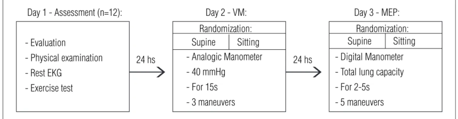
he clinical evaluation, the tests and the experimental pro-cedures were performed in the afternoon, on alternate days, with an interval of 24 hours at the Cardiovascular Physical herapy Laboratory - Nucleus of Research in Physical Exercise - Department of Physical herapy, UFSCar (Figure 1). During the experiments, the room temperature (between 22ºC and 24ºC) and the relative humidity of air (between 40 and 60%) were observed and controlled to not interfere on the tests results. he volunteers were instructed to not drink alcoholic and/or stimulants beverages for at least 12 hours before the test; to avoid strenuous exercises and to have a period of regu-lar sleep with good quality on the night that preceded the data collection.
risk factors for cardiovascular disease, smoking, medications and physical activity level. hey also underwent to a physical exam to identify musculoskeletal disorders, 12-lead electrocar-diogram (ECG) at resting and during test of physical exercise (Bruce protocol). In addition, familiarization procedures were performed with the equipment and experimental protocols in order to reduce the subject anxiety and expectation and to promote the learning of the VM and the MEP. he maneuvers were performed in the supine position with the volunteer po-sitioned with the upper and lower limbs in extension and the head lexed at 45º; and in the sitting position with the volunteer positioned with the trunk leaning on the chair and the feet on the ground, making an angle of approximately 90º between the trunk and the thighs. In both positions, the volunteers were in-structed to remain with their clothes pieces loosened to ensure that the respiratory movements were not limited.
Manometers
he manometers used in the present study (analogical and digital) were previously tested with regards to the measure-ments of the pressure values. here was no diference between the pressure values generated in the pilot study between the researchers, allowing its use. A digital manometer (MVD300, Globalmed, Brazil) connected to a microcomputer (software MVD300, Globalmed, Brazil) was used to collect the pressure values during the maneuvers and the visual control of the pres-sure curve vs time by the examiners.
Valsalva Maneuver
For the execution of VM, an analogical manometer (Dyasist, Brazil) connected to a digital manometer and a mouthpiece through a semi rigid tube was used. he volunteers should be
with the nostrils occluded by a nasal clip and with the mouth-piece irmly attached to the mouth, in order to prevent the air escape. he volunteers were asked to maintain spontaneous ventilation (~15 rpm), to perform a deep inhalation before the maneuver and to execute the voluntary expiratory efort after receiving an audio signal. During the VM the volunteers should maintain the expiratory efort equivalent to 40 mmHg for 15 seconds15,21,22. his maneuver was repeated three times, with a
minimum time interval of ive minutes among them, with the aim of the HR and BP return to the baseline values. In order to guarantee the glottic opening during the performance of the maneuvers a mouthpiece with oriice was used15. To evaluate
the correct performance of the VM, the following items were observed16,23: a) maintenance of pressure on the manometer; b)
facial lushing; c) jugular stasis; d) movements of the chest and e) rapid increase of HR followed by bradycardia. he order that the volunteers performed the maneuvers, i.e. supine or sitting positions, was random.
Maximal Expiratory Pressure (MEP)
he MEP measures were carried out using a digital manom-eter and a mouthpiece with a 2 mm oriice7,8,24. he volunteers
were instructed to take a deep breath from the residual volume to total lung capacity, to maintain the nostrils occluded by a nasal clip and the mouthpiece irmly attached to the mouth, in order to prevent the escape of air, to maintain spontaneous ventilation (~15 rpm) and to execute the expiratory efort after receiving the audio signal. Five attempts of maximal expiratory efort were performed, being at least three of them should be reproduced, that is, they could not difer to each other more than 10%. he volunteer should sustain the expiratory efort for at least two seconds. During this period, the examiner observed the curve generated by the expiratory efort on the microcom-puter screen (software MVD300, Globalmed, Brazil), in order - Evaluation
- Physical examination - Rest EKG
- Exercise test
24 hs 24 hs
Day 2 - VM:
Day 1 - Assessment (n=12): Day 3 - MEP:
- Analogic Manometer - 40 mmHg
- For 15s - 3 maneuvers
Randomization: Randomization:
Supine Sitting Supine Sitting - Digital Manometer - Total lung capacity - For 2-5s
- 5 maneuvers
to identify the development of a plateau. he expiratory pres-sure value observed at the irst second of the plateau after the peak pressure of each maneuver was recorded and compared with the predictive values for the Brazilian population25. If the
highest value was observed in the last attempt, a new measure would be performed, in order to exclude the learning efect7,8,24.
Between the measures, there was an interval of ive minutes in order to reproduce the procedure used in the VM. For the MEP measures, the order of the positions was randomly deined.
RRI Capture
To capture the intervals between two R waves (RRI) a portable heart rate monitor was used (S810i, Polar, Finland). To guarantee the quality of the signal, electrocardiographic signals measured by a one-channel heart monitor (TC-500, ECAFIX, São Paulo, SP, Brazil) were observed. he electrodes were positioned on the derivation MC5, being the negative pole placed at the sternal notch (M) and the positive pole and the ground wire placed at the area of the 5th intercostal space (C5), on the anterior axillary line on left and right, respectively. Prior to the performance of experimental protocols, the individuals remained in rest for 10 minutes for stabilization of vital signs (BP, HR and respiratory rate) and, thereafter the experiments were initiated. he capture of the RRI during the procedures was performed as follows: 60 seconds at rest; the maneuver execution time (15 seconds in the VM and 5 seconds in the MEP) and 300 seconds corresponding to the recovery period, which the individuals should return to the baseline values of HR and BP.
Data analysis
We analyzed the data regards to HRrest, mean HR values
within 60 seconds prior to the execution of each maneuver; HRpeak, the highest HR value obtained during the maneuvers;
HRnadir, the lowest value obtained after the interruption of
the maneuvers; HRrecovery, mean of the inal 180 seconds of
the recovery period of each maneuver26; heart rate variation
of (ΔHR), calculated by the diference between HRpeak
dur-ing each maneuver and HRrest
16,27; ΔHR
isotime, calculated by
subtracting the HR value in the third second after the deep inhalation from the mean HR at rest; the indexes of the Vals-alva maneuver (VI)15,16,18,28,29 and of the MEP maneuver (MEPI),
obtained by the ratio between the greatest RRI of the recovery period and the lowest RRI during the peak of the maneuver. he estimated workload (W) of the maneuvers was calculated
by multiplying the expiratory pressure values observed in the maneuvers by the total time (Wtotal) or by 3 seconds (Wisotime).
To evaluate the values of the workload performed in propor-tion to ∆HR, the ratios Wtotal/ΔHRtotal and Wisotime/ΔHRisotime
were calculated.
Statistical analysis
he sample size calculation was based on the results of ΔHR (VM: 35.80 bpm; MEP: 23.73 bpm and combined stand-ard deviation of 9.78 bpm) obtained in a pilot study (n=5), with β=0.8 and α=0.05. his calculation suggested a sample of 12 participants in each group (VM and MEP). For statistical analysis, the software SigmaPlot 11.0 (Systat, USA, 2011) was used. he Shapiro-Wilk test was used to verify the normality of data distribution and the two-way ANOVA for repeated measures with Holm-Sidak post-hoc to verify the efect of the maneuvers and positions. he signiicance level was 5%. Data are presented as mean and standard deviation.
Results
Table 1 presents age, anthropometric data of the volun-teers, the MEP values obtained and the percentage in relation to the values predicted by Neder et al.25. With regards to the
body mass index (BMI), the subjects were initially subdivided according to their BMI level in normal weight and overweight and tested the inluence of this variable on the HR responses. herefore, as no diference between the subgroups was found all the statistical analyses were performed considering only one group.
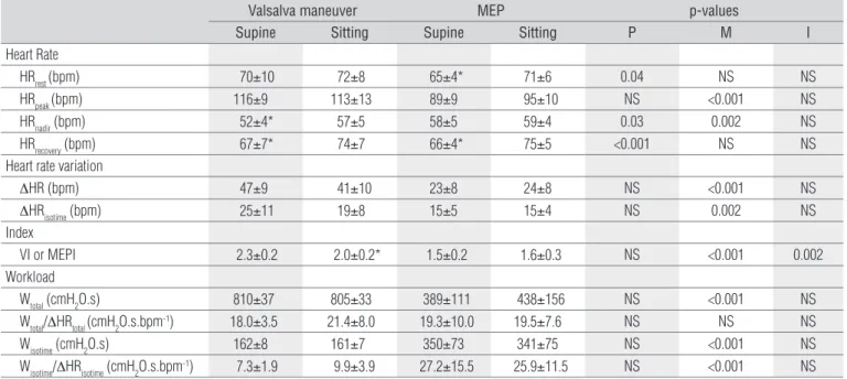
he HR values during the performance of the maneuvers are shown in Table 2. Figure 2 illustrates the HR responses ob-served during the maneuvers. A signiicant diference between the maneuvers for the HRpeak and HRnadir values was observed.
Among the positions, there were diferences in the values of
HRrest, HRnadir and HRrecovery. However, when comparing the
posi-tions intra maneuvers, there were diferences for the values of
HRnadir and HRrecovery in the VM and of HRrest and HRrecovery in the
MEP. here were signiicant diferences between the maneu-vers for the values of ΔHR and ΔHRisotime. he indexes VI and
MEPI were signiicantly diferent among the maneuvers, with interaction between position and maneuver. In the VM, the index VI had higher values in the supine position. he values
of Wtotal, Wisotime and the ratio Wisotime/ΔHRisotime were diferent
Discussion
he main indings of this study are: the performance of MEP does not reproduce the HR responses (ΔHR and indexes) observed in the VM. he results regarding to the workload and its ratios show an inluence of execution time of the maneu-ver. Additionally, the HR responses (ΔHR and indexes), the workload responses and its ratios were not inluenced by the studied positions.
Heart Rate
he HR response may be inluenced by several factors, such as gender, age, genetic and anthropometric characteristics, changes of body posture, itness level etc16,17,19,20. In the
pre-sent study, the body postural change has inluenced the HRrest,
HRnadir and HRrecovery responses, possibly due to the changes in
the sympathetic and parasympathetic modulation promoted by the postural change17,20,21.
However, during the execution of the VM the HR is inlu-enced mainly by the autonomic modulation, being responsible not only for the phase of rapid increase (vagal withdrawal on the sino-atrial node) but also for the decrease of its values after interruption of the maneuver (resumption of the vagal modula-tion on sino-atrial node)17,20,21. hese adjustments associated to
the adjustment of blood pressure and the peripheral vascular resistance occur in response to the activation of the arterial baroreceptors (carotid and aortic) and of the cardiopulmonary receptors14,15,22,23. Moreover, other studies show that the
interac-tion between the sympathetic relex of the arterial barorecep-tors and the vagal relex, probably activated by the pulmonary stretch receptors, is the responsible for the cardiovascular ac-tions during the VM14,22.
In the present study, there were no diferences in the indexes values (VI and MEPI) and ΔHR between supine and sitting positions. Our data agree with other studies, in which the inluence of position on the VM was investigated14,19. hese
authors identiied that the position does not interfere on HR and the HR response to VM is a compensatory mechanism that occurs in order to avoid hypotension due to decreased venous return.
he importance of the venous return on HR responses can be observed during the execution of the VM in the supine position. In this situation, the highest HR values during the maneuver (116±9 bpm) and the lowest HR values after the maneuver (52±4 bpm) were observed, which have resulted in higher values of VI (2.3±0.2). his response is probably due to the cardiovascular adjustments necessary to maintain the car-diac output during and after the interruption of the maneuver. Subjects (n=12)
Age (years) 25±2
Height (m) 1.78±0.06
Body mass (kg) 78±9
Body Mass Index (kg.m-2) 23.4±2.8
Predicted MEP* (cmH2O) 131±2
Supine
MEP(cmH2O) 117±24
MEP(predicted %) 89±18
Sitting
MEP(cmH2O) 114±25
MEP(predicted %) 87±19
Table 1. Age, anthropometric data, predicted and obtained MEP values at supine and sitting position.
*MEP values predicted by Neder et al.25.
HR (bpm)
Valsalva Maneuver
Recovery
Time (s)
Time (s) Rest
140
A
120
100
80
60
40
0 100 200 300 400
0 100 200 300 400
HR (bpm)
140
120
100
80
60
40 Rest Recovery
MEP
B
he increase of HR during the VM is accompanied by increase of peripheral vascular resistance (PVR) in response to the decrease of venous return and decrease of the left ventricular illing, due to the increase of intrathoracic pressure. With the interruption of the maneuver there is an increase in venous return and BP, which stimulates the vagal recovery via barore-ceptors leading to accentuated bradycardia14-16.
he VI is a parameter used in the indirect evaluation of the ANS integrity, characterized by values above 1.428. he VI
values obtained in this study (supine: 2.3±0.2; sitting: 2.0±0.2) are in agreement with values found in other studies16,27,28
in-dicating that the individuals present intact ANS. Although there is no previous report in the literature about the MEPI values, our results (supine: 1.5±0.2; sitting: 1.6±0.3) showed lower values of the MEPI compared to the VI values. his dif-ference (Table 2) is probably due to the highest sympathetic activation (increase of HR and PVR) and parasympathetic (vagal recovery) that happens in response to hemodynamic changes provoked by the increase of intrathoracic pressure during the VM14-16,22.
Another important result of the present study showed that ∆HR is higher during the VM compared to the MEP regardless of the subject position. he ∆HR is inluenced by hemodynamic adjustments that occur during the expiratory efort against resistance present in the maneuvers15,16,22.
Dur-ing the VM, these adjustments seem to happen in greater proportion than in the MEP measures, probably due to the longer duration of execution of this maneuver29. In the
pre-sent study, the execution time of the VM (15 seconds) was
approximately four times longer than in the MEP measures (4 seconds). Besides the execution time of the maneuver29,
others pre-conditions are necessary for the HR response be similar to the values found in the VM, such as high pulmo-nary volume during the maneuver, low expiratory pressure and normal cardiovascular reactivity23.
Accordingly, Elghozi et al.30 evaluated the cardiovascular
responses of tuba players who should: a) to play low, medium and high notes for 15 seconds, and b) to execute the VM at pressures of 10, 40 and 60 mmHg for the same time. During the execution of high notes, the tuba players had similar responses to those found in the VM (40 and 60 mmHg). However, when playing low notes, the cardiovascular responses were minimal, approaching the responses observed in the VM of 10 mmHg. he diference between the performance of high and low notes is the pattern expiratory low adopted, being higher during the low notes. he high expiratory low pattern necessary to play the low notes is similar to the expiratory efort performed dur-ing the MEP measures found in this study.
hus, although the volunteers of the present study have normal cardiovascular reactivity (indicated by the VI values)23
and they have generated high pulmonary volumes (TLC) pre-viously to the MEP measures, during the execution of these measures high expiratory pressure values were generated in very short periods of time. his characteristic, associated to the high expiratory efort low pattern is probably responsi-ble for a lower intrathoracic stress and lower proportion of cardiovascular adjustments compared to those that occur during the VM31.
Valsalva maneuver MEP p-values
Supine Sitting Supine Sitting P M I
Heart Rate
HRrest (bpm) 70±10 72±8 65±4* 71±6 0.04 NS NS
HRpeak (bpm) 116±9 113±13 89±9 95±10 NS <0.001 NS
HRnadir (bpm) 52±4* 57±5 58±5 59±4 0.03 0.002 NS
HRrecovery (bpm) 67±7* 74±7 66±4* 75±5 <0.001 NS NS Heart rate variation
ΔHR (bpm) 47±9 41±10 23±8 24±8 NS <0.001 NS
ΔHRisotime (bpm) 25±11 19±8 15±5 15±4 NS 0.002 NS
Index
VI or MEPI 2.3±0.2 2.0±0.2* 1.5±0.2 1.6±0.3 NS <0.001 0.002
Workload
Wtotal (cmH2O.s) 810±37 805±33 389±111 438±156 NS <0.001 NS Wtotal/ΔHRtotal (cmH2O.s.bpm-1) 18.0±3.5 21.4±8.0 19.3±10.0 19.5±7.6 NS NS NS
Wisotime (cmH2O.s) 162±8 161±7 350±73 341±75 NS <0.001 NS Wisotime/ΔHRisotime (cmH2O.s.bpm-1) 7.3±1.9 9.9±3.9 27.2±15.5 25.9±11.5 NS <0.001 NS Table 2. Heart rate, index and workload responses observed on Valsalva maneuver and MEP measure at supine and sitting position.
Workload
Regarding the estimated values of the workload performed during the maneuvers there was statistical signiicant dif-ferences between the maneuvers for Wtotal, but not for the
Wtotal/∆HRtotal ratio regardless of the position adopted. In
re-lation to Wtotal, it was observed that the highest values were
found during the VM showing the inluence of the execution time of the maneuver (15 seconds) on the comportment of this variable.
On the other hand, there was a statistical diference (p<0.001) between values of Wisotime and Wisotime/∆HRisotime,
re-gardless of the position adopted. In this case, when we analyzed the workload in a same period of maneuver time (3 seconds), the highest values of expiratory pressure generated during the MEP (supine: 117±24 cmH2O; sitting: 114±25 cmH2O) were
re-sponsible for the highest values of Wisotime and Wisotime/∆HRisotime.
hus, the execution form of the maneuver, characterized by the longest time of execution and for the highest expiratory pres-sure generated in the VM and in the MEP, respectively, seem to be determinants of the workload measure.
However, although the highest values of Wisotime and
Wisotime/∆HRisotime occurred during the MEP, it is important to
point out that the greatest variations of HR occurred during the VM. his probably occurs because the MEP is performed in short period of time and with high expiratory pressures that lead to lower intrathoracic stress. hese conditions difer from those described by Looga23 (high pulmonary volume during
the maneuver, low expiratory pressure and normal cardiovas-cular reactivity), as been required for an expiratory maneuver reproduces the cardiovascular responses found in the VM.
Clinical implications and limitations
Although a simple method for evaluation of HR responses has been used, it allowed to formulate inferences about them,
as well as to obtain important results on the comportment of this variable during the VM and the MEP. However, the present study was limited by the unavailability of equipments for con-tinuous measurement of BP and intrathoracic pressure. hese measures could provide additional information about the car-diovascular response during the execution of these maneuvers.
he results of this study allow inferring that the execution of the MEP does not reproduce the HR responses observed in the VM. herefore, we can airm that in relation to the cardiac stress, its application in clinical practice of physical therapy is safe when performed under similar conditions to the present study (subjects and methodology). Besides, the results found in this study can be used as reference for further studies about the HR response during the VM and the MEP measure performed in other age groups and/or clinical conditions.
Conclusion
Based on data concerning the HR responses (VI e ΔHR) and the estimated workload during the maneuvers, it can be concluded that the execution of the measures of MEP in young men, apparently healthy, does not reproduce the responses observed in the execution of the VM. hus, it seems that the application of the MEP measures in this population is a safe evaluation procedure in the studied conditions.
Acknowledgements
To the National Council of Scientiic and Technologi-cal Development (CNPq), Brasília, DF, Brazil - protocol no. 480638/2009-8, and to the Institutional Program of Scientiic Initiation Scholarships - UFSCar (PIBIC-CNPq/UFSCar) - pro-tocol no. 10672/2008-0, for the inancial support.
References
1. Burakgazi AZ, Höke A. Respiratory muscle weakness in peripheral neuropathies. J Peripher Nerv Syst. 2010;15(4):307-13.
2. Wong E, Selig S, Hare DL. Respiratory muscle dysfunction and training in chronic heart failure. Heart Lung Circ. 2011;20(5):289-94.
3. Raupach T, Bahr F, Herrmann P, Lüthje L, Hasenfuss G, Andreas S. Inspiratory resistive loading does not increase sympathetic tone in COPD. Respir Med. 2010;104(1):107-13.
4. Kaminski DM, Schaan BA, da Silva AMV, Soares PP, Plentz RDM, Dall’Ago P. Inspiratory muscle weakness is associated with autonomic cardiovascular dysfunction in patients with type 2 diabetes mellitus. Clin Auton Res. 2011;21(1):29-35.
5. Neves LMT, Karsten M, Neves VR, Beltrame T, Borghi-Silva A, Catai AM. Relationship between inspiratory muscle capacity and peak exercise tolerance in patients post-myocardial infarction. Heart Lung. 2012;41(4):137-45.
6. Dall’Ago P, Chiappa GRS, Guths H, Stein R, Ribeiro JP. Inspiratory muscle training in patients with heart failure and inspiratory muscle weakness: a randomized trial. J Am Coll Cardiol. 2006;47(4):757-63.
7. American Thoracic Society/European Respiratory Society. ATS/ERS statement on respiratory muscle testing. Am J Respir Crit Care Med. 2002;166:518-624.
9. Goldwasser R, Farias A, Freitas EE, Saddy F, Amado V, Okamoto VN. Desmame e interrupção da ventilação mecânica. Rev Bras Ter Intensiva. 2007;19(3)384-92.
10. Fiore Junior FJ, Paisani DM, Franceschini J, Chiavegato LD, Faresin SM. Maximal respiratory pressures and vital capacity: comparison mouthpiece and face-mask evaluation methods. J Bras Pneumol. 2004;30(6):512-20.
11. Fiz JA, Moreira J. Exploración funcional de los músculos respiratórios. Arch Bronconeumol. 2000;36(7):391-410
12. Hughes JMB. Interpreting pulmonary functions tests. Breathe. 2009;6(2):103-10.
13. Bonow RO, Mann DL, Braunwald E, Zipes DP, Libby P. Braunwald’s heart disease: textbook of cardiovascular disease. USA: Saunders/Elsevier; 2011.
14. Liang F, Liu H. Simulation of hemodynamic responses of the Valsalva Maneuver: an integrative computacional model of the cardiovascular system and the autonomic nervous system. J Physiol Sci. 2006;56(1):45-65.
15. Looga R. The Valsalva Manoeuvre – cardiovascular effects and performance technique: a critical review. Respir Physiol Neurobiol. 2005;147(1):39-49.
16. Marães VFRS, Santos MDB, Catai AM, Moraes FR, Oliveira L, Gallo Júnior L, et al. Modulation of autonomic nervous system in the heart rate response to rest and the Valsalva maneuver. Rev Bras Fisioter. 2004;8(2):97-103.
17. Reis AF, Bastos BG, Mesquita ET, Romêo Filho LJM, Nóbrega ACL. Disfunção parassimpática, variabilidade da frequência cardíaca e estimulação colinérgica após infarto agudo do miocárdio. Arq Bras Cardiol. 1998;70(3):193-9.
18. Singer W, Opfer-Ggehrking TL, McPhee BR, Hilz MJ, Low PA. Influence of posture on the Valsalva manoeuvre. Clin Sci (Lond). 2001;100(4):433-40.
19. Gallo Júnior L, Maciel BC, Marin Neto JA, Martins LEB, Lima Filho EC, Golfetti R, et al. Control of heart rate during exercise in health and disease. Braz J Med Biol Res. 1995;28(11-12):1179-84.
20. Maciel BC, Gallo Júnior L, Marin Neto JA, Lima Filho EC, Martins LEB. Autonomic nervous control of the heart rate during dynamic exercise in normal man. Clin Sci (Lond). 1986;71(4):457-60.
21. Hohnloser SH, Klingenhben T. Basic autonomic tests. In: Malik M (ed). Clinical guide to cardiac autonomic tests. London: Kluwer Academic Publishers; 1998. p. 51-65.
22. Freeman R. Assessment of cardiovascular autonomic function. Clin Neurophysiol. 2006;117(4):716-30.
23. Looga R. The bradycardic response to the Valsalva manoeuvre in normal man. Respir Physiol. 2001;124(3):205-15.
24. Parreira VF, França DC, Zampa CC, Fonseca MM, Tomich GM, Birtto RR. Pressões respiratórias máximas: valores encontrados e preditos em indivíduos saudáveis. Rev Bras Fisioter. 2007;11(5):361-8.
25. Neder JA, Andreoni S, Lerario MC, Nery LE. Reference values for lung function tests. II. Maximal respiratory pressures and voluntary ventilation. Braz J Med Biol Res. 1999;32(6):719-27.
26. Leite PH, Melo RC, Mello MF, Silva E, Borghi-Silva A, Catai AM. Heart rate responses during isometric exercises in patients undergoing a phase III cardiac rehabilitation program. Rev Bras Fisioter. 2010;14(5):383-9.
27. Gelber DA, Pfeifer M, Dawson B, Schumer M. Cardiovascular autonomic nervous system tests: determination of normative values and effect of confounding variables. J Auto Nerv Syst. 1997;62(1-2):40-4.
28. O’Brien IA, O’Hare P, Corrall RJ. Heart rate variability in healthy subjects: effect of age and the derivation of normal ranges for tests of autonomic function. Br Heart J. 1986;55(4): 348-54.
29. Farinatti PTV, Soares PPS, Monteiro WD, Duarte AFA, Castro LAV. Cardiovascular responses to passive static flexibility exercises are influenced by the stretched muscle mass and the Valsalva maneuver. Clinics. 2011;66(3):459-64.
30. Elghozi JL, Girard A, Fritsch P, Laude D, Petitprez JL. Tuba players reproduce a Valsalva maneuver while playing high notes. Clin Auton Res. 2008;18(2):96-104.