UV dominant optical emission newly detected from radioisotopes and XRF sources
M. A. Padmanabha Rao
Charak Sadan, Vikaspuri, New Delhi 110018, India
(Received on 21 September, 2009)
The current paper reports first and definite experimental evidence forγ-, X-, or βradiation causing UV dominant optical radiation from (1) radiochemicals such as131I; (2) XRF sources such as Rb XRF source present as salts; and (3) metal sources such as57Co, and Cu XRF sources. Due to low quantum yield a need arose to develop two techniques with narrow band optical filters, and sheet polarizers that helped in the successful detection of optical radiation. The metal57Co spectrum observed at room temperature hinted that it could be optical emission from excited57Co atoms by a previously unknown phenomenon. In order to explain UV emission, it was predicted that some eV energies higher than that of UV, termed temporarily as Bharat radiation are generated within the excited atom, whileγ-, X-, orβradiation passes through core-Coulomb field. In turn, the Bharat energy internally produced within the excited atom causes UV dominant high-energy spectrum by valence excitation. As excited atoms become free from surrounding unexcited atoms by valence excitation, room temperature atomic spectra of solid radioisotopes and XRF sources became a possibility. It implies existence of temporary atomic state of solids. The experimental evidence thatγ-, X-, andβradiations causing UV dominant optical emission from within excited atoms of radioisotopes suggests the possibility for solarγ-, X-, andβ radiations causing EUV by the atomic phenomenon described here.
Keywords: Radioisotopes; XRF sources; Optical emission; Atomic spectra; Atomic phenomenon; Solar EUV
I. INTRODUCTION
Radioisotopes and XRF sources came to be known as ion-izing radiation sources ever since their discoveries a century ago, in the absence of reported literature on any non-ionizing radiation emission from these sources either by an experimen-tal evidence or theoretical prediction. In this difficult situ-ation, the current paper mainly reports first and definite ex-perimental evidence for UV dominant optical radiation, from both radioisotopes and XRF sources. Critical analysis of ex-perimental data strongly suggested that it is nothing but op-tical emission from excited atoms of the radioisotopes and XRF sources. In order to justify that it is truly an emission, the most plausible phenomenological explanation is briefly given at the end of this paper. Truly speaking, a surprise find-ing led to the exhaustive study reported here. There are valid reasons why the emission has evaded all these years from pre-vious scientists. Firstly, the dominant UV not only invisible like ionizing radiations but also follow the later from one and the same source. Secondly, the emission is of low quantum yield such that the conventional atomic spectrometer was of no avail in its detection. However, the two optical techniques specially designed and developed for low light yield doubly ensured optical radiation from both radioisotopes and XRF sources. These techniques helped in distinctly identifying op-tical component from ionizing radiations. There is a valid reason why the emission has evaded from the previous users of Gamma-ray Spectrometer fitted with a bare photomulti-plier tube (PMT) as liquid scintillation detector etc. Success-ful UV detection demands setting gain of the linear amplifier slightly higher than what is normally required to a scintilla-tion detector.
The experimental set up is nothing but a simple Gamma-ray Spectrometer. Instead of scintillation detector, use of a bare photomultiplier tube (PMT) fitted with a preamplifier in the current study helped in detection of light directly from the sources. Previously, we have used in our laboratory a bare photomultiplier tube (9635QB, Thorn EMI) fitted with a preamplifier as a light sensor to test the efficacy of
vari-ous scintillation materials [1]. When bare PMT was used to test locally developed thin film scintillators with beta emit-ters, it has been found to be a beta sensor [2]. However, when gain of the linear amplifier was set to be slightly higher than what is required for a scintillation detector performance of the PMT differed very much with radioisotopes and XRF sources, while background level remained optimum around 12 cps. Of all the radioisotopes and XRF sources tested, Rb XRF source (AMC 2084, U.K.) amazingly showed 125 381 cps instead of the expected 8800 Rb X-ray photon yield sec-1 steradian-1. Although bare PMT is an efficient light sensor, optical radiation was the least expected from an ionizing ra-diation source in the absence of prior theory or any experi-mental study on the subject. Various experiments ensued to get the clue for the unaccountable counts noted. Ultimately, a steep fall to 59 cps from 125 381 cps noticed on interposing a thin black polyethylene sheet in between the source and PMT. The insight has hinted that Rb X-rays might be causing opti-cal radiation from Rb XRF source, demanding confirmation by a full proof method.
Atomic Spectrometer was of no avail in testing Rb XRF source due to its possible low light yield, and the need to test it at room temperature. Therefore, the author had to develop two decisive optical techniques to verify the suspected low light yield from Rb XRF source, other XRF sources, and ra-dioisotopes. As the spectral data obtained with narrow band optical filters provide direct evidence on optical emission, presented first in this paper. Rb XRF source was tested for any optical intensity at 330, 350, 365, 383, 400, 500, 600, 650, 700, 750, 800, and 850 nm peak wavelengths in terms of counts per sec (cps). These filters have provided definite evidence for unprecedented high-energy spectrum with UV dominance as a spectral feature of Rb XRF source present in the form of solid salt (see Fig. 1 in Ref. [3]). Surprisingly, Ba XRF source (AMC 2084, U.K.), and all the radiochemicals tested including22Na,57Co, 60Co, 133Ba, 137Cs,204Tl, and 241Am; and metal57Co also exhibited analogous high-energy
spectra.
radio-chemicals, XRF sources, and metal57Co from measurements of UV (up to 400 nm), visible (400 to 710 nm), and near in-frared (beyond 710 nm) radiation intensities [4-6]. The exper-imental technique described in the current paper is the same in Refs. [4, 6]. Table 1 in Ref. 4 lists all the 21 radioiso-topes and six XRF sources tested showing optical radiation. The99mTc subsequently tested also showed UV dominant op-tical radiation (Table 1 in Ref. [6]). Sheet polarizers have provided key information that the nature of optical spectrum of any source depends upon energy of its abundant ionizing radiation. Polarizers confirmed that both radioisotopes and XRF sources emit mostly UV radiation, as high as 83.36% to 99.62% in the gross light intensity, as spectral feature. All this information helped in explaining optical emission.
In nut shell, both the techniques doubly ensured UV dom-inant optical radiation from all the sources tested. How-ever, a need arose to understand the true nature of the op-tical radiation commonly detected from (1) radiochemicals such as137Cs, (2) XRF sources present as salts, and (3) metal 57Co. These insights can be of fundamental significance to
the subjects of nuclear physics, X-ray physics, atomic spec-troscopy, and Solar physics. However, optical spectra with UV dominance as spectral feature of the radioisotopes and XRF sources differ much from familiar luminescence [7, 8]. From this key information it was realized that the radiation did not arise from materials around the source but directly from the source itself as optical emission. A further clue has come from metal57Co spectrum at room temperature. The insight pinpointed that ionizing radiation might be causing atomic emission spectrum from the parent excited57Co atom
by a previously unknown phenomenon. Since atomic spectra of solids (solid radioisotopes and XRF sources) are new to literature, a valid explanation became absolutely necessary.
First of all, it became necessary to explain the origin of en-ergies responsible for valence excitation causing these atomic spectra. Unlike the basic atomic spectra caused by thermal energies [9, 10], some exciting energies generated internally within excited atom seemed to be causing these room temper-ature UV dominant atomic spectra. Therefore, the author has postulated thatγ-, X-, orβradiation energies at keV or MeV level generate some eV energies higher than that of UV within the parent excited atom. For example, Rb X-rays may gener-ate wavelengths 12.87 to 47.488 nm lying in between X-ray and optical spectra. Since these wavelengths do not belong to either X-rays or light, they were termed temporarily as Bharat Radiation for convenience in Refs. [3, 6]. Citing the author’s research work, Carlos Austerlitz et al recently described that production of light including Bharat energy following X-rays can excite the electrons and enhances the Fricke dosimeter response [11].
Understandably, environment of excited atoms in solid ra-dioisotopes and XRF sources that cause UV dominant opti-cal spectra differs much from that of thermally excited atoms in gaseous phase causing the basic atomic spectra. However, atomic spectra of solid radioisotopes and XRF sources can re-ally happen when excited atoms become free from surround-ing unexcited atoms. It seems, formation of free atoms occurs due to valence excitation by Bharat radiation. All those free atoms constituting a temporary atomic state of solids seemed to be responsible for the room temperature atomic emission spectra of solid radioisotopes and XRF sources [3]. An
ex-cited atom may remain as free atom, during valence excitation resulting into fluorescent light emission, and return to ground state. It is the hope that the current study may prompt de-tailed investigations into the characteristics of this new form of matter and atomic spectroscopy of radioisotopes and XRF sources for further progress.
Unlike the basic atomic spectra caused by thermal energy from an external source, the current spectra are caused by energy higher than that of UV internally produced by ion-izing radiation within excited atom itself. For this reason, UV dominant atomic spectra of ionizing radiation sources widely differed from basic atomic spectra. Analysis of spec-tral data pinpointed that the nature of atomic spectrum of any source depends purely upon its ionizing radiation energy re-gardless of the type of radiation, atomic number Z, and na-ture of source medium whether salt or metal. In nut shell, the current study describes ionizing radiations successively generate two low energy electromagnetic radiation emissions at eV level: Bharat (predicted) and optical emissions from within excited atoms of radioisotopes, and XRF sources. The current paper also briefly describes the most plausible phe-nomenological explanation on generation of exciting energy, which in turn causing UV dominant high- energy spectrum by valence excitation. The current paper has mentioned3H is an exception that did not give rise to any optical emission. According to the phenomenon described in this paper, core electrons should occupy a minimum of two orbits to observe the optical emission. Probably, absence of optical emission from3H might support the validity of the phenomenon. Es-sentially, degradation of keV or MeV energies to eV level taking place within an excited atom might be the reason why atomic optical emission of light was observed from both salts as well as metals.
The current experimental results onγ-, X-, andβradiations causing UV dominant optical emission from excited atoms of radioisotopes suggest a strong possibility of solarγ-, X-, andβradiations causing EUV lines in Solar flares [3]. The UV emission detected from radioisotopes and XRF sources has bearing in radiation biology in contributing more radia-tion dosage to patients of Radiotherapy and Nuclear Medicine than expected [6].
II. EXPERIMENTAL
A. Radioisotopes and XRF sources
B. Equipment
The experimental set up is nothing but a simple Gamma Ray Spectrometer with a difference in its probe [1, 2, 4, 6]. Unexpected detection of UV dominant optical radiation owes to the use of bare PMT (9635QB, Thorn EMI) on which the source was directly kept, setting gain of the linear amplifier relatively higher than the requirement for a scintillation de-tector and time constant at 0.1µs [4, 6]. To avoid spurious counts due to possible light leak to the PMT, kept the probe consisting of a bare PMT tube coupled to a preamplifier in a sealed metal container in turn in a lead castle. Moreover, terminated the high voltage supply prior to opening the metal casing intended for source replacement, and conducted the experiments in darkness. Despite high gain setting, the op-timally low background rate (12 cps) of the PMT noticed throughout the experiments ensured its satisfactory operating condition free from any light leak. Ultimately, 8K MCA dis-played a single pulse height spectrum for simultaneously de-tected optical and ionizing radiations by thin quartz window of the PMT. Ultimately, Table 1 displays counts per second (cps) for the integral counts accrued for 4 min.
Suspected low quantum yield from the calibration sources and the need to test them at room temperature did not allow drawing line spectra by Atomic Spectrometer. Addressing the limitation, the author has developed two decisive techniques useful in the situation of low light yield.
C. Two optical techniques
1. Using narrow band optical filters
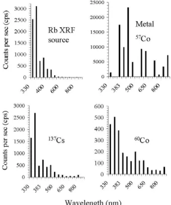
Manual use of narrow band optical filters was opted to ver-ify optical intensities, if any, at 330,350, 365, 383, 400, 450, 500, 550, 600, 650, 700, 750, 800, and 850 nm peak wave-lengths in terms of counts per sec (cps). First, background level of the bare PMT has been just 12 cps on keeping the 600 nm filter directly on its quartz window. Next, on keeping Rb XRF source (AMC 2084, U.K.) on the filter noted a marginal raise of 16 cps to 28 cps, owing to poor efficiency of PMT to Rb X-rays and poor NIR radiation intensity at 600 nm peak wavelength (Fig.1). Likewise, counts remained low at 650, 700, 750, 800, and 850 nm indicating poor efficiency of PMT to Rb X-rays and poor intensity in the spectral near infrared (NIR) region. However, significant rise to 330 cps at 450 nm, and 356 cps at 400 nm provided concrete evidence for low intense emission in the spectral visible (VIS) region. In the spectral UV region, a steep rise to 852 cps at 383 nm, 710 cps at 365 nm, 3095 cps at 350 nm, and 2527 cps at 330 nm provided concrete evidence for UV emission with maximum intensity, while contribution by Rb X-rays remained below 16 cps. On plotting this data as shown in Fig.1, strong peak intensity lines in spectral UV region have indicated an un-precedented UV dominant high-energy spectrum of Rb XRF source, unlike the known strongest air wavelengths of rubid-ium at 424.440 nm (Rb II), and 780.027 nm (Rb I) in basic Rb atomic spectrum [9,10].
Spectral data of137Cs,60Co, Rb XRF source, and metal
57Co in Fig.1 exemplify UV dominance as the spectral
fea-FIG. 1: First and definite experimental evidence for UV dominant optical spectra of Rb XRF source, metal57Co source (notably at room temperature),137Cs, and60Co from peak intensity measure-ments made with narrow band optical filters at 330, 350, 365, 383, 400, 450, 500, 550, 600, 650, 700, 750, 800, and 850 nm peak wave-lengths.
ture of ionizing radiation sources. Like metal57Co, metal Cu XRF source also has shown UV dominance (see Fig.1 in Ref. [3]). Analogous UV dominant high energy spectra were also observed from Ba, and Tb XRF sources (salts); radiochemi-cals such as22Na,57Co,133Ba,204Tl, and241Am. The optical spectra of Rb XRF source, metal57Co, radiochemicals137Cs, and60Co shown in Fig.1 reveal that X-,γ-, orβradiation can independently cause a high-energy spectrum. Precisely, the energy of abundant ionizing radiation is responsible for the differences noticed between any two spectra in Fig.1. At the same time, better understanding of high-energy spectra from three types of ionizing radiation sources: (1) radiochemicals, (2) XRF sources present as salts, and (3) radioisotopes, and XRF sources present as metals demanded further information.
2. Using a pair of sheet polarizers
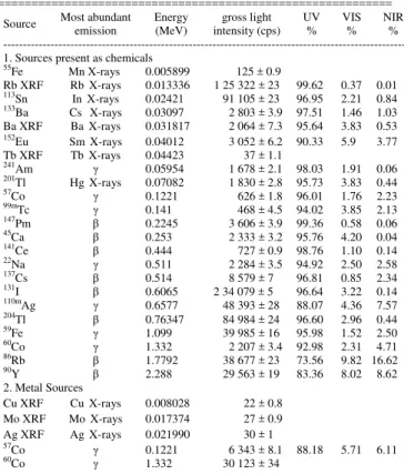
block visible light but transmit NIR radiation beyond 710 nm. These characteristics helped to estimate UV, VIS, and NIR radiation intensities from a radioisotope or XRF source. The following technique demonstrates how the UV, VIS, and NIR radiation intensity estimates could be made from metal57Co.
FIG. 2: Schematic of the technique developed with a pair of sheet polarizers illustrating UV dominant optical emission of metal57Co, kept directly on quartz window of a bare Photomultiplier Tube (9635QB, Thorn EMI). (a). The bare PMT detected 7773±5.7 cps from57Co source, owing to UV (up to 400 nm), VIS (400 - 710 nm), NIR (beyond 710 nm) radiations, andγ–ray as well as Fe X-ray (IR) intensities. (b): On inserting a parallel pair of polarizers in between PMT and source, counts dipped sharply due to blockage of UV from source, yet VIS, and NIR radiation intensities from 400 nm onwards together with IR caused 2180±3.0 cps. (c): When polarizers were reset as crossed pair, counts dipped further due to blockage of even linearly polarized visible (VIS) light, however NIR radiation from 710 nm onwards and IR caused 1818±2.4 cps. (d): On inserting a 0.26 mm thin black polyethylene sheet in between crossed polar-izers and PMT, a further dip in counts was evident due to blockage of NIR radiation, yet IR alone caused 1430±2.4 cps. IR represents ionizing radiations.
UV, VIS, and NIR radiation intensities from57Co were es-timated simply from the differences in counts between four successive steps, considering IRS contribution (1430±2.4 cps) remains nearly the same in all four steps as the dominant gamma rays penetrate well through polarizers and polyethy-lene sheet.
Steps (a) - (b) = UV radiation intensity = 5593±8.7 cps (88.18 %).
Steps (b) - (c) = VIS radiation intensity = 362±5.4 cps (5.71 %)
Steps (c) - (d) = NIR radiation intensity = 388±4.8 cps (6.11%)
Gross light intensity (UV+VIS+NIR intensities) = 6343±
8.1 cps (Table I).
Previously, UV, VIS, and NIR radiation intensities were shown in terms of counts [4, 6]. In Table I here, the intensity
estimates are better shown as percent of the gross light inten-sity to address the problem of unequal source strengths and different standard units of radioisotopes and XRF sources. In metal57Co spectrum, UV intensity was as high as 88.18
% of gross light intensity, while VIS, and NIR radiation in-tensities remained low at 5.71% and 6.11% respectively (Ta-ble 1). Both Fig. 1 and Ta(Ta-ble I doubly ensure high-energy spectra with UV dominance as characteristic emissive feature of radioisotopes and XRF sources. A comparison of spec-tral data shown in Fig.1 and Table I shows that the presence of more number of peak intensity lines in spectral UV re-gion is responsible for raise in %UV intensity of the sources when tested with polarizers. The intensity estimates in Ta-ble I were found reproduciTa-ble with reasonaTa-ble consistency, on repetition of experiments. High-energy spectra of metal Cu, Mo, and Ag XRF sources, Tb XRF source present in the form of Tb salt; and55Fe, could not be ascertained for want of higher strength. It was also not possible to ascertain high-energy spectrum of60Co metal pellet, for want of fixation in a planchet for safe handling.
TABLE I. UV dominant optical emission from (1) Rb and Ba XRF sources, (2) radiochemicals such as113Sn, and (3) metal57Co is evident from the columns on %UV, %VIS, and %NIR intensities in the gross light intensity measured with a pair of sheet polarizers. Likewise, optical emission from metal sources: Cu. Mo, and Ag XRF sources, and60Co is
evident from the gross light intensity.
===========================================================
Source Most abundant emission
Energy (MeV)
gross light intensity (cps)
UV %
VIS %
NIR % --- 1. Sources present as chemicals
55
Fe Mn X-rays 0.005899 125 ± 0.9
Rb XRF Rb X-rays 0.013336 1 25 322 ± 23 99.62 0.37 0.01 113
Sn In X-rays 0.02421 91 105 ± 23 96.95 2.21 0.84 133
Ba Cs X-rays 0.03097 2 803 ± 3.9 97.51 1.46 1.03 Ba XRF Ba X-rays 0.031817 2 064 ± 7.3 95.64 3.83 0.53 152
Eu Sm X-rays 0.04012 3 052 ± 6.2 90.33 5.9 3.77 Tb XRF Tb X-rays 0.04423 37 ± 1.1
241
Am γ 0.05954 1 678 ± 2.1 98.03 1.91 0.06 201
Tl Hg X-rays 0.07082 1 830 ± 2.8 95.73 3.83 0.44 57
Co γ 0.1221 626 ± 1.8 96.01 1.76 2.23 99m
Tc γ 0.141 468 ± 4.5 94.02 3.85 2.13 147
Pm β 0.2245 3 606 ± 3.9 99.36 0.58 0.06 45
Ca β 0.253 2 333 ± 3.2 95.76 4.20 0.04 141
Ce β 0.444 727 ± 0.9 98.76 1.10 0.14 22
Na γ 0.511 2 284 ± 3.5 94.92 2.50 2.58 137
Cs β 0.514 8 579 ± 7 96.81 0.85 2.34 131
I β 0.6065 2 34 079 ± 5 96.64 3.22 0.14 110m
Ag γ 0.6577 48 393 ± 28 88.07 4.36 7.57 204
Tl β 0.76347 84 984 ± 24 96.60 2.96 0.44 59
Fe γ 1.099 39 985 ± 16 95.98 1.52 2.50 60
Co γ 1.332 2 207 ± 3.4 92.98 2.31 4.71 86
Rb β 1.7792 38 677 ± 23 73.56 9.82 16.62 90
Y β 2.288 29 563 ± 19 83.36 8.02 8.62 2. Metal Sources
Cu XRF Cu X-rays 0.008028 22 ± 0.8 Mo XRF Mo X-rays 0.017374 27 ± 0.9 Ag XRF Ag X-rays 0.021990 30 ± 1 57
Co γ 0.1221 6 343 ± 8.1 88.18 5.71 6.11 60
Co γ 1.332 30 123 ± 34
Since both radioisotopes and XRF sources are involved in the study, energy of the most abundant radiation was taken as a common parameter to represent either one of the sources in Table I. It implies that 0.013 336 MeV (of the abundant Rb α1K LII X-ray) is mainly responsible for the 99.62%
β) is mainly responsible for the 96.81% UV, 0.85% VIS, and 2.34% NIR radiation intensities of137Cs, though it also emits
γ-rays and Ba X-rays. The UV, VIS, and NIR radiation inten-sity estimates of the sources provided complimentary infor-mation to what was obtained with optical filters on the nature of optical spectra. Dominant %UV intensity of the sources seen in Table I is due to presence of more number of peak in-tensity lines in spectral UV region (Fig.1). The data in Table I hinted the possibility of a linear relation between the UV, VIS, and NIR radiation intensities of the sources present as salts and the energy of their most abundantγ-, X-, orβ radi-ation, when the sources are listed in increasing order of en-ergy from 0.005 899 to 2.288 MeV. However, on plotting the data of Table 1 as shown in Fig.3, the linear relation between UV, VIS, and NIR radiation intensities and the energy of their most abundantγ-, X-, orβ radiation became more evident. This key information helped ultimately in explaining the op-tical emission phenomenon. Low light yielding metal sources including Cu, Mo, and Ag XRF sources are shown separately as a group in Table I. Despite total opacity of metallic solids to light at room temperature, metal57Co yielding 88.18% UV
intensity close to 96.01% UV of the radiochemical57Co ex-emplifies unusual optical properties of excited atoms situated on the metal surface.
III. RESULTS AND DISCUSSION
A. Atomic spectra of solid metal sources
It was not clearly understood why (1) radiochemicals; (2) Rb, and Ba XRF sources; and (3) metal 57Co commonly caused high-energy spectra. However, optical spectra of metal sources (Fig.1, Table I) provided some vital clues on the nature of spectra of radioisotopes and XRF sources, in general. First of all, the observed optical spectra of metal sources unprecedented at room temperature hinted involve-ment of a previously unknown phenomenon. Furthermore, the observed optical spectra of metal sources opaque to light excluded from being the familiar luminescence. Therefore, optical emission from excited atoms situated on the metal sur-face was thought to be the most likely possibility. In clear words, γ-, X-, or β radiation energy at keV or MeV level generates a low energy at eV level that is optical emission from within the parent excited atom of a radioisotope or XRF source [3-6].
In general, atomic spectra of radioisotopes and XRF sources exhibited two spectral features (1) UV dominance, and (2) dependence upon ionizing radiation energy. UV dom-inance can be evident from the peak values: 99.62% UV at 0.013 336 MeV (Rb XRF), and 98.03% at 0.05954 MeV (γ,
241Am) given in Table I. Though UV is predominant in
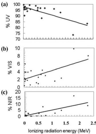
gen-eral from ionizing radiation sources, %UV falls from 99.62% to 83.36% when energy of maximum abundant ionizing ra-diation increases from 0.013 336 MeV (Rb XRF) to 2.288 MeV (ofβ,90Y) as can be seen in Table 1 & Fig.3. a. When %UV attains maximum, %VIS, and NIR radiation intensities will be correspondingly low, as can be seen in the cases of Rb XRF source (0.37%, 0.01%), and241Am (1.91%, 0.06%). However, on plotting the spectral data of Table I in Fig.3, the plots (a, b, and c) provided further clues on the nature of
ob-served spectra. Fig.3 (a) has disclosed a low energy source gives rise to high %UV, regardless of the fact whether it emits
γ, X, orβradiation. Fig.3 (b, and c) shows such a source emits correspondingly low %VIS, and %NIR radiation intensities. In comparison, the %UV dips to a significant level, not be-low 83.36% in any case, from a relatively high energy source. The percent fall in UV is compensated by raise in %VIS, and NIR radiation intensities. Fig.3 provides a set of percent UV, VIS, and NIR radiation intensity estimates at any given ioniz-ing radiation energy. In conclusion, the nature of spectrum of any source is predictable from its ionizing radiation energy.
The UV dominant high-energy atomic spectra were found to be independent of atomic number Z of the radioisotope or XRF source concerned, unlike the case of the basic atomic spectra [9, 10]. For example, 0.1221 MeV could cause 96.01% UV from within excited57Co atom, whereas 1.332 MeV did 92.98% UV from within excited60Co atom, despite both the sources are of cobalt element. Likewise, 0.013 336 MeV could cause 99.62% UV from within excited Rb atom in Rb XRF source, whereas 1.7 792 MeV did just 73.56% UV from within excited 86Rb atom in 86Rb, though both the sources are of rubidium element. Atomic number Z yet plays a pivotal role in causing gross light intensity somewhat comparable to the familiar strong lines of the element con-cerned in basic atomic spectra. Rb XRF source, 131I, and
137Cs proved to be exceptional among the sources tested in
displaying gross light intensities far exceeding the number of ionizing radiation emissions, when tested with a bare PMT and then with a scintillation counter. It could be due to the reason that rubidium, cesium and iodine exceptionally show strongest air wavelengths at 424.440 nm (Rb II), 780.027 nm (Rb I), 460.379 nm (Cs II), 894.347 nm (Cs I), and 804.374 nm (I I), in Ref. [9]. Unlike these strong wavelengths in spec-tral VIS and NIR regions, the current high-energy spectra in general showed strong wavelengths in UV region. Use of rel-atively higher energies than the conventional thermal energies in valence excitation seemed to be responsible for shifting of strongest lines to spectral UV region in the case of radioiso-topes and XRF sources. Futuristic studies of simultaneously detectable X-ray and optical spectra of XRF sources, and pair of nuclear and optical spectra of radioisotopes may enable better understanding of energy levels of excited atoms.
Table 1 suggests that 99.36% UV, 0.58% VIS, and 0.06% NIR radiation intensities in the gross light intensity are spe-cific to 0.2245 MeV (Eβmaxof147Pm). However, Fig.3 clari-fied further that 0.2245 MeV energy is responsible for causing the 99.36% UV, 0.58% VIS, and 0.06% NIR radiation inten-sities, regardless of the type of radiation whetherγ, X-ray or
FIG. 3: The three plots (a, b, and c) made from spectral data of Ta-ble I disclose existence of a linear relation between ionizing radia-tion energy and the percent UV, VIS, and NIR radiaradia-tion intensities in the gross light intensity. In other words, the nature of atomic spec-trum of a radioisotope or XRF source is predictable as it depends upon energy of its abundant ionizing radiation. Fig. 3 (a) shows maximum UV radiation from both radioisotopes and XRF sources, and the %UV attains maximum at very lowγ, X, orβradiation en-ergy. Fig.3 (a, b, and c) shows as %UV decreases, the %VIS, and %NIR radiation intensities correspondingly raise. Extrapolation of the graphs showsγ, X, orβradiation sources with energy 14 MeV and above cause mostly visible (VIS), and NIR radiations. Some scattering of points seen in the graphs is due to ignoring the energies of less abundant emissions for sake of convenience.
B. Atomic state of solid sources
Radioisotopes and XRF sources giving rise to high-energy atomic spectra of excited atoms has been confirmed by both the optical techniques (Figs.1 & 3). On the basis of atomic spectra of radioisotopes and XRF sources it is reasonable to believe the existence of free atoms in analogy to the thermally excited atoms in gaseous phase causing the basic atomic spec-tra [9, 10]. Mainly, during excitation to optical levels the ex-cited atom may act as free atom without any bondage from surrounding unexcited atoms in a radioisotope or XRF source. Most importantly, formation of free atoms at room tempera-ture owes to the generation of exciting energies within ex-cited atoms, in wide contrast to the case of thermal energies from an external source in basic atomic spectra. For exam-ple, the 0.0 080 278 MeV (Cu Kα1X-ray) produced exciting
energy is mainly responsible for formation of free Cu atoms
in Cu XRF source at room temperature. The excited free Cu atoms lying in between unexcited Cu atoms return to ground state not immediately following Cu X-ray emission but after the successive atomic emission of light. Similarly, in metal
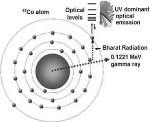
57Co source, the 0.1221 MeV (γ) produced exciting energy
is mainly responsible for formation of free57Co atoms, sur-rounded by unexcited Co atoms, as shown in Fig.4. In com-parison, low abundant (33.4%) Fe Kα1X-ray energy fails to
compete equally with 0.1221 MeV (γ) in generating the free atoms in required number. Excited free 57Co atom returns
to ground state as an unexcited Co atom not immediately af-terγ, and Fe X-ray emissions but after atomic emission of light (Fig.1, Table I). Formation of free atoms within solid ra-dioisotopes and XRF sources implies existence of temporary atomic state of solids, regardless of temperature [3]. The du-ration of free atomic state is limited to valence excitation to optical levels resulting into fluorescence light emission. The free atoms behaving differently from thermally excited atoms in gaseous phase in basic spectroscopy are responsible for a new class of room temperature atomic emission spectra of solid radioisotopes and XRF sources.
FIG. 4: Schematic of 0.1221 MeVγ- rays followed by two succes-sive generations: predicted exciting radiation (Bharat radiation) and the observed UV dominant optical emission at eV level from an ex-cited57Co atom in metal57Co source. Valence excitation byγ- pro-duced Bharat energy enables the excited57Co atoms to be free from surrounding unexcited Co atoms in ground state. The free excited atoms (shown in dark shade) that remain temporarily in atomic state of solids, regardless of temperature are responsible for the atomic spectrum of metal57Co (Fig.1)
IV. PREDICTED EXCITING ENERGY
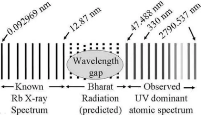
generate some exciting energy to be slightly higher than that of the observed UV at eV level, so that they do valence exci-tation resulting into UV emission. At the same time, it was verified whether the predicted energy really exists in electro-magnetic spectrum. Ifγ-, X-, orβradiation truly generates the predicted exciting energy slightly higher than that of the observed UV, which in turn optical emission from within an excited atom, the exciting energy should occupy in between ionizing radiation and optical radiation in the electromagnetic spectrum. Since 0.010154 nm (0.1221 MeV) of the most abundant γ-emission is mainly responsible for the observed 330 nm peak wavelength of metal 57Co in Fig.1, the excit-ing wavelength should fall in between 0.010154 nm and 330 nm in the electromagnetic spectrum. However, since the ba-sic cobalt spectrum [9] has minimum wavelengths 126.593 nm (Co II) in vacuum, it is very likely that exciting wave-lengths from 57Co exist between 0.010154 to 126.593 nm. As no name exists for the range of wavelengths 0.010154 to 126.593 nm in between X-ray and optical spectral regions [9] of electromagnetic spectrum, the predicted radiation has been temporarily termed as Bharat radiation just for convenience [3, 6]. Citing the author’s research work, Carlos Austerlitz et al described the role of Bharat energy and light produced following X-rays in exciting electron in their paper [11]. The wavelengths in the said range cannot be detected well by the currently available detector like photomultiplier tube. Figure 5 illustrates Rb X-ray spectrum followed by the experimen-tally observed UV dominant optical emission spectrum and the predicted wavelengths in between X-ray and optical spec-tra all from one and the same excited atom of Rb XRF source. Bharat wavelengths differ from one source to another. Most Bharat wavelengths exist between 0.010154 to 126.593 nm from57Co, and between 12.87 nm and 47.488 nm from Rb XRF source (Fig. 5) in the electromagnetic spectrum.
V. ATOMIC PHENOMENON
A brief phenomenological explanation comprising of two postulates is described in the following, so that a detailed mathematical explanation can follow later.
(1) Ionizing radiation, particularlyγ-, X-, orβradiation en-ergy at keV or MeV level loses enen-ergy at eV level while passing through a core-Coulomb field. The loss of en-ergy is reproduced as electromagnetic radiation with the same energy at eV level but higher than that of UV or EUV that the source emits.
(2) The energy causes valence excitation resulting into UV dominant atomic spectrum.
The phenomenon is explained here in simple terms, keep-ing in mind that a detailed mathematical explanation can fol-low based on detailed spectral studies in future. Suppose gamma, X-ray or beta radiation with energy E is passing through M shell Coulomb field in an excited atom of a ra-dioisotope or XRF source. According to the phenomenon de-scribed in Fig. 6, the radiation loses energy only by a few eV, so escapes from excited atom with the rest of the energy E1. The loss of energy (E - E1) reappears as exciting energy
(Bharat energy), EBR. The exciting energy EBRdepends upon
FIG. 5: Schematic of known Rb X-ray spectrum from Rb excited atom of Rb XRF source is shown on the left. The observed UV dominant optical emission spectrum generated by Rb X-rays from the same excited Rb atom is shown on the right. The 330 nm peak observed with narrow band optical filters in Fig.1 is expected to have caused by the highly abundant 0.092 969 nm (α1K LII) Rb X-ray. Therefore, the predicted wavelengths that do valence exci-tation and cause optical emission spectrum thus exist between the wavelengths 0.092 969 to 330 nm of the electromagnetic spectrum. However, since the basic rubidium X-ray spectrum ends at 12.87 nm, whereas optical spectrum begins at 47.488 nm (Rb II) in vacuum [9], Bharat wavelengths from Rb XRF source may exist between 12.87 and 47.488 nm in the electromagnetic spectrum. As these wave-lengths cannot be called as X-rays or EUV, they were given the name Bharat radiation. Moreover, the wavelengths in this range cannot be detected well by the commercially available detector like photomul-tiplier tube
the initial ionizing radiation energy E, as explained already in the case of57Co, and Rb XRF source.
E−E1=EBR
The variation in %UV from one source to another can be explained by the phenomenon shown in Fig. 6. At low en-ergy, the loss at eV level is relatively high while theγ-, X-, orβradiation passes through core- Coulomb field. For exam-ple, while 0.013 336 MeV (α1K LIIRb X-ray) passes through
a M shell Coulomb field in Rb XRF atom, probability ex-ists to lose more energy, say, X eV, resulting in high energy Bharat photons at X eV. Moreover, as probability is high for such events to take place at low energies, the Bharat photons generated from Rb XRF source will be more in number. For these two reasons, the X eV Bharat photons from Rb XRF source have caused more peak intensity lines in spectral UV region, by valence excitation (Fig.1). In comparison, while 1.332 MeV energy passes through M shell Coulomb field in
60Co atom tend to lose relatively less energy, say, Y eV,
result-ing in low energy Bharat photons at Y eV. For this reason, the Y eV Bharat photons have caused more peak intensity lines in spectral VIS and NIR regions as compared to that of UV, by valence excitation (Fig.1). In clear words, X eV Bharat energy from Rb XRF source will be of higher energy than the Y eV Bharat energy from60Co. The Bharat energies far
FIG. 6: Schematic diagram of a 57Co atom illustrating the phe-nomenon that generates some exciting energy higher than that of UV or EUV at eV level (termed temporarily as Bharat radiation). When the abundant 0.1221 MeVγ-ray passes through M shell Coulomb field loses energy at eV level only to reappear as Bharat energy with the same eV energy so as to be slightly higher that of the observed UV or EUV. Typically Bharat energy internally produced within the 57Co excited atom excites the valence electron (4S2) and gives rise
to room temperature UV dominant atomic spectrum shown in Fig.1.
A. Tritium: a source of Bharat radiation
The author was able to verify the validity of the phe-nomenon, when 3H did not show any optical emission on keeping a3H ampoule directly on the quartz window of the bare PMT (9635QB Thorn EMI). The reason being 3H has
only one electron, which is in K-shell. Passage ofβ-emission through K-shell Coulomb field generates a Bharat photon. However, in the absence of an electron in L–shell, the Bharat photon simply escapes from3H atom without producing any light photon by valence excitation. Hopefully, this insight might prompt others to verify the author’s experimental find-ing on 3H. Likewise, Bharat radiation emission alone takes place from highly ionized radionuclides left with a singly filled K shell that can happen in a situation like nuclear fis-sion. Confirmation of this newly predicted Bharat energies higher than that of UV or EUV needs development of a PMT or some other detector sensitive enough in this energy region.
B. The phenomenon explains solar EUV
Findings of the current experimental study have a direct bearing on solar emissions. There is a similarity in theγ-, X-,
β, UV, VIS, and NIR radiation emissions from radioisotopes, XRF sources, and solar flares [14-19]. Therefore, the author has preliminarily reported [3] that solar γ-, X-, or β radia-tions cause EUV regardless of temperature by the atomic phe-nomenon described here. On the basis of the current study, it may be worthy of a review of interpretation of solar EUV lines to be of highly ionized atoms at high temperatures, and estimation of solar temperature from EUV line ratios [20-21].
An overall view of published reports suggesting presence of
235U,238U, and radioisotopes in solar flare indicate that the
phenomenon described here could be the most likely cause for Solar EUV emission. For example, as detection ofγ, and neutron fission counts helps in finding the presence of ura-nium [22-23], simultaneous detection of X- rays,γ-rays, and neutrons reported in solar flares [24-27] suggests the presence of uranium in Sun. The hypothesis on likely presence of nium in Sun derive further strength from the report on ura-nium content of solar salts [28] and traditional wisdom that
235U and238U metals in the solar system are formed from
pre-vious supernovae. Moreover, presence of activation products such as 56Co, and24Na in solar flare [29-32] and presence of7Be in open air after a strong solar wind [33] need to be critically examined to see whether any possibility exists for Uranium fission in Sun. If fission truly happens, the fission fragments left over at the site of fission might constitute dark matter [3].
In the context of solar flare, the predicted Bharat radiation causing UV dominant optical radiation from radioisotopes and XRF sources by valence excitation seemed to be the fa-miliar dark radiation from cosmic sources [3]. As Bharat en-ergies produced internally within an excited atom cause non-thermal valence excitation resulting into UV emission from radioisotopes at room temperature, solar EUV may take place by valence excitation of dark energies from within excited atoms of radioisotopes present in solar flare regardless of tem-perature. As in the case of the current study, theγ-, X-, orβ
radiation emissions from radioisotopes formed by fission re-action in Sun cause two more generation of emissions: the predicted dark radiation, which is the same as Bharat radi-ation followed by EUV. Any how the current experimental study may prompt to examine all these possibilities.
C. UV in radiation dose data
Radiation dose data may need entry of UV as one more component, besides ionizing radiations in giving radiation dose to Nuclear Medicine patients administered with radio-pharmaceuticals such as99mTc,131I, and201Tl for diagnostic and therapeutic purposes [6]. Since UV follows X-rays ac-cording to the current study, UV from diagnostic X-ray tubes may subject the patients to higher skin dose than previously thought. UV emission from metal60Co may also contribute for the skin erythema noticed in cancer patients during60Co Teletherapy treatment.
VI. CONCLUSIONS
predicted Bharat radiation or dark radiation requires improve-ment of PMT or some other detector to be sensitive enough to detect these energies between characteristic X-rays and EUV.
ACKNOWLEDGEMENTS
The author, former Head of the Radiation Safety Group (Sc.E) at the Defence Laboratory (DRDO), Jodhpur,
Ra-jasthan, India gratefully acknowledges his colleagues Dinesh Bohra, and Arvind Parihar for associating in initial experi-ments with black polyethylene sheet.
[1] M. A. Padmanabha Rao, M. R. Patel, Beta excitation of a or-ganic scintillator in powder form in Luminescence: Phenom-ena Materials and Devices, (Ed) R.P. Rao, Nova Science Pub-lishers Inc., Commack, New York, 1992.
[2] D. Bohra, A. Parihar, and M. A. Padmanabha Rao, Nucl.Instrum.Methods.Phys.Res. A320, 393 (1992).
[3] M. A. Padmanabha Rao, in Proc. 7th Int. Conf. on Human Ecology and Nature (HEN 2008), Moscow-Ples, Russia, 2008, edited by Vladimir V.Zaitsev (Moscow Scientific and industrial Association “Radon”) p. 45.
[4] M. A. Padmanabha Rao, Technical Report No: DLJ/IL/97/7*, Defence Laboratory, Jodhpur- 342011, Rajasthan, India, 1997. [5] M. A. Padmanabha Rao, in Proc. 12th National Symposium on Radiation Physics, Defence Laboratory, Jodhpur, Rajasthan, India, 1998, edited by P. K. Bhatnagar and A .S. Pradhan (Hin-dusthan Enterprises: Jodhpur-342003, India, 1998) p. 273. [6] M. A. Padmanabha Rao. in Proc. Symposium on Low Level
Electromagnetic Phenomena in Biological Systems, 1999, School of Environmental Sciences, Jawaharlal Nehru Univer-sity, New Delhi, India, edited by Jitendra Behari, and Editors of Indian Journal of Biochemistry and Biophysics (Printed at National Institute of Science Communication, New Delhi- 110 012, 1999) p. 68.
[7] H. Becquerel, Competes rendus de l’ Academie des Sci-ences, Paris,122, 501, (1890), translation in Nuclear Chem-istry, Benchmark papers in Physical Chemistry and Chemical Physics, edited by G. T. Seaborg and W. Loveland (Hutchinson Ross Publishing Company, Pennsylvania, 1896) V.5, p. 23. [8] G. F. Knoll, Radiation Detection and Measurement (John
Wi-ley & Sons, New York, 1979) p. 745.
[9] David R. Lide and H. P. R Frederikse, C.R.C. Handbook of Chemistry and Physics, 74th Edition (CRC Press Inc, London, 1993-1994).
[10] Basic Atomic Spectroscopic data (National Institute of Stan-dards and Technology, Gaithersburg, U.S.A).
[11] Carlos Austerlitz, Vivianne L´ucia Bormann de Souza, Diana Maria Tavares Campos, Cristina Kurachi, Vanderley Bagnato, and Cl´audio Sibata, Braz. arch. biol. technol. 51, n.2, p. 271 (2008).
[12] C. Michael Lederer and Virginia S. Shirley, Table of Isotopes, 7th Edition, (John Wiley & Sons, INC, New York, 1978). [13] Radionuclide Transformations, Energy and Intensity of
Emis-sions, Report of a task Group of Committee 2, ICRP Publica-tion 38 (Pergamon Press: Oxford, 1983).
[14] S.R. Kane, in Proc. the IAU Symposium No.68 on Solar Gamma, X-, and EUV radiation, Buenos Aires, Argentina,
1974 (D. Reidel Publ. Co, Dordrecht, Holland, 1975) p. 446. [15] G. A. Doschek, in Proc. IAU Symposium No.68 on Solar
Gamma, X-, and EUV radiation, Buenos Aires, Argentina, 1974 (D. Reidel Publ. Co, Dordrecht, Holland, 1975) p. 165. [16] Leon Golub, Rev. Sci. Instrum, 74, 4583 (2003)
[17] Jeffrey W. Brosius and Kenneth J. H. Phillips, Astrophys. J., 613, 580 (2004)
[18] Alphonse C. Sterling and Ronald L. Moore, Astrophys. J., 602, 1024 (2004).
[19] Alphonse C. Sterling, in Proc. Int. Astronomical Union 226 on Coronal and Stellar Mass Ejections, Beijing, edited by K. Dere, J. Wang, and Y. Yan, 27, 2004 (Published Online by Cambridge University Press, 2005).
[20] B. N. Dwivedi and Anita Mohan, Solar Physics, 156, no.1, 81 (1995),
[21] E. E. Benevolenskaya, Advances in Space Research, 39, 1860 (2007).
[22] Caldwell, Richard L, United States Patent 4568510, Dallas, TX, 1986.
[23] E. E. Clark, Technical Report: K-2021-Suppl., Oak Ridge Gaseous Diffusion Plant, TN,USA,1981.
[24] M. Yoshimori, K. Okudaira, Y. Hirasima, T. Igarashi, M. Akasaka, Y. Takai, K. Morimoto, T.Watanabe, K. Ohki, J. Nishimura, T. Yamagami, Y. Ogawara, and I.Kondo, Solar Phys., 136, 69 (1991).
[25] K. Watanabe, M. Gros, P. H. Stoker, K. Kudela, C. Lopate, J. F. Vald´es Galicia, A. Hurtado, O. Musalem, R. Ogasawara, Y. Mizumoto, M. Nakagiri, A. Miyashita, Y. Matsubara, T. Sako, Y. Muraki, T. Sakai, and S. Shibata, Astrophys. J., 636, 1135 (2006).
[26] T. Sako et al., ApJ, 651, L69 (2006).
[27] R. J. Murphy, Benzion Kozlovsky, G.H. Share, X-M Hua, and R.E. Lingenfelter, ApJS, 168, 167 (2007).
[28] K. Ogiwara, T. Oi, T. Ossaka, M. Mukaida, and T. Honda, J.Radioanal.Nucl.Chem, 191, No.2, 273 (1995).
[29] E. L. Chupp, D. J. Forrest, P. R. Higbie, A. N. Suri, C. Tsai, and P. P. Dumphy, Nature, 241, 333 (1973).
[30] H. Nutley and M. D. Voth, J.Geophy.Res, 92:A10, 11179 (1987).
[31] R.J. Murphy, X-M Hua, B Kozlovsky, and R. Ramaty, Astro-phys. J., 351, 299 (1990).
[32] V. Tatischeff, B. Kozlovsky, J. Kiener, and R.J. Murphy, astro-ph/0604325, 2006.