r e v a s s o c m e d b r a s .2 0 1 3;5 9(6):622–628
Revista da
ASSOCIAÇÃO MÉDICA BRASILEIRA
w w w . r a m b . o r g . b r
Review article
Homocysteine: cardiovascular risk factor in children
and adolescents?
夽
Adriana Amorim De Farias Leal
a,∗, Ástrid Camêlo Palmeira
a,
Gabriella Menezes Almeida De Castro
b, Mônica Oliveira Da Silva Simões
b,
Alessandra Teixeira Ramos
b, Carla Campos Muniz Medeiros
baPost-graduation Program in Public Health, Universidade Estadual da Paraíba, Campina Grande, PB, Brazil
bDepartment of Pharmacy, Universidade Estadual da Paraíba, Campina Grande, PB, Brazil
a r t i c l e
i n f o
Article history: Received 23 May 2012 Accepted 27 May 2013
Available online 30 October 2013
Keywords: Homocysteine
Cardiovascular diseases Child
Adolescent
a b s t r a c t
The aim of this study was to identify publications in literature that investigated Homocys-teine (He) as a risk factor for CVD among children and adolescents. An active search for information in LILACS, IBECS, Science Direct, Medline and Cochrane Library databases was conducted using the following combination of keywords “homocysteine”, “cardiovascular diseases”, “child” and “adolescent”. Fifteen articles were analyzed showing direct relation-ship with increasing age (8 studies) and male gender (10 studies), and an inverse relationrelation-ship with serum vitamins B6, B12 and folate levels. Thus, the results suggest that more research must be carried through in order to determine in a more coherent way the causes of the hiperhomocisteinemia in the pediatric population, guiding for an adequate diet, rich in nutrients necessary to favor the metabolism of the He.
© 2012 Elsevier Editora Ltda. All rights reserved.
Homocisteína: fator de risco cardiovascular em crianc¸as e adolescentes?
Palavras-chave: Homocisteína
Doenc¸as cardiovasculares Crianc¸as
Adolescentes
r e s u m o
O objetivo do estudo foi identificar na literatura publicac¸ões que investigaram a Homocis-teína (He) como um fator de risco para doenc¸as cardiovasculares na faixa etária de crianc¸as e adolescentes. Realizou-se uma busca ativa de informac¸ões nas bases de dados LILACS e IBECS, Science Direct, Medline e Biblioteca Cochrane, utilizando-se a combinac¸ão dos descritores “homocysteine”, “cardiovascular diseases”, “child” e “adolescent”. Foram anal-isados 15 artigos, os quais apontaram relac¸ão direta dos níveis de He com o sexo masculino (10 estudos) e com o aumento da idade (8 estudos), e uma relac¸ão inversa com os níveis séricos das vitaminas B6, B12 e folatos (10 estudos). Sugere-se que sejam realizadas mais
夽
Study conducted at Centro de Obesidade Infantil, Instituto de Saúde Elpídio de Almeida, Campina Grande, PB, Brazil.
∗ Corresponding author.
E-mail: [email protected] (A. Amorim De Farias Leal)
pesquisas a fim de determinar de maneira mais coerente as causas da hiperhomocisteine-mia na populac¸ão pediátrica, orientando para uma dieta adequada e rica em nutrientes necessários para favorecer o metabolismo da He.
© 2012 Elsevier Editora Ltda. Todos os direitos reservados.
Introduction
Cardiovascular diseases (CVD) are a major public health prob-lem for being the leading cause of death and disability, affecting adults in full productive age, resulting in loss of potential years of life and producing high costs for the public health system.1
Some risk factors for the development of CVD are well known, such as age, male gender, dyslipidemia, smoking, systemic hypertension, diabetes mellitus, obesity, seden-tary lifestyle and genetic factors or parental history of atherosclerotic diseases.2 Currently, high levels of plasma homocysteine (hyperhomocysteinemia) have been associated with increased cardiovascular mortality rates, especially in the adult population.3
The pathogenesis of the vascular lesion caused by hyper-homocysteinemia (HHe) includes endothelial cell lesion, vascular smooth muscle growth, increased platelet adhesive-ness, increased LDL-cholesterol oxidation with deposition on the vascular wall and direct activation of the coagulation cascade.4
In this context, interest in homocysteine (He) as a causal risk factor for CVD in childhood was stimulated by the obser-vation that over 50% of children with genetic disorder of homocysteinuria died prematurely from vascular diseases, as well as the fact of high levels of He being associated with physiologic and nutritional factors.5
Thus, to determine the prevalence of cardiovascular risk factors in early childhood should be a priority among preven-tive measures, because atherogenesis may precede by many years its clinical manifestations, such as acute myocardial infarction (AMI) and stroke.6Therefore, this study is a system-atic literature review of publications that have investigated He as a risk factor for CVD in the age group composed of children and adolescents.
Methods
The study methodology was the active search for information in LILACS and IBECS (Virtual Health Library), Science Direct, Medline and Cochrane Library databases, using the follow-ing combination of keywords “homocysteine”, “cardiovascular diseases”, “child” and “adolescent”. There was a query to the DeCS service (Keywords in Health Sciences) of the Virtual Health Library for standardization of keywords used in the search. Studies conducted in the last 15 years were screened (from 1997 to 2011), in Brazil and abroad, without restriction as for the language of publications, in which the assessment of plasma homocysteine concentrations was an independent variable of interest in the analysis of risk for cardiovascular diseases by restricting the age group of children and/or ado-lescents (0-19 years).
The inclusion criteria were: (1) only original studies, full text available online; (2) samples that included children and adolescents (0-19 years); (3) cross-sectional, case-control and/or cohort studies. Theses/dissertations, review articles, meta-analyzes and experimental studies with animal mod-els were excluded. These criteria were used to increase the comparability of findings between studies.
Data were extracted independently by three of the authors. Disagreements were resolved by consensus among authors. The first screening was performed including the following combination of keywords “homocysteine” and “cardiovascular diseases” and “child” and “homocysteine” and “cardiovascu-lar diseases” and “adolescent”, in which the authors sought to identify abstracts in duplicate, delete references of abstracts without full article available, literature reviews and disser-tations. In a second step, only publications of studies that investigated the topic of interest with samples composed of children and adolescents were selected. In the third step, the references of articles selected were reviewed in order to cap-ture manuscripts not found in the search.
Results
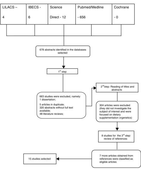
A total of 678 studies were identified and obtained by the first screening. In the previous analysis, 663 studies were excluded (Fig. 1). After the other two steps of the search strategy adopted, 15 studies were selected, which met the pre-established inclusion and exclusion criteria, and, upon completing their reading, all were included in the final sample of this study.
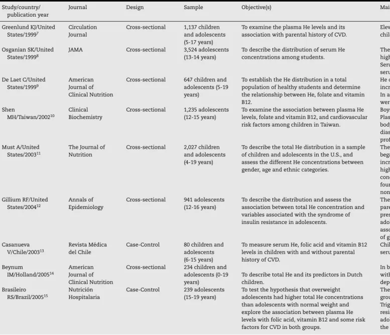
Table 1 shows the characterization of articles according to the first author’s name, country where the study was con-ducted, publication year, journal name, study type, sample, objectives and main results.
All studies showed He as a dependent variable, whose plasma concentration was compared to folate, vitamin B12 and cobalamin concentrations, and to lipid and glucose pro-files, as well as to other demographic and clinical variables and those related to lifestyle (age, gender, BMI, blood pressure, genetic polymorphism and parental history of CVD).
In most studies, boys had total He values higher than girls8–11,15–17,19–21 with mean values of He for boys ranging from 5.22-13.30mol/L, and for girls from 4.84-10.4mol/L. Nevertheless, in only one study, no differences were observed between genders in relation to He concentrations.18
624
rev
assoc
med
bras.
2013
;
5
9(6)
:622–628
Table 1 – Characteristics of screened studies. Study/country/
publication year
Journal Design Sample Objective(s) Main results
Greenlund KJ/United States/19997
Circulation Journal
Cross-sectional 1,137 children and adolescents (5-17 years)
To examine the plasma He levels and its association with parental history of CVD.
Elevated He levels have been observed in children with positive parental history of CVD
Osganian SK/United States/19998
JAMA Cross-sectional 3,524 adolescents (13-14 years)
To describe the distribution of serum He concentrations among students.
The average He concentration was significantly higher for boys;
Serum He was not significantly associated with serum lipids or parental history of CVD; De Laet C/United
States/19999
American Journal of Clinical Nutrition
Cross-sectional 647 children and adolescents (5-19 years)
To establish the He distribution in a total population of healthy students and determine the relationship between He, folate and vitamin B12.
He concentrations were lower in children and increased proportionally with age;
In adolescents aged 15 years, He concentrations were higher in boys;
Shen
MH/Taiwan/200210
Clinical Biochemistry
Cross-sectional 1,235 adolescents (12-15 years)
To examine the association between plasma He levels, folate and vitamin B12, and cardiovascular risk factors among children in Taiwan.
Boys had higher plasma He levels than girls; Plasma He levels were positively associated with body weight and height and to systolic and diastolic blood pressure, but not related to lipid profile;
Must A/United States/200311
The Journal of Nutrition
Cross-sectional 2,027 children and adolescents (4-19 years)
To describe the total He distribution in a sample of children and adolescents in the U.S., and assess the different He concentrations between gender, age and ethnic categories.
The total He concentrations for boys and girls began to diverge at the age of 10 years and increased in adolescence, and boys showed higher concentrations;−Differences in total He concentrations between ethnic categories were found in the group of girls and were higher in non-Hispanic African American girls; Gillium RF/United
States/200412
Annals of Epidemiology
Cross-sectional 941 adolescents (12-16 years)
To describe the distribution and assess the association between total He concentration and variables associated with the syndrome of insulin resistance in adolescents.
There was association between total He and parental history of stroke and high blood pressure or systolic blood pressure in male adolescents;−There was no significant association between total He and the percentage of glycated hemoglobin A1c in boys and girls; Casanueva
V/Chile/200313
Revista Médica del Chile
Case-Control 80 children and adolescents (6-15 years)
To measure serum He, folic acid and vitamin B12 levels in children with and without parental history of CVD.
Children with parental history of CVD had higher serum He levels than those without such history;
Beynum
IM/Holland/200514
American Journal of Clinical Nutrition
Cross-sectional 234 children and adolescents (0-19 years)
To describe total He and its predictors in Dutch children.
In both sexes, total He concentrations increased with age;−Plasma folate was considered a dependent predictor of total He
Brasileiro RS/Brazil/200515
Nutrición Hospitalaria
Case-Control 239 adolescents (15-19 years)
To test the hypothesis that overweight adolescents had higher total He concentrations than adolescents with normal weight and explore the association between plasma He levels with folic acid, vitamin B12 and some risk factors for CVD in both groups.
The averages of total He were elevated in both groups and were higher in boys than in girls;− Triglyceride, LDL cholesterol and insulin resistance were higher in overweight
rev
assoc
med
bras.
2013
;
5
9(6)
:622–628
625
Study/country/ publication year
Journal Design Sample Objective(s) Main results
Papandreou D/Greece/200616
Clinical Nutrition Cross-sectional 524 children and adolescents (6-15 years)
To investigate the distribution and determinants of total serum He levels in healthy Greek children.
Homocysteine levels were increasing with age and boys showed total He levels higher than girls;−Age and folate levels were the most significant and independent determinants associated with total He;
Papandreou D/Greece/200617
British Journal of Nutrition
Cross-sectional 520 children and adolescents from 6-15 years of age
To provide a set of specific data for total He levels and determine the relationship between He and folic acid, vitamin B12, age, BMI, blood pressure and diet in a Greek pediatric population.
The highest mean Hct concentration was found in boys;
He concentrations were lower in children and increased in proportion to age;
There was correlation between He levels and age, serum folate, BMI and systolic blood pressure; Huemer
M/Austria/200618
Pediatrics Research
Cross-sectional 264 healthy children and adolescents aged from 2-17 years
To investigate total He concentrations and the relationship between He and folate, cobalamin, genetic polymorphisms and other clinical variables.
He concentrations were significantly influenced by age and folate and cobalamin concentrations; −No differences were observed between genders in relation to He concentrations; Villarreal
E/Colombia/200819
Biomédica Cross-sectional 600 children aged from 5-14 years
To determine the lipid profile, He and PCR, and identify the relationship between these markers with gender, age and type of school.
The prevalence of high He levels was higher in boys than in girls;−The prevalence of high serum lipids was higher in girls than in boys; Kerr
MA/England/200920
Pediatrics Cross-sectional 2,127 youth aged from 4-18 years
To investigate age, gender, and lifestyle factors as determinants of folate, vitamin B12 and He in British children and adolescents and propose reference ranges by age for these biomarkers.
He concentrations increased progressively with age and were higher in boys than in girls;
Akanji
AO/Kuwait/201021
Nutrition, Metabolism & Cardiovascular Diseases
Cross-sectional 774 healthy adolescents (316 boys, 458 girls) aged from 10-19 years
To investigate age, gender and body mass as determinants of folate, vitamin B12 and He levels in Arab adolescents and propose reference ranges by age and gender for these biomarkers.
There was a relationship between age and increased He levels;
626
r e v a s s o c m e d b r a s .2 0 1 3;5 9(6):622–628LILACS –
4
IBECS -
6
Science
Direct - 12
Pubmed/Medline
- 656
Cochrane
- 0
678 abstracts identified in the databases selected
663 studies were excluded, namely: 1 dissertation;
5 articles in duplicate; 320 abstracts without full text available;
48 literature reviews;
7 more articles obtained from references were classified as eligible articles 1st step
8 studies for the 3rd step:
review of references
15 studies selected
304 articles were excluded (they did not investigate the subject of interest and were focused on dietary supplementation orgenetics)
2ndstep: Reading of titles and
abstracts
Fig. 1 – Search strategy flowchart.
Discussion
The aim of this study was to make a literature review on the relationship between He and cardiovascular risk among chil-dren and adolescents. Epidemiological studies have shown that HHe is an important risk factor for the development of vascular disease.22Noteworthy is the fact that 30-35% of individuals with CVD present normocolesterolemia, but more than 40% of patients with primary disease of the coronary artery, cerebrovascular or peripheral vascular have Hhe.23
Nevertheless, none of the articles evaluated pointed He as an independent cardiovascular risk factor in children and ado-lescents. The interest in He as a causal factor began from the observation that over 50% of children with genetic disorder of homocysteinuria died prematurely from vascular diseases.24 The study by Huemer et al.18found a significant association in subjects who had high He concentrations and the MTHFR 677T allele. In adults, genotypes MTHFR 677T and heterozy-gous MTHFR 677T/1298C are associated with HHe and CVD.25 In addition to genetic disorders, folate and vitamin B12 levels are inversely related to plasma homocysteine concentration.26. Intracellular He metabolism occurs through
two-way remethylation, which is responsible for the conver-sion of He into methionine, and a one-way transsulfuration, which converts He into cysteine. In the remethylation process, folate and vitamin B12 act as coenzymes, or co-substrates, and in the transsulfuration process, in turn, vitamin B6 act as coenzyme.2,27Accordingly, the studies included in this review assessed He and these substrates, and found an inverse rela-tionship.
Eight of the 15 studies analyzed showed that the He lev-els in adolescents (aged over 10 years) were higher than in children. Homocysteine levels increase with age, regardless of gender. This is secondary to the decreased levels of vita-min cofactors, resulting in reduced enzymatic activity in the metabolic pathway or to the coexistence of renal disease.31 In 1998, the study by Bydlowski et al.32reported that children tend to have lower He levels, and these levels tend to increase with age. The decrease in production or in enzymatic activ-ity for the He metabolism, renal dysfunction, or decreased bioavailability of vitamins (B6, B12 and folate), may explain this phenomenon.27
In this review, three studies have evaluated the associ-ation between He and a positive parental history for CVD, which emphasized the history of stroke and high systolic and/or diastolic blood pressure. This is alarming, considering the strong relationship between He and atherogenic mech-anisms, since in all the articles analyzed, He was identified as an independent risk factor for cardiovascular diseases, regardless of age group in the three continents covered by the studies (America, Europe and Asia). A systematic review with meta-analysis on He and cardiovascular risk developed by Humphrey et al.24concluded that high He levels may inde-pendently and moderately increase by about 20% the risks of developing CVD. In addition, other studies, also using meta-analysis techniques, assessed the benefits of reducing serum He concentrations and found that the decrease of 3-5mol/L in serum He levels can reduce the incidence of deep vein thrombosis and stroke.33,34
Thus, the study by Brasileiro et al.15 stands out, whose sample consisted of adolescents with and without excess weight and whose results showed higher He levels in the overweight ones. Therefore, it could be inferred that, despite the pathophysiological mechanisms by which HHe can pro-mote atherothrombosis are not well defined, vascular lesion defined by the action of He includes endothelial cell lesion, vascular smooth muscle growth, increased oxidation of LDL cholesterol with direct deposition on the vascular wall and increased platelet adhesiveness, phenomena also observed in overweight individuals.35,36
On the other hand, the findings of this review indicate the need for further studies, correlating He with other vari-ables such as renal function and insulin resistance. Only one article proposed assessing the relationship between He and glycated hemoglobin A1c levels; however, it found no sig-nificant association. Nevertheless, it would be necessary to investigate insulin levels which have an inverse relationship with He levels, since insulin contributes to decrease the serum He levels because it stimulates the biosynthesis of the hepatic cystathionine -synthase enzyme, which is responsible for He degradation in the transsulfuration pathway.37 Finally, it is also important to evaluate the renal function, since kid-neys contain significant amounts of enzymes involved in the transsulfuration and remethylation process, playing an impor-tant role in He metabolism and clearance.38
Conclusions
The results of the reviewed articles were quite homogeneous, emphasizing the relationship between high He levels and
increased age and male gender. However, high levels of He were not related to the determination of cardiovascular risk in this population at none of the studies evaluated. Thus, the results of this review suggest that further studies should be conducted, especially in Brazil, since it has high morbidity and mortality rates from cardiovascular causes, especially case-control or cohort studies on the issue proposed, in order to more accurately determine the causes of HHe in the pediatric population.
Conflicts of interest
The authors declare no conflicts of interest.
r e f e r e n c e s
1. Farias Júnior JC, Mendes JKF, Barbosa DBM, Lopes AS. Fatores de risco cardiovascular em adolescentes: prevalência e associac¸ão com fatores sociodemográficos. Rev Bras Epidemiol. 2011;14:50–62.
2. Venâncio LS, Burini RC, Yoshida WB. Tratamento dietético da hiper-homocisteinemia na doenc¸a arterial periférica. J Vasc Bras. 2010;9:28–41.
3. Amorim KS, Lopes AS, Pererira IA. Impacto do exercício físico nos níveis de homocisteína, um fator de risco para
aterosclerose: revisão sistemática. Rev Bras Ativ Fís Saúde. 2011;16:70–5.
4. Vannucchi H, Melo SS. Hiper-homocisteinemia e risco cardiometabólico. Arq Bras Endocrinol Metab. 2009;53:540–9. 5. Amorin FG, Rezende LCD, Coitinho LB, Freitas JV, Scher JÁ,
Dettogni RS. Bioquímica clínica da aterosclerose provocada por hiperhomocisteinemia. Rev Eletrônica Farmácia. 2011;8:36–59.
6. Beck CC, Lopes AS, Giuliano ICB, Borgatto AF. Fatores de risco cardiovascular em adolescentes do município do sul do Brasil. Rev Bras Epidemiol. 2011;14:36–49.
7. Greenlund KJ, Srinivasan SR, Xu JH, Dalferes Jr R, Myers L, Pickoff A, et al. Plasma homocysteine distribution and its association with parental history of coronary artery disease in black and white children. Circulation. 1999;99:2144–9. 8. Osganian SK, Stampfer MJ, Speigelman D, Rimm E, Cutler JA,
Feldman HA, et al. Distribution of and factors associated with serum homocysteine levels in children. JAMA.
1999;281:1189–96.
9. De Laet C, Wautrecht JC, Brasseur D, Dramaix M, Boeynaems JM, Decuyper J, et al. Plasma homocysteine concentrations in a Belgian school-age population. Am J Clin Nutr.
1999;69:968–72.
10. Shen MH, Chu NF, Wu DM, Chang JB. Plasma homocyst(e)ine, folate and vitamin B12 levels among school children in Taiwan: The Taipei Children Heart Study. Clin Biochem. 2002;35:495–8.
11. Must A, Jacques PF, Rogers G, Rosenberg IH, Selhub J. Serum total homocysteine concentrations in children and adolescents: results from the Third National Health and Nutrition Examination Survey (NHANES III). J Nutr. 2003;133:2643–9.
12. Gillium RF. Distribution of total serum homocysteine and its association with parental history and cardiovascular risk factors at ages 12-16 years: The Third National Health and Nutrition Examination Survey. Ann Epidemiol.
2004;14:229–33.
628
r e v a s s o c m e d b r a s .2 0 1 3;5 9(6):622–628Relación con historia familiar de enfermedad cardiovascular. Rev Méd Chile. 2003;131:997–1002.
14. Beynum IM, Heijer M, Thomas CMG, Afman L, Emmerzaal DO, Blom HJ. Total homocysteine and its predictors in Dutch children. Am J Clin Nutr. 2005;81:1110–6.
15. Brasileiro RS, Escrivão MAMS, Taddei JAAC, Almeida VD, Ancona-Lopez F. Plasma total homocysteine overweight and non-overweight adolescents: a case-control studt. Nutr Hosp. 2005;20:313–9.
16. Papandreou D, Mavromichalis I, Makedou A, Rousso I, Arvanitidou M. Total serum homocysteine, folate and vitamin B12 in a Greek school age population. Clin Nutr.
2006;25:797–802.
17. Papandreou D, Mavromichalis I, Makedou A, Rousso I, Arvanitidou M. Reference range of total serum homocysteine level and dietary indexes in healthy Greek schoolchildren aged 6-15 years. Br J Nutr. 2006;96:719–24.
18. Huemer M, Vonblon K, Födinger M, Krumpholz R, Hubmann M, Ulmer H, et al. Total homocysteine, folate, and cobalamin, and their relation to genetic polymorphisms, lifestyle and body mass index in healthy children and adolescents. Pediatr Res. 2006;60:764–9.
19. Villarreal E, Forero Y, Poveda E, Baracaldo C, López E. Marcadores de riesgo cardiovascular em escolares de cinco departamentos de la región oriental em Colombia. Biomédica. 2008;28:38–49.
20. Kerr MA, Livingstone B, Bates CJ, Bradbury I, Scott JM, Ward M, et al. Folate, related B vitamins, and homocysteine in childhood and adolescence: potential implications for disease risk in later life. Pediatrics. 2009;123:627–35.
21. Akanji AO, Thalib L, Al-Isa AN. Folate, vitamin B12 and total homocysteine levels in Arab adolescent subjects: reference ranges and potential determinants. Nutr Metab Cardiovasc Dis. 2012;22:900–6.
22. Pinto WJ, Areas MA, Marialva JE, Cardoso SMG, Pinto EG. Homocisteína e risco cardiovascular. Rev Ciênc Campinas. 2009;18:259–68.
23. Neves LB, Macedo DM, Lopes AC. Homocisteína J Bras Patol. 2004;40:3110–20.
24. Humphrey LL, Fu R, Rogers K, Freeman M, Helfand M. Homocysteine level and coronary heart disease incidence: a systematic review and meta-analysis. Mayo Clin Proc. 2008;83:1203–12.
25. Prengler M, Sturt N, Kywawych S, Surtees R, Liesner R, Kirkhan F. Homozygous thermolabile variant of the methylenetetrahydrofolate reductase gene: a potential risk
factor for hiperhomocysteinaemia. CVD, and strok in childhood Rev Med Child Neurol. 2001;43:220–5.
26. Diniz-Santos DR, Andrade GCF. A homocisteína como fator de risco para a aterosclerose. Rev Ciênc Méd Biol. 2005;4:158–66. 27. Zacarias-Castillo R, Hernández-Rebollar AE,
Zajarias-Rabchinskey A, González-Bárcena D.
Hiperhomocisteinemia. Um nuevo factor de riesgo coronário. Gac Méd Méx. 2001;137:335–45.
28. Fukagawa NK, Martin JM, Wurthmann A, Prue AH, Ebenstein D, O’rourke B. Sex-related differences in methionine metabolism and plasma homocysteine concentrations. Am J Clin Nutr. 2000;72:22–9.
29. Harboe-Gonc¸alves L, Vaz LS, Buzzi M. Associac¸ão entre níveis plasmáticos de homocisteína e acidente vascular cerebral isquêmico Arq Neuropsiquiatr. 2005;63:97–103.
30. Venâncio LS, Burini RC, Yoshida WB. Concentrac¸ão de homocisteína em pacientes portadores de doenc¸a arterial periférica atendidos em um servic¸o público de saúde. J Vasc Bras. 2009;8:318–26.
31. Dias PMT, Mezzomo A, Peteffi C, Pezzi DR. Homocisteína: um fator de risco vascular. Rev Cient AMECS. 2001;10:53–8. 32. Bydlowski SP, Magnanelli AC, Chamone DAF.
Hiper-homocisteinemia e Doenc¸as Vaso-Oclusivas. Arq Bras Cardiol. 1998;71:69–76.
33. Ueland PM, Refsum H, Beresford AS, Mollset SE. The controversy over homocysteine and cardiovascular disease. Am J Clin Nutr. 2000;72:324–32.
34. Van Guldener C, Nanayakkara PW, Stehouver CD. Homocysteine and blood pressure. Curr Hypertens R. 2003;5:26–31.
35. Moura MSB, Martins MCC, Souza Filho MD.
Hiper-homocisteinemia como fator de risco cardiovascular. ConSientiae Saúde. 2011;10:181–5.
36. Santos MG, Pegoraro M, Sandrini F, Macuco EC. Fatores de risco no desenvolvimento da aterosclerose na infância e adolescência. Arq Bras Cardiol. 2008;90:
301–8.
37. Rodrigues LEA. Hiper-homocisteínemia como fator de risco no diabetes. Diabetes Clínica. 2003;6:434–40.