Abstract
Objective: To determine the time of diagnosis of typical orofacial clefts in different Brazilian regions and its inluence on age at surgical correction.
Method: This was a prospective, descriptive, cross-sectional study conducted in medical centers in the Southeast, South, and Northeast of Brazil. Trained speech therapists and geneticists interviewed the parents of affected children using a previously validated questionnaire. Epi-Info and SPSS were used for data analysis. Signiicance level was set at 5% (p ≤ 0.05).
Results: The sample consisted of 215 interviews conducted in the following regions: 21.9% (47) in the Southeast, 51.1% (110) in the South, and 27% (58) in the Northeast. Monthly family income was higher in the Southeast (p ≤ 0.05). Cleft lip and palate were found in 61.4% (132) of cases, cleft palate in 20.9% (45), and cleft lip in 17.7% (38). Diagnosis occurred in the maternity ward in 75.3% (162) of cases, during the prenatal period in 14% (30), and after hospital discharge in 10.2% (22). The Southeast had a higher frequency of prenatal diagnosis (27.7%), possibly related to greater purchasing power in this region and greater availability of prenatal investigation. Of all cases diagnosed in the maternity ward, 74.4% occurred in the Northeast. However, no signiicant difference was found when comparing time of diagnosis, region, and age at irst surgery.
Conclusion: Considering that diagnosis is more common in the maternity ward, local health care teams should be trained in order to effectively improve the initial care of these patients. Although time of diagnosis did not affect age at surgery, it favors the planning of neonatal care and treatment of affected infants.
J Pediatr (Rio J). 2011;87(3):225-230: Cleft lip, cleft palate, diagnosis, public health.
O
RiginAlA
RtiCle Copyright © 2011 by Sociedade Brasileira de Pediatria225
time of diagnosis of oral clefts: a multicenter study
lívia g. Amstalden-Mendes,1 Ana Carolina Xavier,2 Denise K. Antunes,3
Ana Carolina R. g. Ferreira,4 Rita tonocchi,5 Agnes C. Fett-Conte,6
Raquel n. Silva,7 Vera H. V. leirião,8 lázara P. C. Caramori,9
luiz A. Magna,10 Vera lúcia gil-da-Silva-lopes11
1. Fonoaudióloga. Doutoranda, Faculdade de Ciências Médicas (FCM), Universidade Estadual de Campinas (UNICAMP), Campinas, SP, Brazil. 2. Fonoaudióloga, Centrinho Prefeito Luiz Gomes, Joinville, SC, Brazil.
3. Mestre. Fonoaudióloga, Núcleo de Atenção Médica Integrada (NAMI), Fortaleza, CE, Brazil.
4. Fonoaudióloga, Universidade Estadual de Ciências da Saúde de Alagoas (UNCISAL), Maceió, AL, Brazil.
5. Mestre. Fonoaudióloga, Centro de Atendimento Integral ao Fissurado (CAIF), Associação de Reabilitação e Promoção Social ao Fissurado Labiopalatal (AFISSUR), Curitiba, PR, Brazil.
6. Doutora. Geneticista, Hospital de Base, Fundação Faculdade Regional de Medicina de São José do Rio Preto (FUNFARME), São José do Rio Preto, SP, Brazil.
7. Fonoaudióloga, Hospital Infantil Albert Sabin (HIAS), Fortaleza, CE, Brazil.
8. Doutora. Fonoaudióloga, Hospital de Reabilitação em Anomalia Craniofacial (HRAC), Bauru, SP, Brazil. 9. Doutora. Geneticista, Centro Universitário Filadélfia (UNIFIL), Londrina, PR, Brazil.
10. Professor titular, Departamento de Genética, FCM, UNICAMP, Campinas, SP, Brazil.
11. Professor associado, Departamento de Genética Médica, FCM, UNICAMP, Campinas, SP, Brazil. No conflicts of interest declared concerning the publication of this article.
Financial support: Fundação de Amparo à Pesquisa do Estado de São Paulo (FAPESP), Brazil.
Suggested citation: Amstalden-Mendes LG, Xavier AC, Antunes DK, Ferreira AC, Tonocchi R, Fett-Conte AC, et al. Time of diagnosis of oral clefts: a multicenter study. J Pediatr (Rio J). 2011;87(3):225-230.
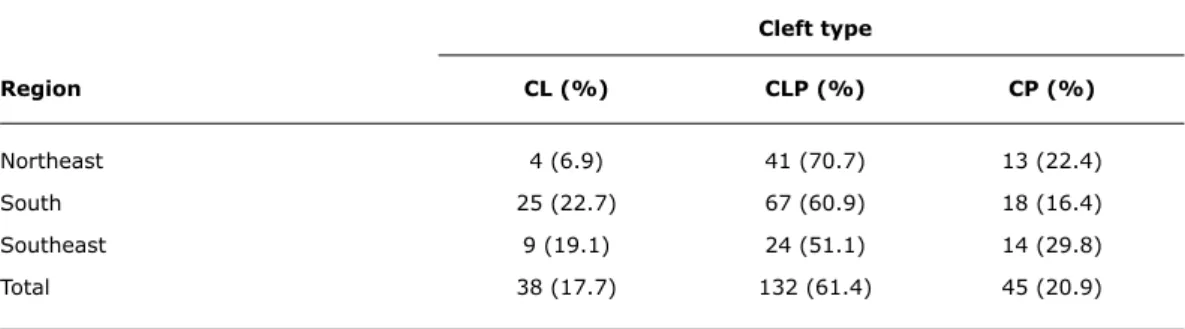
Cleft type
Region Cl (%) ClP (%) CP (%)
Northeast 4 (6.9) 41 (70.7) 13 (22.4)
South 25 (22.7) 67 (60.9) 18 (16.4)
Southeast 9 (19.1) 24 (51.1) 14 (29.8)
Total 38 (17.7) 132 (61.4) 45 (20.9)
table 1 - Distribution of subjects with cleft lip and/or palate according to Brazilian regions*
CL = cleft lip; CLP = cleft lip and palate; CP = cleft palate. * Chi-square test (p = 0.040).
introduction
Cleft lip and/or palate, also called typical orofacial clefts (TOCs), are the most common craniofacial birth defects and their formation occurs during embryonic development.1,2 TOCs affect one in every 600 newborn
babies3,4 and manifest alone or associated with other birth
defects.5 About 300 syndromes appear to have TOC as
one of their characteristics.6-8
Early diagnosis of TOCs makes it possible to investigate other defects and prevent and/or minimize complications.9-12
Dificulties in feeding are the most common complications, such as insuficient sucking, presence of milk within the nasal cavity, suction, and decreased food intake, affecting the infant’s nutritional status and resulting in poor weight gain.13,14
Neonatal care is a complex approach and the involvement of the health care team since diagnosis may help parents understand the implications of that birth defect and the potential for esthetic and functional correction, in addition to taking measures to minimize comorbidities.15-17
Immediate postnatal pediatric management requires critical decisions, such as measures regarding infant hygiene and feeding practices, investigation of associated anomalies, and referral to clinical and genetic assessment and to surgical correction in reference centers.16,18,19
The objective of the present study was to determine the time of diagnosis made in different Brazilian regions.
Method
This prospective, descriptive, cross-sectional study was approved by the Research Ethics Committee (no. 438/2002). Parents or legal guardians of children with TOCs, aged 0 to 12 years, from eight health care facilities were invited to participate in the study. Of these facilities, ive were specialized centers (three in the South, one in the Northeast, and one in the Southeast) and three were non-specialized centers (two in the Northeast and one in the Southeast). Specialized centers were considered as those in which cleft
care was provided by a multidisciplinary team of specialists, regardless of the specialist skills of all team members.
Data were collected using a validated structured interview and through voluntary statements. The interviews were recorded; a single researcher listened to all recordings and tabulated the data.
Statistical analysis was performed using Epi-Info (version 6.04d, Centers for Disease Control and Prevention, Atlanta, USA) and SPSS version 16.0 (SPSS Inc., Chicago, USA). The chi-square test and analysis of variance (ANOVA) were used to compare mean values. Signiicance level was set at 5% (p ≤ 0.05).
Results
Of 230 interviews received for analysis, 15 were excluded. The inal sample consisted of 215 (100%) valid interviews: 51.1% (110) from the South, 27% (58) from the Northeast, and 21.9% (47) from the Southeast. There were no participating centers from the North and Midwest. Specialized centers accounted for 86.51% (186) and non-specialized centers for 13.49% (29) of interviews.
Of all cases, 62.8% (135) were male and 37.2% (80) were female. The patients’ mean age at interview was 4 years, and 75% were under 8 years of age.
According to data reported by the families, associated anomalies were investigated in 89.3% (192) of cases. Clefts alone were found in 85.42% (164) of cases, and in 14.58% (28) of cases clefts were associated with other anomalies. In the Northeast, in 10.7% (23) of cases the diagnosis had yet to be established.
time of diagnosis
Prenatal care Maternity ward After hospital total
Region (%) (%) discharge (%) (%)
Northeast 4 (7.1) 43 (74.4) 10 (17.6) 57 (100)
South 13 (11.8) 90 (81.8) 7 (6.4) 110 (100)
Southeast 13 (27.7) 29 (61.7) 5 (10.6) 47 (100)
Cleft type
time of diagnosis Cl (%) ClP (%) CP (%)
Prenatal care 11 (29) 19 (63.3) 0 (0)
Maternity ward 27 (71) 110 (84) 25 (55.5)
After hospital discharge 0 (0) 2 (1.5) 20 (44.4)
Total 38 (100) 131 (100) 45 (100)
table 3 - Distribution of subjects with cleft lip and/or palate according to Brazilian regions and time of diagnosis*
* Chi-square test (p = 0.005).
table 2 - Distribution of subjects with cleft lip and/or palate according to time of diagnosis*
CL = cleft lip; CLP = cleft lip and palate; CP = cleft palate. * Chi-square test (p < 0.0001).
Regarding time of diagnosis, clefting was diagnosed after birth in the maternity ward in 75.3% (162) of cases; during the prenatal period in 14% (30); and after hospital discharge in 10.2% (22).
Data analysis demonstrated signiicant differences between time of diagnosis and cleft type (p < 0.0001) (Table 2).
The diagnosis of CL occurred mainly during the prenatal period, and that of CP mainly in the maternity ward or after hospital discharge (p = 0.007). Among cases detected in the maternity ward and after hospital discharge, in the latter, almost all patients had a diagnosis of CP (p < 0.0001).
Regarding socioeconomic status, the average monthly family income was 3.72 times the minimum wage in the Southeast, 2.22 in the South, and 1.57 in the Northeast. Participants from the Southeast reported higher incomes than those interviewed in the Northeast and in the South (p < 0.001). The Northeast showed a trend toward lower incomes than the South, but without statistical signiicance (p = 0.080).
An analysis of the effect of monthly family income on the time of diagnosis revealed that the average monthly income of cases diagnosed during the prenatal period was signiicantly higher than that of cases diagnosed in the maternity ward or after discharge (p < 0.0001). However,
among cases of postnatal diagnosis, there was no difference between cases diagnosed in the maternity ward and after discharge (p = 0.6223).
In 81.9% (176) of cases the physician was the professional to disclose the presence of oral clefts to the parents; the nurse in 8.8% (19); family members or friends in 4.7% (10); and other professionals in 4.2% (9). In 0.5% (1) of cases, the information was not disclosed.
Regarding time of diagnosis per region, TOCs were diagnosed in the maternity ward in 81.8% (90) of cases in the South, in 74.4% (43) in the Northeast, and in 61.7% (29) in the Southeast (Table 3). No signiicant differences were observed when time of diagnosis and regions were correlated (p = 0.005). Prenatal diagnosis was signiicantly higher in the Southeast, whereas diagnosis in the maternity ward was higher in the Northeast (p = 0.007). There was no signiicant difference (p = 0.094) between regions when diagnosis in the maternity ward and after discharge were correlated.
surgery between groups at different times of diagnosis was not signiicant (p = 0.185).
Palate surgery was performed in 69.5% (123) out of 177 cases with cleft palate (45) or cleft lip and palate (132). Half of the cases diagnosed during the prenatal period, in the maternity ward, and after hospital discharge underwent surgery, on average, at 14, 19, and 16 months of age, respectively. There was no signiicant difference in age at palate surgery between groups at different times of diagnosis (p = 0.937).
Of 312 clefts eligible for surgical correction in individuals who, according to the parents, had the minimum age to undergo surgery (162 between CL and CLP; 150 between CP and CLP), 51.11% (160) were not corrected within the scheduled time. The reasons were: other diseases (mainly ear infections and pneumonia) in 22.5% (36) of cases, service problems (scheduling delays, lack of beds) in 20.62% (33), anemia in 18.12% (29), and poor weight gain in 18.12% (29). The centers followed different surgical protocols; thus, delay to surgery was based on information obtained from parents or legal guardians, according to surgical planning at each service.
Discussion
This is the irst multicenter study conducted within the Skull/Face Project Brazil, which aims to contribute to the improvement of care delivered to individuals with craniofacial anomalies. Health care facilities located in the Southeast, South, and Northeast of Brazil participated in this study. Centers located in the North and Midwest were not interested in participating despite attempts to contact them. Coincidentally, these are the regions providing less specialized care.19
There was a prevalence of CLP in the three regions, which is consistent with data published by Mossey & Little.3
Similar results were found by Loffredo et al.20 and by Nunes
et al.,21 who also detected a larger proportion of cases
among men,3 in agreement with our indings.
There was a prevalence of clefts alone, and in 14.58% of cases clefts were associated with other anomalies. This result is consistent with the indings by Cohen et al.7 and
Mossey & Little,3 who revealed a mean of 15% of syndromic
clefts. A Brazilian study conducted in the city of São José dos Campos, southeastern Brazil, found 9.1% of syndromic clefts.22
In the Northeast, there were 23 cases without an established diagnosis. Dificult access to a geneticist has already been documented by a previous study within the Skull/Face Project Brazil.19 That health care facility provides
care to the population of the state of Ceará, northeastern Brazil, and has a geneticist on staff, but staff members have no direct access to genetic laboratory tests, factors that
delay diagnosis. Moreover, dysmorphologic evaluation is an evolutionary process and may require investigation of other organs and systems and monitoring of neuropsychomotor conditions before a diagnostic conclusion is reached.11,18
TOCs were diagnosed in the maternity ward in 75.3% of cases. A similar result was reported by Di Ninno et al.,23
who found 80% of postnatal diagnosis in a sample from the city of Belo Horizonte, southeastern Brazil.
Only 17.64% of individuals with CL and CLP were diagnosed during the prenatal period, and 10.2% of affected subjects, mainly with CP, were diagnosed after hospital discharge. Thus, such inding reinforces the need for attention to an ultrasound diagnosis during prenatal care, particularly of CL and CLP, and pediatric evaluation during the irst examination of the newborn.17
Examination of oral structures is essential, especially due to dificulties in feeding experienced by individuals with TOCs. Such indings require prompt intervention, such as appropriate feeding practices and guidance on posture and oral hygiene, thus ensuring adequate nutrition and weight gain.13
The prenatal diagnosis found in only a few cases in our sample is consistent with that found by Di Ninno et al.23
Jones24 believes that this diagnosis may cause emotional
problems for parents. Di Ninno et al.23 and Johnson &
Sandy25 concluded that prenatal diagnosis is beneicial
and that families want to be informed of the results. This diagnosis allows parental counseling and effective postnatal surgical, feeding, and treatment planning.26
Bunduki et al.11 state that, after the diagnosis is conirmed
by ultrasound, parents should be referred to speciic follow-up programs in reference centers. According to Chitty & Grifin,16 indings of structural or chromosomal abnormalities
determine prognosis and indications for chromosome investigation and fetal echocardiography.
CL can be easily diagnosed by ultrasound, whereas CP is diagnosed more often after birth by clinical examination of the newborn.9,10,12 These indings were conirmed in
our study, with signiicance between cleft type and time of diagnosis.
Although there was an overall prevalence of diagnosis in the maternity ward across the three regions, prenatal diagnosis was more frequent in the Southeast. The average monthly family income of cases diagnosed during the prenatal period was signiicantly higher than that of cases diagnosed in the maternity ward or after discharge. This inding suggests that the higher income of southeastern families allowed greater access to prenatal care and ultrasound examination.
In our study, the physician was the main person responsible for delivering information on the presence of clefts to parents, followed by the nurse. Di Ninno et al.27
level of knowledge on TOCs. As a result, many parents return home with unanswered questions. These aspects were also considered deicient by Schardosim et al.,28 who
suggested that health professionals should have greater commitment to provide comfort and helpful guidance to parents. Diagnosis disclosure by family members, friends, and other professionals found in 19 cases in our sample relects the lack of training among health care teams to address a common birth defect associated with high morbidity. In addition to technical clariication by the health care team, emotional support to family members is also of paramount importance.18
The delay to surgery observed in our sample was mostly due to factors related to the health status of affected subjects (anemia, diseases, poor weight gain), in addition to service problems (scheduling delays, lack of beds). Amstalden-Mendes et al.13 showed similar results, with poor weight
gain as the main factor leading to delay to surgery in their sample, reinforcing that attention to nutritional status is crucial to surgical correction within appropriate time. Other important aspects in approaching subjects with TOCs include the correct indication of feeding practices, care provided by specialized staff, and referral of patients to reference centers.13,29
In fact, a Brazilian study of normal newborn infants found that preventive measures are likely to minimize interruption of exclusive breastfeeding in the infant’s irst month of life.30
These measures, together with a longitudinal follow-up performed until the establishment of feeding practices and other resources tailored to the peculiarities of each infant with clefts, may promote appropriate weight gain.
Age of affected individuals at corrective surgery was not dependent on early diagnosis of TOCs. Although necessary, surgical correction is not the only treatment. Early diagnosis allows contact of parents with specialists, favors early introduction of different feeding practices, preventing weaning whenever possible, and facilitates overall treatment planning and neonatal care tailored to the peculiarities of this congenital defect.
Despite the need for a highly complex structure for rehabilitation treatment, child care and treatment of morbid complications should be carried out in primary and secondary health care facilities,13 which justiies staff training and
organization of the public health system in order to improve attention to this important group of congenital defects.
Across Brazilian regions, clefts appear to be more often diagnosed in the maternity ward. Therefore, hospital health care teams should be able to provide initial care, thus contributing to future health care required. Considering the prevalence and morbidity associated with TOCs, staff training is suggested for multidisciplinary management of affected subjects as part of health care policies, starting at the maternity unit.
References
1. Wyszynski DF. Cleft lip and palate: from origin to treatment. New York: Oxford University Press; 2002.
2. Moore KL, Persaud TV. The Pharyngeal (Branchial) Apparatus. In: Moore KL, Persaud TV. The Developing Human Clinically Oriented Embryology. 6th ed. Philadelphia, Pa: WB Saunders; 1998. p. 215-56.
3. Mossey PA, Little J. Epidemiology of oral clefts: an international perspective. In: Wyszynski DF, editor. Cleft Lip and palate: from origin to treatment. New York: Oxford University Press; 2002. p. 127-58.
4. Word Health Organization. Global strategies to reduce the health-care burden of craniofacial anomalies. Report of WHO meetings on International Collaborative Research on Craniofacial Anomalies. Geneva: WHO; 2002.
5. Stoll C, Alembik Y, Dott B, Roth MP. Associated malformation in
cases with oral clefts. Cleft Palate Craniofac J. 2000;37:41-7. 6. Marazita ML, Mooney MP. Current concepts in the embryology
and genetics of cleft lip and cleft palate. Clin Plast Surg. 2004;31:125-40.
7. Cohen Junior MM, Gorlin RJ, Fraser FC. Craniofacial Disorders. In: Rimoin DL, Connor JM, Pyeritz RE, Korf BR, editors. Emery and Rimoin’s Principles and Practice of Medical Genetics. New York: Churchill Livingstone; 1997. p. 1121-48.
8. Shprintzen RJ, Siegel-Sadewitz VL, Amato J, Golberg RB. Anomalies associated with cleft lip, cleft palate, or both. Am J Med Genet.
1985;20:585-95.
9. Offerdal K, Jebens N, Syvertsen T, Blaas HG, Johansen OJ, Eik-Nes SH. Prenatal ultrasound detection of facial clefts: a prospective study of 49,314 deliveries in a non-selected population in Norway. Ultrasound Obstet Gynecol. 2008;31:639-46.
10. Russell KA, Allen VM, MacDonald ME, Smith K, Dodds L. A population-based evaluation of antenatal diagnosis of orofacial
clefts. Cleft Palate Craniofac J. 2008;45:148-53.
11. Bunduki V, Ruano R, Sapienza AD, Hanaoka BY, Zugaib M. Diagnóstico pré-natal de fenda labial e palatina: experiência de 40 casos. RBGO. 2001;23:561-6.
12. Grandjean H, Larroque D, Levi S. The performance of routine ultrasonographic screening of pregnancies in the Eurofetus Study.
Am J Obstet Gynecol. 1999;181:446-54.
13. Amstalden–Mendes LG, Magna LA, Gil-da-Silva-Lopes VL. Neonatal care of infants with cleft lip and/or palate: feeding orientations and evolution of weight gain in non-specialized Brazilian hospital.
Cleft Palate Craniofacial J. 2007;44:329-34.
14. Reid J. A review of feeding interventions for infants with cleft
palate. Cleft Palate Craniofac J. 2004;41:268-78.
15. Shaw WC, Semb G, Nelson P, Brattström V, Molsted K, Prahl-Andersen B, et al. The Eurocleft Project 1996-2000: overview. J Cranio-maxillofacial Surgery. 2001;29:131-40.
16. Chitty LS, Grifin DR. Anormalidades do lábio e do palato fetal: diagnóstico ultra-sonográico. In: Watson AC, Sell DA, Grunwell P. Tratamento de issura labial e fenda palatina. São Paulo: Editora Santos; 2005. p. 107-116.
17. Habel A. O papel do pediatra. In: Watson AC, Sell DA, Grunwell P. Tratamento de issura labial e fenda palatina. São Paulo: Editora Santos; 2005. p. 123-35.
18. Ribeiro-Roda S, Gil-da-Silva-Lopes VL. Aspectos odontológicos das fendas labiopalatinas e orientações para cuidados básicos. Rev Cienc Med. 2008;17:95-103.
19. Monlleó IL, Gil-da-Silva-Lopes VL. Anomalias craniofaciais: descrição e avaliação das características gerais da atenção no
Sistema Único de Saúde. Cad Saude Publica. 2006;22:913-22. 20. Loffredo LC, Freitas JA, Grigolli AA. Prevalência das issuras orais
de 1975 a 1994. Rev Saude Publica. 2001;35:571-5.
22. Cerqueira MN, Teixeira SC, Naressi SC, Ferrreira AP. Ocorrência de issuras labiopalatais na cidade de São José dos Campos-SP. Rev Bras Epidemiol. 2005;8:161-6.
23. Di Ninno CQ, Santos PG, Bueno MG, Syrio IM. A inluência da época do diagnóstico das issuras labiopalatinas. Rev Soc Bras Fonoaudiol. 2006;11:75-81.
24. Jones MC. Prenatal diagnosis of cleft lip and palate: detection rates, accuracy of ultrasonography, associated anomalies and strategies for counseling.Cleft Palate Craniofac J. 2002;39:169-73. 25. Johnson N, R Sandy J. Prenatal diagnosis of cleft lip and palate.
Cleft Palate Craniofac J. 2003;40:186-9.
26. Bradbury E, Bannister P. Aconselhamento pré-natal, perinatal e pós-natal. In: Watson AC, Sell DA, Grunwell P. Tratamento de issura labial e fenda palatina. São Paulo: Editora Santos; 2005. p. 117-122.
27. Di Ninno CQ, Gomes RO, Santos PG, Bueno MG, Galvão DA, Meira AL, et al. O conhecimento de proissionais da área da saúde sobre issura labiopalatina. Rev Soc Bras Fonoaudiol. 2004;9:93-101.
Correspondence:
Vera Lúcia Gil da Silva Lopes
Depto de Genética Médica, FCM, UNICAMP Rua Tessália Vieira de Camargo, 126 CEP 13083-887 – Campinas, SP – Brazil Tel.: +55 (19) 3521.8909
Fax: +55 (19) 3521.8909 E-mail: [email protected]
28. Schardosim LR, Nogueira DA, Bosco VL, Pereima MJ. Bebês portadores de issura labiopalatal: satisfação dos pais com as orientações recebidas dos proissionais. JBP rev Ibero-am Odontopediatr Odontol Bebe. 2004;7:568-73.
29. Amstalden-Mendes LG, Gil-da-Silva-Lopes VL. Fenda de lábio e ou palato: recursos para alimentação antes da correção cirúrgica. Rev Cienc Med (Campinas). 2006;15:437-48.