Adjustment of oxygen use by means of pulse oximetry:
an important tool for patient safety
Adequação do uso do oxigênio por meio da oximetria de pulso:
um processo importante de segurança do paciente
Telma de Almeida Busch Mendes1, Paola Bruno de Araújo Andreoli2, Leny Vieira Cavalheiro3, Claudia Talerman4,
Claudia Laselva5
ABSTRACT
Objective: To assess patient’s level of oxygenation by means of pulse oximetry, avoiding hypoxia (that causes rapid and severe damage), hyperoxia, and waste. Methods: Calculations were made with a 7% margin of error and a 95% confidence interval. Physical therapists were instructed to check pulse oximetry of all patients with prescriptions for physical therapy, observing the scheduled number of procedures. Results: A total of 129 patients were evaluated. Hyperoxia predominated in the sectors in which the patient was constantly monitored and hypoxia in the sectors in which monitoring was not continuous. Conclusions: Professionals involved in patient care must be made aware of the importance of adjusting oxygen use and the risk that non-adjustment represents in terms of quality of care and patient safety.
Keywords: Anoxia; Oximetry; Oxygen therapy/adverse effects
RESUMO
Objetivo: Avaliar a oxigenação dos pacientes por meio da oximetria de pulso, evitando a hipóxia (cujo dano é rápido e grave) e a hiperóxia, evitando o desperdício. Métodos: Foi realizado um cálculo de amostra com margem de erro em 7% e intervalo de confiança de 95%. Os fisioterapeutas foram orientados a checar a oximetria de pulso de todos os pacientes com prescrição de fisioterapia, respeitando o número de atendimentos programados. Resultados:
Foram avaliados 129 pacientes. A hiperóxia predominou nos setores que o paciente permaneceu monitorado e a hipóxia nos setores em que a monitorização não era contínua. Conclusões: Faz-se necessária
a conscientização dos profissionais envolvidos na assistência ao paciente sobre a importância da adequação do oxigênio e o risco que sua inadequação representa na qualidade do atendimento e na segurança do paciente.
Descritores: Anoxia; Oximetria; Oxigenoterapia/efeitos adversos
INTRODUCTION
Oxygen therapy is a treatment in which the partial pressure of oxygen in arterial blood is increased by means of a higher concentration of oxygen in the inhaled air. It is an effective treatment, which is indicated in respiratory failure, i.e., when the respiratory system is unable to maintain normal values of arterial oxygen
pressure (PaO2) and/or arterial carbon dioxide
pressure (PaCO2). Oxygen administration is indicated
in cases of acute respiratory failure (RF), when PaO2
is lower than 60 mmHg or SaO2 is lower than 88-90%
as per the oxyhemoglobin dissociation curve in room
air (FiO2 21%).
In cases of chronic hypoxemia in which there is greater tolerance to hypoxemia, a 55 mmHg threshold
level of PaO2 may be used. Under these conditions,
oxygen should always be administered, particularly in Type I RF (hypoxemic or alveolocapillary), comprising conditions that primarily affect vessels, alveoli, and pulmonary interstitium, such as acute respiratory
Study carried out at Hospital Israelita Albert Einstein – HIAE, São Paulo (SP), Brazil.
1 Physiotherapist; PhD in Health Sciences, Coordinator of Graduate Program in Gerontology of Hospital Israelita Albert Einstein - HIAE; Full Professor of Geriatrics at Universidade São Camilo,
São Paulo (SP) Brazil.
2 Psychologist, PhD in Health Sciences and Manager of Healthcare Practice at Sociedade Beneficente Israelita Brasileira Albert Einstein – SBIBAE, São Paulo (SP), Brazil. 3 Physiotherapist, Master in Rehabilitation and Advisor of Healthcare Practice at Sociedade Beneficente Israelita Brasileira Albert Einstein – SBIBAE, São Paulo (SP), Brazil. 4 Physiotherapist at Hospital Israelita Albert Einstein – HIAE, São Paulo (SP), Brazil.
5 Nurse, Manager of Surgery and Clinical Medicine at Sociedade Beneficente Israelita Brasileira Albert Einstein – SBIBAE, São Paulo (SP), Brazil.
Corresponding author: Telma de Almeida Busch Mendes – Rua Deputado João Sussumo Hirata, 770 – apto. 12 – Morumbi – CEP 05715-010 – São Paulo (SP), Brasil – Tel.: 11 3742-7565 – e-mail: [email protected]
distress syndrome (ARDS), pneumonias, atelectasis, pulmonary edema, pulmonary embolism, quasi-drowning, exacerbated chronic obstructive pulmonary disease (COPD), severe asthma and pneumothorax. In
these cases, there is a drop in PaO2 with normal values
of PaCO2(1) .
In cases of Type II RF, there is an elevation of carbon dioxide levels due to ventilatory failure (with central nervous system – CNS – alterations, neuromuscular and peripheral alterations, dysfunction of the thoracic wall and pleura, obstruction of upper airways). Additionally, hypoxemia is common in patients who breathe room air. The administration of oxygen to the patient has clinical indications described in the literature that must be rigidly followed by the multiprofessional team.
The objective is to maintain adequate levels of oxygenation in order to avoid suspected or confirmed acute hypoxemia that causes rapid and severe damage, to reduce symptoms associated with chronic hypoxemia, and to reduce the work load that hypoxemia imposes on the cardiopulmonary system (pulmonary hypertension, arrhythmia, and myocardial ischemia) and on the CNS.
Confirmation of the presence of RF is only made by analysis of blood gases. A rapid indication of the gas
exchanges conditions is given by pulse oximetry(1).
An interesting fact that must be considered is that respiratory failure is not characterized by an exclusive clinical finding. Dyspnea may be the primary symptom and its intensity, sudden appearance in addition to progression help in the diagnosis. When present, cyanosis is an important sign of hypoxemia, but it may go unnoticed. In cases of even mild anemia, it will only be present when the reduced hemoglobin concentration exceeds 5 g/dl. As hypoxemia is accentuated, some manifestations such as decreased cognitive function, deterioration of good judgment capacity, aggressiveness, motor incoordination, and even coma and death may occur. A similar clinical picture may occur when CO2 is elevated. In the case of chronic hypoxemia, patients may present with somnolence, lack of concentration, apathy, fatigue, and delayed reaction
time(1). The nervous system is the most vulnerable of all,
followed by the kidneys, heart, and liver, and this is why neurological symptoms predominate in RF.
But there are other clinical manifestations that may be minimal or absent even in the presence of significant hypoxemia, which reinforces the need for pulse
oximetry monitoring. Oximetry (SPO2) is considered
to be the best non-invasive monitoring method. With the use of oximetry, it is possible to assess if the level of oxygen in arterial blood is adequate for tissue needs. It is a useful measurement for evaluating acute changes in the patient’s clinical status and for adjusting the flow of oxygen according to recommended levels.
Nevertheless, one cannot forget the limitations of pulse oximetry(2).
Saturation values are equivalent to arterial gases.
Gasometrically, acute RF (ARF) corresponds to PaO2
< 60 mmHg, SaO2 < 90%, and PaCO2 > 50 mmHg);
except for patients who are chronic CO2 retainers.
Literature confirms the correlation between the
reading done by pulse oximeter (SPO2), which is
hemoglobin oxygen saturation in peripheral arterial blood, and the saturation measured in arterial blood
(SaO2). There is a variation of precision that should be
taken into consideration (Chart 1)(3).
Monitoring by pulse oximetry reduces preoperative hypoxemia, enables the detection and treatment of hypoxemia related to respiratory events, and promotes
significant changes in patient care(4). The Brazilian
Society of Anesthesiology, along with the Federal Council of Medicine, recommends the use of pulse oximetry during anesthesia, sedation, in intensive care units, and in transporting critically ill patients(5-6).
The patient’s oxygenation reflects changes in the patient’s clinical status and may be altered for a number of reasons: accumulation of secretions, changes in decubitus with alteration of the V/Q (ventilation/ perfusion) ratio, therefore involving the entire team
associated with direct patient care(7).
In a prior study carried out at the Hospital Israelita Albert Einstein (HIAE), in 2006, a total of 1,092 inpatients were accompanied; they were undergoing respiratory physical therapy, and 11% of them were found to be in hypoxia, i.e., with oxygen saturation lower than 92%, a substantial value when considering accuracy variation (data not published).
The physical therapy routine included assessment of oximetry only at each respiratory physical therapy procedure. Based on the results found in 2006, a new routine was implemented and some recommendations were made to the multiprofessional team regardless of the type of care to be given:
- systematically assess vital signs (heart rate, respiratory rate, temperature, blood pressure). Oximetry is the fifth vital sign;
- always correlate oximetry with clinical data;
- assess the patient’s clinical picture. Note if there are alterations in consciousness and motor instability, which are signs of neurological alterations;
SaO2 (%) Accuracy range (%)
> 90 ± 2%
80-90 ± 5%
< 80 ± 12%
- determine whether the patient displays tachycardia
and arterial hypertension. Remember that
bradycardia occurs later;
- do not wait for the presence of cyanosis to exclude hypoxemia, which will only occur when the levels of
PaO2 are lower than 50 mmHg;
- check respiratory rate. If the patient presents with tachypnea (> 20 breaths/min, in adults) or bradypnea (incapacity to generate or conduct respiratory stimuli), check pulse oximetry and monitor frequency to accompany progression; - determine whether the oximeter was placed correctly,
avoiding excessive pressure on the fingers. In children, depending on the type of oximeter, the installation site should be inspected to avoid lesions;
- check the oximeter cable if the reading is not consistent with the patient’s clinical status;
- in case of doubt as to a result, oximetry should be check on you in order to verify if there is a problem with the device;
- maintain oximetry monitoring after the removal of oxygen during rest and throughout handling of the patient, including during bathing;
- install oxygen and repeat the reading if the oximetry shows altered values. Saturation should
be maintained at ≥92%, considering the difference
described in literature;
- record the results ascertained on the patient’s clinical chart;
- suggest to the physician the collection of arterial gases if the patient presents with a critical clinical picture associated with change in saturation, since oximetry is unable to detect hypercapnia or acidosis.
Other acts of patient care were recommended in relation to the saturation reading:
- individuals of the black race: try to place the sensor in areas where the skin is lighter and, when the result is lower than 90%, apply the sensor turned towards the palm of the hand or sole of the foot;
- exaggerated environmental light may falsely elevate oximetry;
- smokers or individuals who live in large urban areas (taxi drivers) may present with high levels
of carboxyhemoglobin leading to higher SPO2
readings(8);
- individuals who use sodium nitroprusside, local anesthetics, nitroglycerin, methochlopramide and medications containing sulfa may also present with higher readings(9);
- intravenous dyes, such as methylene blue, indigo carmine, and indocyanine green, as well as nail polish in colors red, black, blue and green also modify readings; therefore, avoid readings with nail polishes;
- in case of bacteremia, the drop in saturation is a result of inadequate perfusion. In this case, consider the general aspect of the patient and check blood pressure: if there is associated hypotension, it may be an indication of imminent septic shock;
- remember that hypothermia may disguise the reading.
Additionally, other actions were reinforced with the physical therapy team, such as pulse oximetry monitoring, the values recommended in literature, the importance of recording clinical progress of the levels identified in clinical charts, as well as establishing a more systemic assessment of oxygen saturation when the patient shows changes in clinical picture or a borderline saturation measurement.
Some barriers had to be broken down in order to implant these actions:
- non-compliance and non-involvement of professionals implicated in direct patient care, especially physical therapists and nurses who should record saturation results in the clinical progress notes;
- the need for nurses to assess pulse oximetry in patients who do not receive oxygen at least once a day and are not accompanied by physical therapy.
This second action impacted costs, since as routine in the medical clinic (patients not seriously ill) there should be medical prescription of oximetry measurements and a daily rate was charged by the nursing staff for the use of the oximeter, regardless of the number of times the saturation was checked.
OBJECTIVE
To verify oxygen saturation levels in patients with prescriptions for physical therapy and the justifications given in the medical chart by the Physical Therapy team about patients with alterations.
METHODS
Calculation of the sample was based on the number of patients hospitalized without mechanical ventilation and seen by physical therapists over a period of 15 days, with a 7% margin of error and 95% confidence interval (CI).
increase or drop in saturation, should also be recorded, as well as the justification for its non-correction, whenever necessary. Each and every alteration was to be followed and recorded, and the professionals involved in patient’s care were to be made aware of the status. In daily practice, the nurse centralizes the information and should transmit it to the physician and all involved, also being aware of each and every change detected by the team. When there was no justification in the physical therapist progress notes, the nurses’ notes and the control sheet with the licensed practicing nurse records of the times and values of saturation should be checked, as well as the clinical progress report made by the speech therapist when involved in the process. A second progress report was audited to verify if the oxygen saturation was maintained adequate as per the notes in the patient’s clinical chart.
RESULTS
In analyzing adequate use of oxygen, it is clear that out of the total 129 measurements audited in the first progress report, 72 displayed normal values and 57 were altered (Table 1). Of the total number of measurements found to be altered, 46 were due to hyperoxia and 11 were due to hypoxia (Table 2).
Among the measurements that did not satisfy criteria of normality (in hyperoxia or hypoxia: n = 57),
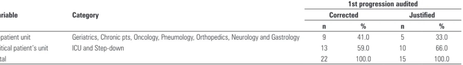
22 were corrected by the physical therapist and 15 were not corrected, but were justified (Table 3).
In the second progress report, 69 clinical progress records were evaluated, and 37 of them satisfied criteria of normality and 32 showed alterations (Table 4).
Among the abnormal measurements, 30 were found as hyperoxia in the second progress report, and only 2 in hypoxia (Table 5). It is worth pointing out that the latter were identified in sectors where patients were not continually monitored and depended on evaluations of the physical therapist at the time of the procedure.
Hyperoxia predominated in all assessed sectors, except in two units where only one measurement was evaluated, since the patients only had physical therapy once a day (data not shown on the table).
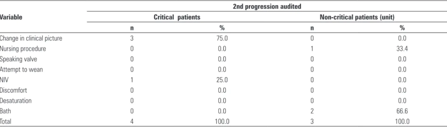
Of this total number of alterations found in the second progress report, ten were corrected by physical therapy. Among the 22 cases which were not corrected, only 8 were justified (Table 6).
Among the justifications encountered in units with patients bearing greater severity and risk, the change in clinical picture predominated as the most frequent justification in both clinical progress reports audited, regardless of the severity of the case. A lower percentage of justifications was found in sectors with patients presenting with less severe conditions (Tables 7 and 8).
Variable Category
1st progression audited
Normal (≥92%) Altered
n % n %
In-patient unit Geriatrics, Chronic pts, Oncology, Pneumology, Orthopedics, Neurology and Gastrology 42 58.4 15 26.3
Critical patient’s unit ICU and Step-down 30 41.6 42 73.7
Total 72 100.0 57 100.0
Table 1. Oxygen saturation values measured in patients not using non-invasive ventilation and monitored by physical therapy in the first progression note audited according to standardized measure criterion
Variable Category
1st progression audited
Hyperoxia Hypoxia
n % n %
In-patient unit Geriatrics, Chronic pts, Oncology, Pneumology, Orthopedics, Neurology and Gastrology 6 13.0 9 82.0
Critical patient’s unit ICU and Step-down 40 87.0 2 18.0
Total 46 100.0 11 100.0
Table 2. Oxygen saturation values measured in patients not using non-invasive ventilation and monitored by physical therapy, which did not meet the normalcy criterion in the 1st progression notes in the chart according to the criterion defined as hyperoxia and hypoxia
Variable Category
1st progression audited
Corrected Justified
n % n %
In-patient unit Geriatrics, Chronic pts, Oncology, Pneumology, Orthopedics, Neurology and Gastrology 9 41.0 5 33.0
Critical patient’s unit ICU and Step-down 13 59.0 10 66.0
Total 22 100.0 15 100.0
Variable Category
2nd progression audited
Normal (≥92%) Altered
n % n %
In-patient unit Geriatrics, Chronic pts, Oncology, Pneumology, Orthopedics, Neurology and Gastrology 20 54.1 9 28.0
Critical patient’s unit ICU and Step-down 17 45.9 23 72.0
Total 37 100.0 32 100.0
Table 4. Oxygen saturation values measured in patients not using non-invasive ventilation and monitored by physical therapy in the 2nd progression notes in the chart audited according to the standardized measurement criterion
Variable Category
2nd progression audited
Hyperoxia Hypoxia
n % n %
In-patient unit Geriatrics, Chronic pts, Oncology, Pneumology, Orthopedics, Neurology and Gastrology 7 23.3 2 100.0
Critical patient’s unit ICU and Step-down 23 76.6 0 0,0
Total 30 100.0 2 100.0
Table 5. Oxygen saturation values measured in patients not using non-invasive ventilation and monitored by physical therapy which did not meet the normalcy criterion in the 2nd progression notes in the chart according to the criterion defined as hyperoxia and hypoxia
Variable Category
2nd progression audited
Corrected Justified
n % n %
In-patient unit Geriatrics, Chronic pts, Oncology, Pneumology, Orthopedics, Neurology and Gastrology 5 50.0 2 25.0
Critical patient’s unit ICU and Step-down 5 50.0 6 75.0
Total 10 100.0 8 100.0
Table 6. Measures found in hyperoxia and hypoxia in patients not using non-invasive ventilation and monitored by physical therapy, according to the criterion adopted as adjustment (correction) and justified in the chart by the professional 2nd progression evaluation
Variable
1st progression audited
Critical patients Non-critical patients (unit)
n % n %
Change in clinical picture 5 50.0 1 33.4
Nursing procedure 0 0.0 1 33.3
Speaking valve 1 10.0 0 0.0
Attempt to wean 1 10.0 0 0.0
NIV 1 10.0 0 0.0
Discomfort 1 10.0 0 0.0
Desaturation 1 10.0 0 0.0
Bath 0 0.0 1 33.3
Total 10 100.0 3 100.0
Table 7. Justifications given to no correction of oxygen saturation according to the criterion adopted in patients not using non-invasive ventilation and monitored by physical therapy, in the 1st progression notes in the chart audited according to criterion of severity of patient
NIV: non invasive ventilation
Variable
2nd progression audited
Critical patients Non-critical patients (unit)
n % n %
Change in clinical picture 3 75.0 0 0.0
Nursing procedure 0 0.0 1 33.4
Speaking valve 0 0.0 0 0.0
Attempt to wean 0 0.0 0 0.0
NIV 1 25.0 0 0.0
Discomfort 0 0.0 0 0.0
Desaturation 0 0.0 0 0.0
Bath 0 0.0 2 66.6
Total 4 100.0 3 100.0
Table 8. Justifications given to no correction of oxygen saturation according to the criterion adopted in patients not using non-invasive ventilation and monitored by physical therapy, in the 2nd progression notes in the chart audited according to criterion of severity of patient
DISCUSSION
The decision of when to monitor pulse oximetry should be made as any other clinical decision, i.e., based on therapeutic objectives.
The clinical assessment of hypoxia and hypoxemia revealed inconsistencies in the definition of terms and of the root cause, as well as of clinical indicators used to evaluate the need for oxygen supplementation.
Some patients, even without the need for oxygen supplementation, may be at risk for development of hypoxia.
Many studies discuss the criteria for indication of oxygen in specific situations, but no comparative study was found assessing the criteria for risk of hypoxia and the oxygen monitoring routine.
The risk of hypoxia under specific conditions is well
discussed in literature, such as in acute and chronic RF(1),
in the immediate postoperative period(10-13), principally
in the postoperative phases of large operations(14),
among them abdominal surgery(13,15).
Other situations offer risk of hypoxia, as the case of
patients who suffered encephalic vascular accident
(14,16-18) and when it occurs due to various factors such
as alterations in the regulation of the respiratory center(17), by bronchoaspiration(17-21), or due to muscular
weakness(12,22), and by possible modifications related
to sleep even in patients who display normal daytime oximetry(21-26).This same risk occurs in patients with
pulmonary and cardiovascular problems(27), respiratory
infections(23-28),pulmonary embolism, acute edema
(29-31), and in obese patients due to respiratory problems
associated with obesity(31).
Despite clinical indications described in literature, oxygen administration to the patient often is not rigorously followed by the multiprofessional team. Checking oximetry is routine when recording clinical progress in physical therapy regardless of the use of oxygen by the patient, but it is not a routine adopted by the nursing staff when the patient is not on continuous oxygen. The consequences of inadequate use or non-use of oxygen lead to a continuous concern about the safety and quality of the care given to patients. It is necessary to evaluate the standard of saturation found in patients submitted to physical therapy in order to adjust the peripheral saturation values and standardize a routine of oximetry assessment in hospitalization units where the risk of hypoxia is higher, considering the fact that the patient is not continuously monitored. In this study, we point out that, in closed units, such as the Semi-Intensive Unit, Coronary Unit, and Intensive Care Unit, where the patient is monitored or receives physical therapy care more intensively, the value of hypoxemia was low, with a prevalence of alterations due
to hyperoxia. The lack of correction of the alterations found was motivated by the change in clinical picture as per the most frequent justification, and in a smaller percentage of cases, the attempt to wean and the use of the speaking valve. Thus, involvement of the nursing team and the speech therapy team in compliance with the routine of assessment of patient oximetry is mandatory, reinforcing the importance of this monitoring.
CONCLUSION
Awareness of all the professionals involved in direct patient care as to the importance of adequate use of oxygen, of vigilance, and of recording values in the patients’ progress charts as well as justification for non-correction of oxygenation deviations is necessary. This project revealed a gap in communication among the teams involved, and brought to light the need to define even more important criteria as to the risk of hypoxia, since pulse oximetry measurement represents costs for the patient and for the healthcare service.
REFERENCES
1. Fishman AP. Acute respiratory failure. In: Fishman AP. Pulmonary disease and disorders. New York: MC Graw-Hill; 1988. p. 2185-201.
2. West JB. Fisioterapia respiratória moderna. 3ª ed. São Paulo: Manole; 1990. 3. Jensen LA, Onyskiw JE, Prasad NG. Meta-analysis of arterial oxygen saturation
monitoring by pulse oximetry in adults. Heart and Lung. 1998;27(6):387-408. 4. Pedersen T, Dyrlund Pedersen B, Møller AM. Pulse oximetry for perioperative
monitoring (Cochrane Review). In: The Cochrane Library.Issue 1; 2007.
5. Moura Fé, Liberal HSP. Resolução do Conselho Federal de Medicina nº1363- Diário Oficial da União, 22 de março de 1993, seção 1. p. 3439.
6. Braz JRC. Monitorização da oxigenação e ventilação. Rev Bras Anestesiol. 1996;46(3):223-40.
7. Stetter K. Safety issues that should be considered when mobilizing critically ill patients. Crit Care Clin. 2007;23(1):35-53.
8. Pinheiro BV, Oliveira JCA. IRpA. [Internet]. [citado 2010 Out 28]. Disponível em: www.pneumoatual.com.br.
9. Barker SJ, Tremper KK. The effect of carbon monoxide inhalation on pulse oximetry and tracutaneous PO2. Anesthesiology. 1987;66(5):667-79. 10. Barker SJ, Tremper KK, Hyatt J. Effects of methemoglobinemie on pulse
oximetry and mixed venous oximetry. Anesthesiology. 1989;70(1):112-7. 11. Powell MB, Menon DK, Jones JG. The effects of hipoxaemia and
recommendations for postoperative oxygen therapy. Anaesthesia. 1996;51(8):769-72.
12. Russell GB, Graybeal JM. Hypoxemic episodes of patients in a post anesthesia
care unit. Chest.1993;104(3):899-903.
13. Caplan BA, Ward RJ, Posner K, Cheney FW. Unexpected cardiac arrest during spinal anesthesia: a closed claims analysis of predisposing factors. Anesthesiology. 1988;68(1):5-11.
14. Pierce EC, Cooper JB. Analysis of anesthetic mishaps. Anesthesiol Clin. 1984;22(1):1-16.
16. Khedr EM, El Shimawy O, Khedr T, Aziz A, Awad EM. Assessment of the corticodiaphragmaic pathway and pulmonary function in acute ischemic stroke patients. Eur J Neurol. 2000;7(3):323-30.
17. Davenport RJ, Dennis MS, Wellwood I, Warlow CP. Complications after acute stroke. Stroke. 1996;27(3):415-20.
18. Langhorne P, Stott DJ, Robertson L, MacDonald JL, McALpine C, Dick F, et al. Medical complications after stroke. Stroke. 2000;31(6):1223-9.
19. Stausholm K, Rosemberg-Adamsen S, Edvardsen L, Kehlet H, Rosemberg J. Validation of pulse oximetry for monitoring of hypoxaemic episodes in the late postoperative period. British J Anaesth. 1997;78(1):86-7.
20. Roffe C, Sills S, Halim M, Wilde K, Allen MB, Jones PW, Crome P. Unexpected nocturnal hypoxia in patients with acute stroke. Stroke. 2003;34(11):2641-5. 21. Roffe C. Hypoxemia and stroke. Rev Clin Gerontol. 2001;11(4):323-35. 22. Nachtmann A, Siebler M, Rose G, Sitzer M, Steinmetz H. Cheyne-Stokes
respiration in ischemic stroke. Neurology. 1995;45(4):820-1.
23. Rowat AM, Wardlaw JM, Dennis MS, Warlow CP. Does feeding alter arterial oxygen saturation in patients with acute stroke? Stroke. 2000;31(9):2134-40. 24. Arzt M, Young T, Finn L, Skatrud JB, Bradley TD. Association of sleep-disordered breathing and the occurrence of stroke. Am J Respir Crit Care Med. 2005;172(11):1447-51.
25. Bassetti C, Aldrich MS. Sleep apnea in acute cerebrovascular diseases: final report on 128 patients. Sleep. 1999;22(2):217-23.
26. Morrell MJ, Finn L, Kim H, Peppard PE, Badr MS, Young T. Sleep fragmentation, awake blood pressure, and sleep-disordered breathing in a population-based study. Am J Respir Crit Care Med. 2000;162(6):2091-6.
27. Smith HA, Lee SH, O’Neill PA, Connolly MJ. The combination of bedside swallowing assessment and oxygen saturation monitoring of swallowing in acute stroke: a safe and humane screening tool. Age Ageing. 2000;29(6):495-9.
28. Houston JG, Morris AD, Grosset DG, Lees KR, McMillan N, Bone I. Ultrasonic evaluation of movement of the diaphragm after acute cerebral infarction. J Neurosurg Psychiatry. 1995;58(6):738-41.
29. Nieto FJ, Young TB, Lind BK, Shahar E, Samet JM, Redline S, et al. Association of sleep-disordered breathing, sleep apnea, and hypertension in a large community-based study. Sleep Heart Health Study. JAMA. 2000;283(14):1829-36.
30. Sulter G, Elting JW, Stewart R, den Arend A, De Kayser J. Continuous pulse oximetry in acute hemiparetic stroke. J Neurol Sci. 2000;179(S 1-2):65-9. 31. Reed C. Care of postoperative patients with pulmonary edema. J Perianesth