INTERICTAL HYPOSEXUALITY IN MALE
PATIENTS WITH EPILEPSY
Diosely C. Silveira
1, Elizabeth A. P. de Souza
2, José F. Carvalho
3, Carlos A. M. Guerreiro
4ABSTRACT - The purpose of this study was to compare the serum levels of androgens between hyposexual and non-hyposexual patients with epilepsy. Adult male patients with epilepsy were investigated. Serum levels of testosterone (T) and free-T, estradiol, and sex hormone binding globulin (SHBG) were measured and the free androgen index (FAI) was calculated. While there were no differences between hyposexual and non-hyposexual patients in the serum levels of T, free-T, and estradiol, or to the FAI, the serum levels of SHBG were significantly higher in hyposexual patients than in non-hyposexual patients. Thus, the effects of increased SHBG upon serum levels of testosterone biologically active in patients with epilepsy and hyposexuality were not detected by the methods used in this study. Four (44%) of nine hyposexual patients who were re-evaluated after two years follow-up improved sexual performance. Thus, clinical treatment that results in good seizure control may improve sexual performance in some patients with epilepsy.
KEY WORDS: epilepsy, sexual dysfunction, testosterone, and androgens.
Hipossexualidade interictal em homens com epilepsia
RESUMO - O objetivo deste estudo foi comparar os níveis séricos de andrógenos e estradiol entre pacientes do sexo masculino com e sem hipossexualidade. Níveis séricos de testosterona e testosterona porção livre, estradiol e globulina ligadora de hormônio sexual (SHBG) foram dosados e calculado o índice de andrógeno livre (FAI). Não houve diferença significante de testosterona, porção livre de testosterona, estradiol e de FAI nos pacientes com e sem hipossexualidade. Os níveis de SHBG foram significantemente maiores nos pacientes hipossexuais quando comparados com os pacientes sem hipossexualidade. Deste modo, os efeitos da SHBG aumentada nos níveis séricos de testosterona biologicamente ativa, em pacientes com epilepsia e hipossexualidade, não foram detectados pelo método usado neste estudo. Quatro (44%) pacientes, reavaliados após dois anos de seguimento, tiveram melhora significativa do desempenho sexual. Portanto, o tratamento medicamentoso, com melhor controle das crises epilépticas, pode melhorar o comportamento sexual de alguns pacientes com epilepsia.
PALAVRAS-CHAVE: epilepsia, disfunção sexual, testosterona, andrógenos.
1Research fellow, Neurology Department, Childrens Hospital and Harvard Medical School, Boston, MA, USA; 2PhD. Professor Assistente,
Departamento de Neurologia da Faculdade de Ciências Médicas (FCM), Universidade Estadual de Campinas (UNICAMP); 3PhD. Professor
Assistente, Departamento de Matemática e Estatística, FCM/UNICAMP; 4MD, PhD. Professor Assistente, Departamento de Neurologia,
FCM/UNICAMP.
Received 16 August 2000, received in final form 3 October 2000. Accepted 3 October 2000.
Diosely C. Silveira, MD, PhD - Childrens Hospital - 300 Longwood Avenue, Enders # 320 - Boston, MA 02115 - USA. Fax: 1 617 734 1646. E-mail: [email protected]
Hyposexuality is one of the most frequent (28%-67%) interictal abnormalities observed in patients with epilepsy1-5. Several factors may disrupt normal sexual function in patients with epilepsy, including psychological influences, hormonal disorders, antie-pileptic drugs (AEDs), and the disease itself6.
Androgens are important for the maintenance of libido and sexual potency7,8. Decreased serum levels of free-testosterone (free-T) have been associated with diminished libido and erectile dysfunction9. The majority (98%) of plasma testosterone (T) is linked to proteins, 43% to 45% to albumin, 53% to 55% to sex hormone binding globulin (SHBG), and 2%
cir-culate under free form10. The free androgen index (FAI) may indirectly estimate the serum levels of T bound to albumin11, which is also considered to be biologically active.
can-didates to temporal lobe surgical resection. The ef-fects of less severe seizures on sexual function are still unclear.
The purpose of this study was to compare the se-rum levels of androgens and estradiol between male hyposexual and non-hyposexual outpatients with epi-lepsy. Patients were inquired about sexual function at the beginning of this study and after two years of follow-up. Clinical variables, including the age of sei-zure onset, seisei-zure type and frequency, and serum lev-els of androgens and estradiol were compared among hyposexual and non-hyposexual patients.
METHOD
Subjects
The Committee of Ethics in Medicine of the General Hospital, Campinas University (São Paulo, Brazil) approved this study. Written informed consent was obtained from both patients and healthy individuals.
Forty-two outpatients from the Epilepsy Unit, Neurol-ogy Department, Campinas University, were included dur-ing the period of six months. Two patients refused to par-ticipate in this study and one patient, who had severe difficulties in relationship with his wife, was excluded. Thus, 39 patients were investigated. Thirty-nine healthy individuals, who used to come with patients to the same hospital, were interviewed about sexual function but not submitted to hormonal dosages. Both patients and con-trols were selected because of gender (male patients), age (from 20 to 45 years), and marital state (married). In all cases, other associated pathologies, as well as heavy alco-hol consumption had been ruled out.
Interviews
Interviews about sexual function were carried out blinded by a second independent investigator. Patients were interviewed at the beginning of this study and after two years of follow-up. The classification of sexual dys-function was based on Diagnostic and Statistical Manual of Mental Disorders19. During the interviews, we followed
a routine of anamnesis of sexual function as follows: 1) Libido, including self-report of sexual desire and frequency of search of sexual intercourse; 2) Sexual potency, includ-ing frequency of sexual intercourse that culminate in gasm, frequency of masturbation that culminate in or-gasm, capacity to obtain and/or maintain penile erections, approximate duration of penile erections, and frequency of complete erections; 3) Self-report of sexual satisfac-tion, from completely satisfied to completely unsatisfied with their sexual experiences and performance. Some of the questions about sexual activity were based on Thornes Sexual Inventory20, including the frequency of search for
sexual intercourse and the degree of satisfaction in sexual experiences.
Libido was considered decreased or absent when the subject reported diminishment or absence of sexual
de-sire and had frequency of search for sexual intercourse at least less than once a month. Potency and sexual pleasure were considered reduced or absent when the frequency of sexual intercourse that culminate in orgasm were at least less than once a month, and/or when there was a failure persistent or recurrent, partial or complete in ob-tain and/or mainob-tain penile erections until the end of sexual activity. Sexual satisfaction was investigated in all cases. Those who were satisfied were not considered hyposexual, even with low frequency of sexual activity or decreased sexual desire.
Only the sexual dysfunction that occurred persistently or recurrently during a period longer than six months was considered for analysis. We did not consider temporary or occasional complains about sexual function. In addition, premature ejaculation, which is commonly seen in the general population21, was not considered hyposexuality.
Those who presented decreased or absent sexual potency and/or libido lasting a period greater than six months and were completely unsatisfied with their sexual experiences were considered hyposexual.
Seizure frequency
The frequency of seizures was divided as follows: a) Very frequent: seizures occurring several times a day or at intervals shorter than seven days; Frequent: seizures at intervals longer than seven days but shorter than 30 days; Occasional: seizures at intervals longer than 30 days but shorter than one year; Rare: seizures at intervals longer than one year.
Hormonal dosages
Blood samples were taken between 9am to 10 am af-ter 30 minutes of rest. Patients were seizure free for at least 48 hours before the blood drawn. The samples were left to coagulate at room temperature, centrifuged, and stored in a freezer at -20°C for further hormonal dosages. Serum levels of T and free-T were determined by radio-immunoassay, serum levels of estradiol and SHBG were determined by immunoradiometric method. The sensitiv-ity of the T-assay was 0.22 ng/ml; the intraassay coeffi-cient of variation was 2.63%; the interassay coefficoeffi-cient of variation was 1.2%. The sensitivity of the free-T-assay was 0.15 pg/ml; the intraassay coefficient of variation was 3.15%; the interassay coefficient of variation was 4.2%. The sensitivity of the SHBG-assay was 0.1 nmol/ml; the intraassay coefficient of variation was 3.58%; the interassay coefficient of variation was 5.1%. The sensitivity of the estradiol-assay was 0.9 pg/ml; the intraassay coefficient of variation was 1.55%; the interassay coefficient of varia-tion was 5.56%. FAI was calculated by multiplying the serum levels of T (nmol/L) by 100 and dividing the serum levels of SHBG (nmol/L) as follows: FAI = TTx100/SHBG.
Data analysis
selected for comparisons regarding the presence of sexual dysfunction. Subsequently, two groups of patients were compared as follows: a) hyposexual group: patients with epilepsy and sexual dysfunction, b) non-hyposexual group: patients with epilepsy and normal sexual function. It was observed clinical variables related to epilepsy, AEDs, hor-monal dosage, and variables related to sexual perfor-mance. The Grizzle, Starmer, Koch22 model was used for
comparisons. It was used IBM-PC computer under Win-dows, with statistical software SAS (SAS Institute, Cary, N.C.). For analysis of sexual interviews, a table of frequency of the conditions before and after was used. Pearsons Q-square was used for tests of independence.
RESULTS
First interview about sexual performance in pa-tients with epilepsy and control group:
Eleven (28.2%) patients were considered hyposexual. Ten patients had decreased or absent libido and sexual potency and one had decreased sexual potency and unaltered libido. All patients were completely unsatisfied with their sexual experiences and performance. The hyposexuality began after epileptic seizures in all patients. The age of onset of hyposexuality ranged from 26 to 40 years (mean: 32.2). One (2.6%) individual of the control group was considered hyposexual. He had decreased libido and sexual potency. This individual was completely unsatisfied with his sexual experiences and perfor-mance. Hyposexuality was more frequently (p=0.002; x2=9.848) observed in patients with epi-lepsy than in the control group.
Clinical data
The mean ages of patients with or without sexual dysfunction were identical. Hyposexual patients were between 28 and 45 years old (mean: 35.4) and non-hyposexual patients were between 24 and 44 years old (mean: 35.4). Similarly, no significant differences in education were found between groups. The ma-jority of patients from both groups had not
com-pleted secondary school. All hyposexual (100%) pa-tients and 21 (75%) non-hyposexual papa-tients had partial seizures with or without secondary genera-lization, and seven (25%) non-hyposexual patients had generalized tonic-clonic seizures. Nine (81.8%) of 11 hyposexual patients and 12 (42.8%) of 28 non-hyposexual patients had temporal lobe epilepsy (TLE; Table 1). TLE was significantly (p = 0.028; x2 = 4.824) more commonly observed among hyposexual than non-hyposexual patients.
No significant differences were found between groups in relation to the seizure frequency. The fre-quency of seizures in hyposexual patients was as follows: Four (36.4%) patients had very frequent seizures, one (9.1%) patient had frequent seizures, three (27.3%) patients had occasional seizures, and three (27.3%) patients had rare seizures. The fre-quency of seizures in non-hyposexual patients was as follows: Seven (25%) patients had very frequent seizures, five (17.8%) patients had frequent seizures, six (21.4%) patients had occasional seizures, and 10 (35.7%) patients had rare seizures. In addition, no evidences of significant differences were found be-tween groups in relation to the age of seizure on-set, and type or serum levels of AEDs.
Brain computed tomography (CT) was normal in 29 (74.3%) patients and abnormal in 10 (25.7%) patients. Seven patients with abnormal CT scan ful-filled-up the criteria of possibility for the diagnosis of calcified form of intracerebral neurocysticercosis (23), with multiple or single nodular calcifications. The other three patients had sequelae of head trauma as follows: two hyposexual patients had left frontal and right temporal areas of hypodensity on CT scan, respectively, and one non-hyposexual pa-tient had bilateral frontal areas of hypodensity on CT scan. No significant differences were found be-tween hyposexual and non-hyposexual patients in relation to brain lesions detected by CT scan. Mag-netic resonance imaging (MRI) was not performed
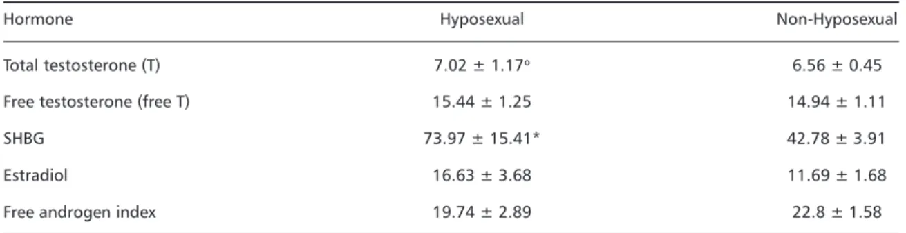
Table 1. Mean serum levels of androgens, estradiol, and SHBG, and the mean of free androgen index.
Hormone Hyposexual Non-Hyposexual
Total testosterone (T) 7.02 ± 1.17o 6.56 ± 0.45
Free testosterone (free T) 15.44 ± 1.25 14.94 ± 1.11
SHBG 73.97 ± 15.41* 42.78 ± 3.91
Estradiol 16.63 ± 3.68 11.69 ± 1.68
Free androgen index 19.74 ± 2.89 22.8 ± 1.58
in all patients in this study and consequently not considered for analysis.
Serum levels of T, FT, SHBG, and FAI (Table 1)
There was no evidence of significant differences between hyposexual and non-hyposexual patients in relation to serum levels of T, free-T, or estradiol. Similarly, we did not find evidences of significant differences in the FAI between hyposexual and non-hyposexual patients. However, the serum levels of SHBH were significantly (p<0.05) higher between hyposexual than non-hyposexual patients.
Second interview about sexual performance in patients with epilepsy after two-years follow-up period
Thirty-six (92.3%) patients could be re-evaluated at this time. Among the remaining three patients, two did not return to the routine visits and one de-veloped systemic arterial hypertension and was ex-cluded. Among those patients previously considered hyposexual, nine were re-evaluated. Four (44.4%) had improvement in their sexual performance and were considered non-hyposexual, whereas five (55.6%) continued hyposexual. Among those patients previously considered non-hyposexual, 27 (96.4%) were re-evaluated and one (3.7%) developed hyposexuality. Thus, during the second interview, six (16.7%) patients were considered hyposexual.
All four patients that improved sexual performance during the follow-up period were taking AEDs and had concomitant improvement in seizure frequency as follows: two had no seizures and two had one seizure during the year that preceded the second interview. Among those patients that continued or developed hyposexuality, three had very frequent seizures that were refractory to AEDs treatment and three had occasional seizures. Two of these hyposexual patients with low seizure frequency had sequelae of head trauma in the left frontal and right temporal lobes, respectively.
The analysis of hyposexuality among the first and second interviews about sexual performance was done using table of frequency of conditions before and after. It was used Pearsons Q-square for test of independence. The result (p = 0.000; x2 = 13.067) shows evidence of significant differences among the first and second evaluations. Although limited by the small sample of hyposexual patients, the results ob-tained in this study may indicate the benefits of clini-cal treatment and seizure control on sexual perfor-mance in patients with epilepsy.
DISCUSSION
Hyposexuality was more frequently observed among patients with epilepsy than in healthy indi-viduals. We observed hyposexuality in almost 30% of men with epilepsy. The majority of hyposexual patients showed global diminishment of libido and potency. We compared sexual activity before and after two years of follow-up in more than 90% of our patients. The analysis of hyposexuality between the first and second interviews about sexual performance showed evidence of improvement in sexual performance between the first and second evaluations.
Although we did not find any differences between seizure frequency and impairment or improvement of sexual function, our results suggest that some patients with good outcome regarding seizure con-trol improve sexual activity. In this study, 44% of patients that were considered hyposexual improved sexual performance during the two-year follow-up period. In contrast, five patients that persisted hyposexual or developed hyposexuality during the two-year follow-up had either seizures refractory to treatment with AEDs (n=3) or sequelae of temporal (n=1) or frontal (n=1) head trauma. In the former patients, the cause of sexual dysfunction is prob-ably the structural brain lesion in the temporal and frontal lobes, respectively. Previous studies demon-strated that patients with refractory epilepsy im-proved sexual performance after temporal lobec-tomy2,3,5,24. Based on the fact that both clinical and surgical treatment for epilepsy that result in better seizure control may influence sexual performance, it is possible that the disease itself contributes to hyposexuality.
sexual activity while human temporal lobe epilepsy usually inhibits sexual function. However, the mecha-nisms underlying the association between limbic epilepsy and sexual dysfunction remain unclear.
While serum levels of T and free-T were not sig-nificantly different among hyposexual and non-hyposexual patients, serum levels of SHBG were in-creased in the hyposexual group. Previous stud-ies12,15,17,18,28-30 showed high serum levels of SHBG in patients treated with AEDs. Increased SHBG would expect to produce sexual dysfunction by decreasing serum levels of free-T and/or albumin-bound test-osterone, which was indirectly estimated by FAI in this study. However, the effects of increased serum levels of SHBG upon free-T and FAI were not signifi-cantly different between groups. Although FAI has been used in several studies, it is controversial whether FAI is an appropriate measurement for males31,32. Based on the facts that increased SHBG did not elicit marked differences between hyposexual and non-hyposexual patients in relation to the se-rum levels of free-T measured by RIA and that FAI may not be reliable for male individuals31,32, further study with more accurate measurements of the bioavailable androgens are necessary to elucidate whether increased SHBG contribute to the sexual dysfunction frequently observed in patients with epilepsy.
The serum levels of estradiol did not differ be-tween hyposexual and non-hyposexual patients in this study. The results about serum levels of estra-diol in patients with epilepsy and sexual dysfunc-tion are controversial. While Murialdo et al.33 found increased serum levels of estradiol, Duncan et al.34 did not find altered levels of estradiol in patients with sexual dysfunction and epilepsy.
Our goal was not to report the complete evalua-tion for sexual dysfuncevalua-tion in patients with epilepsy. However, all patients in this study were submitted to psychological and urological evaluations. Previ-ously, Souza et al.35 reported a significant associa-tion between sexual dysfuncassocia-tion, anxiety, and de-pression in male and female patients with epilepsy. Depressive disorders, which are frequently observed in patients with epilepsy36, may cause sexual dys-function. In addition, previous study37 found vascu-lar impotence in two of 11 male patients with epi-lepsy and sexual dysfunction. Thus, it is important to empathize that the evaluation of patients with epilepsy and sexual dysfunction requires a multidis-ciplinary approach.
CONCLUSION
The results obtained in this study may indicate the benefits of clinical treatment and seizure control on sexual performance in some patients with epilepsy. The serum levels of SHBG were higher in hyposexual than in non-hyposexual patients with epilepsy. However, the effects of increased SHBG on bioavailable androgens, including free-T and albumin-bound T were not detected by the methods used in this study.
Acknowledgments - Authors thank Marcos A. Tambascia, MD, PhD, Gilberto DAssuncao Fernandes, MD, PhD, Antonio R. Amaretto, PhD, and Marilda E.N. Lipp, PhD for their valuable suggestions. We also thank Laurione C. Oliveira for technical support.
REFERENCES
1. Gastaut H and Collomb H. Etude du comportment sexuel chez les epileptiques psychomoteurs. Ann Med Psychiat 1954;II(5):657-696. 2. Hierons R and Saunders M. Impotence in patients with temporal lobe
lesions. Lancet 1966;2:761-763.
3. Taylor DC. Sexual behavior and temporal lobe epilepsy. Arch Neurol 1969;21:510-516.
4. Shukla GD, Srivastava ON, Katiyar BC. Sexual disturbances in tempo-ral lobe epilepsy. Br J Psychiatry 1979;134:288-292.
5. Pritchard PB. Hyposexuality: a complication of complex partial epi-lepsy. Trans Am Neurol Assoc 1980;105:193-195.
6. Morrell MJ. Sexual dysfunction and epilepsy. Epilepsia 1991;32(Suppl. 6):S38-S45.
7. Davidson JM, Camargo CA, Smith ER. Effects of androgen on sexual behav-ior in hypogonadal men. J Clin Endocrinol Metab 1979;48:955-958. 8. Davidson JM, Kwan M, Greeleaf WJ. Hormonal replacement and
sexu-ality in men. Clin Endocrinol Metab 1982;11:599-623.
9. Pirke KM, Kockott G. Endocrinology of sexual dysfunction. Clin Endocrinol Metab 1982;11:625-637.
10. Sodergard R, Backstrom T, Shanbhag V, Carstensen H. Calculation of free and bound fractions of testosterone and estradiol-17 beta to human plasma proteins at body temperature. J Steroid Biochem 1982;16:801-810. 11. Manni A, Pardridge WN, Cefalu W, et. al. Bioavailability of
albumin-bound testosterone. J Clin Endocrinol Metab 1985;61:705-710. 12. Barragry JM, Makin HL, Trafford DJH, Scott DF. Effects of
anticonvul-sants on plasma testosterone and sex hormone binding globulin lev-els. J Neurol Neurosurg Psychiatry 1978;41:913-914.
13. Dana-Haeri J, Oxley J, Richens A. Reduction of free testosterone by antiepileptic drugs. Br Med J 1982;284:85-86.
14. Toone BK, Wheeler M, Nanjee M, Fenwick P, Grant R. Sex hormones, sexual activity, and plasma anticonvulsant levels in male epileptics. J Neurol Neurosurg Psychiatry 1983;46:824-826.
15. Connell JM, Rappeport WG, Beastall GH, Brodie MJ. Changes in circu-lating androgens during short term carbamazepine therapy. Br J Clin Pharmacol 1984;17:347-351.
16. Toone BK, Edeh J, Nanjee MN, Wheeler M. Hyposexuality and epi-lepsy: a community survey of hormonal and behavioral changes in male epileptics. Psychol Med 1989;19:937-943.
17. Isojarvi JI, Pakarinen AJ, Ylipalosaari PJ, Myllyla VV. Serum hormones in male epileptic patients receiving anticonvulsant medication. Arch Neurol 1990;47:670-676.
18. Isojarvi JI, Repo M, Pakarinen AJ, Lukkarinen O, Myllyla VV. Carbamazepine, phenytoin, sex hormones, and sexual function in men with epilepsy. Epilepsia 1995;36:366-370.
19. American Psychiatric Association, Committee on Nomenclature and Statistics. Williams JBW (ed). Diagnostic and statistical manual of
men-tal disorders. Revised 3. Ed. Washington DC: American Psychiatric Association, 1987:307-313.
20. Thorne FC. Scales for rating sexual experience. J Clin Psychol 1966;22:404-407.
22. Grizzle LE, Starmer CF, Koch GG. Analysis of categorical data by lin-ear models. Biometrics 1969;25:489-504.
23. Quagliatto EMAB. Forma epiléptica da neurocisticercose encefálica. Thesis. UNICAMP. Campinas, 1987.
24. Cogen PH, Antunes JL, Correll JW. Reproductive function in temporal lobe epilepsy: the effect of temporal lobectomy. Surg Neurol 1979;12:243-246.
25. Feeney DM, Gullotta FP, Gilmore W. Hyposexuality produced by tem-poral lobe epilepsy in the cat. Epilepsia 1998;39:140-149.
26. Kluver H, Bucy PC. Preliminary analysis of the functions of temporal lobes in monkeys. Arch Neurol Psychiatry 1939;42:979-1000. 27. Anson JA, Kuhlman DT. Post-ictal Kluver-Bucy syndrome after
tem-poral lobectomy.J Neurol Neurosurg Psychiatry 1993;56:311-313.
28. Victor A, Lundberg PO, Johansson ED. Induction of sex hormone bind-ing globulin by phenytoin. Br Med J 1977;2:934-935.
29. Toone BK, Wheeler M, Fenwick P. Sex hormone changes in male epi-leptics. Clin Endocrinol 1980;12:391-395.
30. MacPhee GJ, Larkin JG, Butler E, Beastall GH, Brodie MJ. Circulating hormones and pituitary responsiveness in young epileptic men receiv-ing long-term antiepileptic medication. Epilepsia 1988;29:468-475.
31. Kapoor P, Luttrell BM, Williams D. The free androgen index is not valid for adult males. J Steroid Biochem Mol Biol 1993;45:325-326. 32. Vermeulen A, Verdonck L, Kaufman JM. A critical evaluation of simple
methods for the estimation of free testosterone in serum. J Clin Endocrinol Metab 1999; 84:3666-3672.
33. Murialdo G, Galimberti CA, Fonzi S, et al. Sex hormones and pituitary function in male epileptic patients with altered or normal sexuality. Epilepsia 1995;36:360-365.
34. Duncan S, Blacklaw J, Beastall GH, Brodie MJ. Antiepileptic drug therapy and sexual function in men with epilepsy. Epilepsia 1999; 40:197-204.
35. Souza EAP, Keiralla, DMB, Silveira DC, Guerreiro, CAM. Sexual dys-function in epilepsy: identifying psychological variables. Arq Neuropsiquiatr 2000;58:214-220.
36. Kanner AM, Rivas Nieto JC. Depressive disorders in epilepsy. Neurol-ogy 1999;53(Suppl 2):S26-S32.