Lateral cephalometric radiograph
versus
lateral nasopharyngeal radiograph for quantitative
evaluation of nasopharyngeal airway space
Suelen Cristina da Costa Pereira1, Rejane Targino Soares Beltrão2, Guilherme Janson3, Daniela Gamba Garib3
Objective: This study compared lateral radiographs of the nasopharynx (LN) and lateral cephalometric radiographs (LC) used to assess nasopharyngeal airway space in children. Material and Methods: One examiner measured the nasopha-ryngeal space of 15 oral breathing patients aged between 5 and 11 years old by using LN and LC. Both assessments were made twice with a 15-day interval in between. Intergroup comparison was performed with t-tests (P < 0.05). Results:
Comparison between LN and LC measurements showed no significant differences. Conclusion: Lateral cephalometric radiograph is an acceptable method used to assess nasopharyngeal airway space.
Keywords:Radiology. Nasopharynx. Orthodontics.
How to cite this article: Pereira SCC, Beltrão RTS, Janson G, Garib DG. Lat-eral cephalometric radiograph versus lateral nasopharyngeal radiograph for quantita-tive evaluation of nasopharyngeal airway space. Dental Press J Orthod. 2014 July-Aug;19(4):89-93. DOI: http://dx.doi.org/10.1590/2176-9451.19.4.089-093.oar
Submitted: June 09, 2012 - Revised and accepted: January 09, 2013
» Patients displayed in this article previously approved the use of their facial and intraoral photographs.
Contact address: Suelen Cristina da Costa Pereira E-mail: [email protected]
» The authors report no commercial, proprietary or financial interest in the prod-ucts or companies described in this article.
1 PhD Orthodontic Resident, School of Dentistry — University of São Paulo/
Bauru.
2 PhD in Orthodontics, School of Dentistry — University of São Paulo/Bauru.
3 Full professor, Department of Orthodontics, School of Dentistry — University
of São Paulo/Bauru.
DOI: http://dx.doi.org/10.1590/2176-9451.19.4.089-093.oar
Objetivo: comparar a radiografia da nasofaringe (RN) e a radiografia cefalométrica lateral (RCL) para avaliação do espaço aéreo nasofaríngeo em crianças. Métodos: um examinador mediu o espaço da nasofaringe de 15 pacientes respi-radores bucais, com idade entre 5 e 11 anos, utilizando a RN e a RCL. Ambas as avaliações foram realizadas duas vezes, com um intervalo de 15 dias. A comparação intergrupos foi realizada por meio do teste t (p < 0,05). Resultados: a com-paração entre as medições na RN e na RCL mostrou não haver diferença significante. Conclusão: Concluiu-se que a radiografia cefalométrica lateral é um método aceitável para avaliar o espaço aéreo da nasofaringe.
INTRODUCTION
Two modalities of conventional and extraoral radio-graphs are used to assess nasopharyngeal airway space: lateral nasopharyngeal radiograph (LN), also known as cavum radiography, and lateral cephalometric radio-graph (LC). The former is requested most frequently by physicians to assess the nasopharynx of patients with nasal obstruction, whereas the latter has been used for several years in Orthodontics to assess the morphology and development of dental occlusion, including sot and skeletal tissues of the face.1,2 Moreover, several authors show that lateral cephalometric radiograph allows one to assess adenoid and dimension of nasopharynx.1-11
With the aim of establishing a baseline for measur-ing nasopharyngeal space on lateral radiographs, Mc-Namara Jr7 deined it as the shortest distance between the convex surface of the adenoid (or posterior wall of nasopharynx) and the dorsal surface of the sot palate. Patients with nasopharynx width less or equal to 5 mm reveal apparent airway obstruction. It is used only as an indicator of possible airway impairment. A more accu-rate diagnosis can be made only by an otorhinolaryn-gologist during clinical examination.
According to Kohler12 and Almeida et al,3 lateral cephalometric radiograph and lateral nasopharyngeal radiograph can be used by orthodontists and otorhino-laryngologists as integrated medical-dental examina-tions. Moreover, they can be obtained during the same procedure, which eliminates the need for additional radiographic exposure.
Ikino et al2 assessed the degree of nasopharynx ob-struction by means of applying Cohen and Konak21 score to both lateral cephalometric radiograph and lateral naso-pharyngeal radiograph. His results revealed that similar outcomes were produced by both radiographs in 73.1% of children. The author stated that lateral cephalometric radiograph yields better results in comparison to lateral nasopharyngeal radiograph, since patient’s head position-ing is standardized in the former. Head position is ixed in LC, which avoids variation in the sagittal and transverse planes and allows a more secure airway analysis without the artifacts produced by head rotation. This information is important since children hardly remain in the desired position. Based on these indings, the authors concluded that LC is the radiograph of choice for assessing nasal obstruction due to equally showing nasopharynx airway and minimizing changes in head positioning.
According to Almeida et al,3 computed tomogra-phy (CT) is also used in diagnosis of nasopharyngeal obstruction; however, despite being more accurate, it is also more expensive. Montgomery et al13 evaluated the results obtained by tomography and concluded that ra-diographic examination is poor in information. The au-thors suggest that CT should be used as the gold stan-dard. Conversely, cephalometric radiography should be used to determine whether a more detailed tracking is necessary or not, bearing in mind that this is a two-dimensional and, therefore, limited examination.
No previous study compared lateral nasopharyngeal radiograph with lateral cephalometric radiograph used for quantitative evaluation of nasopharynx. For this rea-son, the objective of this study was to compare lateral cephalometric radiograph and lateral nasopharyngeal ra-diograph for a quantitative evaluation of nasopharyngeal airway space.
MATERIAL AND METHODS
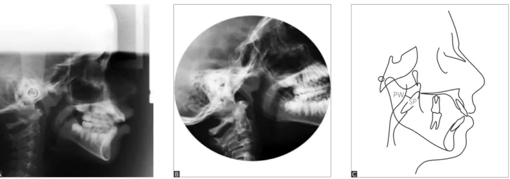
This research was approved by the Federal Uni-versity of Paraíba (UFPB) Institutional Review Board under protocol 574/06. All research subjects signed an informed consent form. This study assessed the orth-odontic records of patients from the School of Dentistry of the University of São Paulo. In selecting the sample, the following inclusion criteria were applied: patients aged between 5 and 11 years old; recent lateral cepha-lometric radiograph of good quality (Fig 1A); signs of mouth breathing including open mouth posture, short upper lip and everted lower lip; large, varying degrees of narrow face; small nostrils, and poorly developed, deep, narrow palate which demonstrated the need for otorhinolaryngologist analysis.
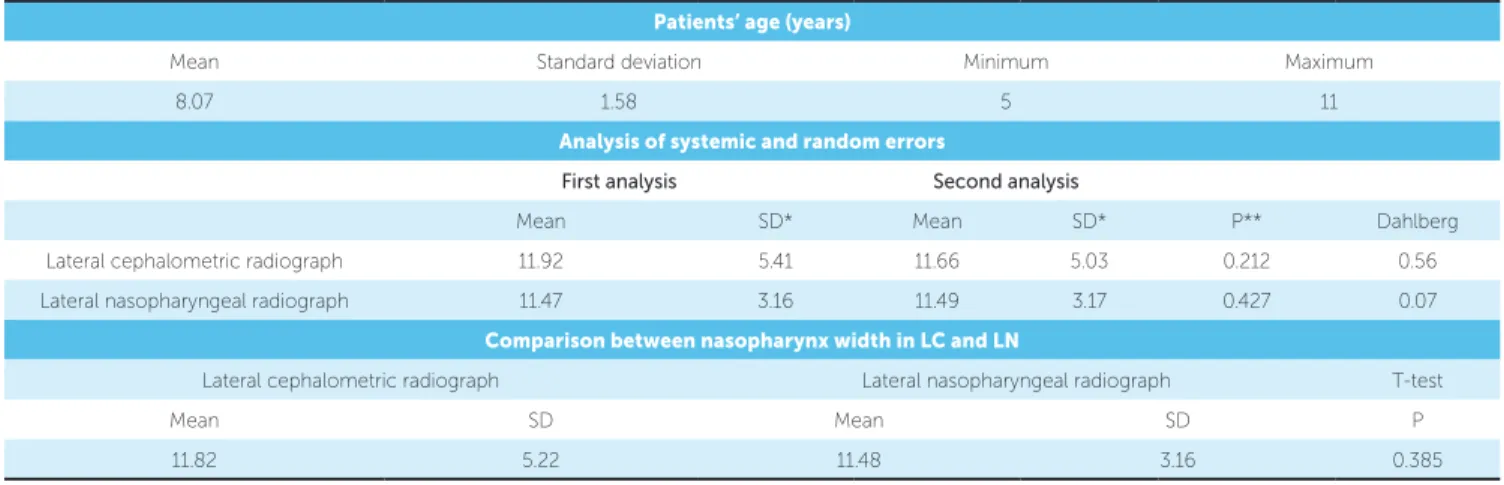
The inal sample comprised 15 patients who were referred to an otorhinolaryngologist for examination of the nasopharynx. A lateral nasopharyngeal radiograph (Fig 1B) was requested for all patients as a supplemen-tary diagnostic tool. The interval between LC and LN was less than three months. Seven patients (46.7%) were males, whereas 8 (53.3%) were females. Patients had a mean age of 8.07 ± 1.58 years (varying from 5 to 11 years) as shown in Table.
Figure 1 - A) Nasopharyngeal space measurement expressed in millimeters from the point of the anterior half of the contour of the soft palate (SP) to the nearest point of the pharyngeal wall (PW). B) Lateral nasopharyngeal radiograph (LN). C) Lateral cephalometric radiograph (LC).
of the contour of the sot palate to the nearest point of the nasopharyngeal posterior wall (Fig 1C).
Rotograph Plus (Villa Sistemi Medicali, Buccinasco, Italy) was used for lateral cephalometric radiograph, while Siemens (AG, Munich, Germany) was used for lateral nasopharyngeal radiograph under 10% and 20% image magniication, respectively. Magniication fac-tors were corrected before comparison.
LC was obtained with the patient positioned in a cephalostat with the Frankfurt Horizontal Plane (FHP) parallel to the ground, lips at rest and in cen-tric occlusion. Focus-sagittal midplane distance was 1.52 m and the exposure parameters were 85 KVp, 10 mA and 0.5 to 1 second of exposure time, depending on patient’s age.
LN was performed with the child standing in pro-ile with the head horizontally oriented and the mouth closed during inspiration. Focus-sagittal midplane dis-tance was 1.42 m and the exposure parameters were 64 kV and 3.5 mA.
Data were processed in a statistical program (SPSS 11.0) for descriptive and inferential analyses. To calcu-late error of the method, all radiographs were remea-sured ater a 15-day interval. The formula proposed by Dahlberg14 (Se2 = Σd2/2n) was used to estimate the order of magnitude of casual errors, while systematic errors were analyzed by paired t-tests, as advocated by Hous-ton.15 Independent t-tests were used for intergroup comparison of nasopharynx width in LC and LN (sig-niicance level was set at 5%).
RESULTS
Casual errors were 0.56 and 0.07 for LN and LC, respectively. No statistically signiicant systematic errors were observed (Table 1).
There were no signiicant diferences between naso-pharynx measurements in LN and LC (Table 1).
DISCUSSION
Intragroup analyses showed no signiicant errors when the irst and second measurements for lateral na-sopharyngeal radiograph and lateral cephalometric ra-diograph were compared, thus demonstrating good pre-cision and reproducibility of measurements.
Previous studies reported that nasopharynx evaluation is important to diagnose adenoid size and permeability of airway space.16,17,18 This study aimed at comparing naso-pharynx width in LC and LN. Correction of image mag-niication of both types of radiograph allowed comparison of nasopharynx measurements. No signiicant diferences were observed between LC and LN (Table 1), corrobo-rating the study by Ikino et al.2 These authors conducted a qualitative analysis of nasopharynx and found an agree-ment in the degree of airway space obstruction for both types of radiographs, an important factor to consider when dealing with patients who will undergo orthodontic treat-ment and are likely to have lateral cephalometric radio-graph requested for diagnosis, regardless of their respira-tory condition. For these patients, evaluation of nasophar-ynx by means of LC can avoid unnecessary inancial and biological costs of taking an extra radiographic exam.1,19,20
A B C
Table 1 - Patients’ age, analysis of systemic and random errors and comparison between nasopharynx width in LC and LN.
SD*= Standard deviation P** = 0.05.
The technique used to obtain LC yields better re-sults in comparison to LN, since patient’s head posi-tioning is standardized in the former. For this reason, it avoids variations in the sagittal, frontal and transverse planes. Rotation of the head may produce undesired ef-fects, especially in children who do not always remain in a desired position.2 In addition, LC has the advantage of having a ixed distance between focal point and ilm.21
Major et al22 assessed the capability of lateral cephalo-metric radiographs to diagnose hypertrophied adenoids and obstructed nasopharyngeal airway. They conducted a systematic literature review and concluded that LC performed reasonably well in evaluating adenoid size. Quantitative measures of adenoid area and subjective grading of adenoid size on LCs had reasonable correla-tions with actual adenoid size.
Barbosa et al1 compared the use of LC and endosco-py of nasopharynx to evaluate nasopharynx obstruction.
Patients’ age (years)
Mean Standard deviation Minimum Maximum
8.07 1.58 5 11
Analysis of systemic and random errors
First analysis Second analysis
Mean SD* Mean SD* P** Dahlberg
Lateral cephalometric radiograph 11.92 5.41 11.66 5.03 0.212 0.56
Lateral nasopharyngeal radiograph 11.47 3.16 11.49 3.17 0.427 0.07
Comparison between nasopharynx width in LC and LN
Lateral cephalometric radiograph Lateral nasopharyngeal radiograph T-test
Mean SD Mean SD P
11.82 5.22 11.48 3.16 0.385
They concluded that LC allows visualization of sot and hard tissue structures , in addition to assessing location, coniguration and growth of nasopharynx and adenoid tissue. Moreover, it allows structures closely related to oral cavity and nasopharynx to be visualized. Although this type of radiograph has greater limitations in com-parison to two-dimensional interpretation of nasophar-ynx, it has proved to be efective as a diagnostic tool. This fact was evidenced by the strong correlation be-tween LC results and nasal endoscopy.
CONCLUSION
1. Barbosa MC, Knop LAH, Lessa MM, Araujo TM. Avaliação da radiografia cefalométrica lateral como meio de diagnóstico da hipertrofia da adenoide. Rev Dental Press Ortod Ortop Facial. 2009;14(4):83-91.
2. Ikino CMY, D’Antonio WEPA, de la Cortina RAC, Lessa M, Castilho AM, Goto EY, et al. Tele-radiografia lateral de crânio e radiografia de cavum: estudo comparativo em crianças com obstrução nasal. Rev Bras Otorrinolaringol. 2000;66(6):592-6.
3. Almeida RCC, Artese F, Carvalho FAR, Cunha RD, Almeida MAO. Comparison between cavum and lateral cephalometric radiographs for the evaluation of the nasopharynx and adenoids by otorhinolaryngologists. Dental Press J Orthod. 2011;16(1):32-3.
4. David AF, Castilho JCM. Estudo comparativo entre os traçados manual e computadorizado da análise do espaço aéreo nasofaríngeo em radiografias cefalométricas laterais. Ortodontia. 1999;32(2):88-93.
5. Chami FAI. Avaliação nasofibroscópica e radiológica de pacientes com hiperplasia da amígdala faríngea. Rev Bras Atual Otorrinolaringol. 1998;5(4):118-24.
6. Cohen LM, Koltai PJ, Scott JR. Lateral cervical radiographs and adenoid size: do they correlate? Ear Nose Throat J. 1992;71(12):638-42.
7. McNamara JA Jr. A method of cephalometric evaluation. Am J Orthod. 1984;86(6):449-69.
8. Poole MN, Engel GA, Chaconas SJ. Nasopharyngeal cephalometrics. Oral Surg Oral Med Oral Pathol. 1980;49(3):266-71.
9. Holmberg H, Linder-Aronson S. Cephalometric radiographs as a means of evaluating the capacity of the nasal and nasopharyngeal airway. Am J Orthod. 1979;76(5):479-90.
10. Fujioka M, Young LW, Girdany BR. Radiographic evaluation of adenoidal size in children: adenoidal-nasopharyngeal ratio. AJR Am J Roentgenol. 1979;133(3):401-4.
11. Pruzansky S. Roentgencephalometric studies of tonsils and adenoids in normal and pathologic states. Ann Otol Rhinol Laryngol. 1975;84(2 pt 2 Suppl 19):55-62.
REFERENCES
12. Köhler GI. Mensuração dosimétrica e otimização imageológica de exames radiológicos na face infantil e adolescente. Departamento de Engenharia Biomédica [dissertação]. Curitiba (PR): Centro Federal de Educação Tecnológica do Paraná; 2005.
13. Montgomery WM, Vig PS, Staab EV, Matteson SR. Computed tomography: a three-dimensional study of the nasal air way. Am J Orthod. 1979;76(4):363-75. 14. Dahlberg G. Statistical methods for medical and biological students. New York:
Interscience; 1940.
15. Houston WJB. The analysis of errors in orthodontic measurements. Am J Orthod. 1983;83(5):382-90.
16. Paradise JL, Bernard BS, Colborn DK, Janosky JE. Assessment of adenoidal obstruction in children: clinical signs versus roentgenographic findings. Pediatrics 1998;101(6):979-86.
17. Weimert TA. Evaluation of the upper airway in children. Ear Nose Throat J. 1987;66:196-200.
18. Jeans WD, Fernando DC, Maw AR. How should adenoidal enlargement be measured? A radiological study based on interobserver agreement. Clin Radiol. 1981;32(3):337-40.
19. Cassano P, Gelardi M, Cassano M, Fiorella ML, Fiorella R. Adenoid tissue rhinopharyngeal obstruction grading based on fiberendoscopic findings: a novel approach to therapeutic management. Int J Pediatr Otorhinolaryngol. 2003;67(12):1303-9.
20. Kubba H, Bingham BJ. Endoscopy in the assessment of children with nasal obstruction. J Laryngol Otol. 2001;115(5):380-4.
21. Cohen D, Konak S. The evaluation of radiographs of the nasopharynx. Clin Otolaryngol Allied Sci. 1985;10(2):73-8.