Dement Neuropsychol 2016 June;10(2):165-167
165
Vieira et al. Progressive multifocal leukoencephalopathy
Neuroimaging through
clinical cases
This study was conducted at the Department of Radiology and Imaging of AC Camargo Cancer Center, São Paulo SP, Brazil.
Gislaine Cristina Lopes Machado Porto. Rua Castro Alves 612 / apto 143 – 01532-000 São Paulo SP – Brasil. E-mail: [email protected]
Disclosure: The authors report no conflicts of interest. Received March 05, 2016. Accepted in final form May 12, 2016.
Progressive multifocal leukoencephalopathy
in non-HIV patient
Diego Rosseman Vieira, Diogo Zanella, Eurípedes Barsanulfo de Paula Avelino,
Renato Batista Soares Moura, Gislaine Cristina Lopes Machado Porto, Liao Shin Yu, Rubens Chojniak
LEUCOENCEFALOPATIA MULTIFOCAL PROGRESSIVA EM PACIENTE NÃO-HIV
Key words: progressive multifocal leukoencephalopathy, non-HIV, lymphoma, JC virus,
Palavras-chave: Leucoencefalopatia multifocal progressiva, HIV negativo, linfoma, vírus JC.
W
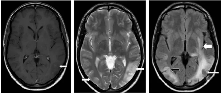
e report the case of a 42-year-old manwith an indolent non-Hodgkin lym-phoma (stage IV) diagnosed 15 months ear-lier. Since diagnosis, he had remained asymp-tomatic and in clinical follow-up, under no speciic therapy. During a follow-up visit, he reported speech and visual diiculties. Cerebrospinal luid analysis was normal and serologic tests for herpes virus, syphilis, toxo-plasmosis and HIV were all negative. MRI dis-closed multifocal asymmetric areas of hypoin-tensity on T1 (Figure 1) and hyperinhypoin-tensity on T2/FLAIR (Figure 2), afecting both temporal and parietal lobes, with no mass efect, dif-fusion restriction or contrast enhancement. Stereotactic biopsy was performed and the anatomopathological examination revealed areas of intense demyelination with presence of large oligodendrocytes Immunohistochem-istry staining on brain biopsy demonstrated JC virus DNA, characterizing progressive mul-tifocal leukoencephalopathy (PML). Despite the support therapy, the patient evolved with clinical and radiological deterioration (Figure 3).
PML is a severe demyelinating disease of the central nervous system (CNS). It is caused by the reactivation of the JC virus (genus: Polyomavirus), which infects nearly 80% of the human population prior to adulthood. he virus remains latent mainly in the kid-neys and lymphoid organs and is reactivated and spreads to the brain, almost exclusively
in the setting of advanced immunosuppres-sion. Once reactivated in the CNS, the virus infects and destroys oligodendrocytes, which are responsible for the formation and mainte-nance of myelin sheaths.1-3
PML is frequently associated with HIV/ AIDS, presenting in up to 80% of cases. How-ever, other causes of immunosuppression such as organ transplantation, chemotherapy, immunotherapies with monoclonal antibod-ies, autoimmune diseases and lymphoma, may also be associated with PML. he clinical picture of PML varies according to the pat-terns of demyelination. Patients may experi-ence changes in cognition, language, behavior and personality. Memory loss, visual deicits such as hemianopsia and cortical blindness, sensory-motor alterations, and generalized neurological decline, may also occur. he pres-ence of seizures, vertigo, headache and apha-sia is less frequent. Cerebellar symptoms may appear in the case of lesion in the posterior fossa. Rarely, there is involvement of the spi-nal cord.1,3 he employment of PCR technique
for the detection of virus DNA in cerebrospi-nal luid, associated with imaging exams, has become an alternative to anatomopathological study, which conirms the diagnosis.1
In general, demyelination occurs in a multifocal and asymmetric manner. he lesions typically start at the cortical junction between the gray and white matter, with con-centric dissemination. hey can be variable
Dement Neuropsychol 2016 June;10(2):165-167
166 Progressive multifocal leukoencephalopathy Vieira et al. Figure 1. T1-weighted image post-contrast reveals cortical-subcortical hypointensity in left temporal lobe, without enhancement (white arrow).
Figure 2. T2-weighted image reveals asymmetri-cal areas of hyperintensity, more marked in the left temporal region (white arrows).
Figure 3. FLAIR image reveals multifocal and asymmetric progression of PML, with involvement of the left temporal-occipital lobes (white arrow) and of the subcortical region of the right temporal lobe (U-fibers) (black arrow). Previous biopsy area (thin white arrow).
in size and may coalesce. here is a predilection for the parieto-occipital lobes.1,3 MRI is the most sensitive
imag-ing modality, particularly in sequences with long repeti-tion time. he lesion appears as hypersignal areas on T2 and FLAIR in the subcortical and periventricular white matter. he areas are small in the beginning, increas-ing progressively, with a tendency for conluence. It is commonly multifocal and has an asymmetric pattern of demyelination, uni- or bilaterally, without mass efect, and invariably without contrast enhancement. he subcorti-cal lesions tend to involve U-ibers. Any lobe of the brain may be afected, but there is preference for the frontal and parieto-occipital regions. here may be involvement of deep gray structures, by afection of myelin ibers that pass through basal ganglia. In about one third of cases, there is involvement of the posterior fossa and compro-mise may be limited to this area in 10% of cases.1,5,6
he diferential diagnosis of PML includes other CNS diseases that can occur in immunocompromised patients, such as toxoplasmosis and primary lymphoma of the CNS. PML can be diferentiated from these dis-eases by its slower temporal evolution, fewer systemic manifestations, absence of fever, preservation of con-science and lack of mass efect, perilesional edema or
contrast enhancement on imaging tests. Herpes virus encephalitis should be considered in the initial phase, but white matter involvement is frequently temporal, bilat-eral and asymmetrical, with greater contrast
enhance-ment than that found in PML.5,6 Another important
diferential is HIV-associated demyelination. It may be diicult to distinguish between the demyelinating lesions caused by PML or by HIV, in patients with AIDS. PML lesions are more often multifocal and asymmetric, with greater predilection for the subcortical white matter and are associated with more pronounced signal change on T1-weighted images, while in HIV-associated demy-elination, lesions are often isointense on T1, and may not be visible. HIV encephalitis lesions are commonly periventricular, difuse, symmetric, and tend to spare subcortical U-ibers. Clinically, HIV-associated demyelin-ation presents as global cognitive disorder and dementia, while in PML motor, sensory or cognitive focal deicits predominate.1,5,6
Dement Neuropsychol 2016 June;10(2):165-167
167
Vieira et al. Progressive multifocal leukoencephalopathy
REFERENCES
1. Tan CS, Koralnik IJ. Progressive multifocal leukoencephalopathy and other disorders caused by JC virus: clinical features and pathogenesis. Lancet Neurol 2010;9:425.
2. Huang D, Cossoy M, Li M, et al. Inflammatory progressive multifocal leukoencephalopathy in human immunodeficiency virus-negative patients. Ann Neurol 2007;62:34.
3. Amend KL, Turnbull B, Foskett N, et al. Incidence of progressive multi-focal leukoencephalopathy in patients without HIV. Neurology 2010;75: 1326.
4. Carson KR, Evens AM, Richey EA, et al. Progressive multifocal
leukoen-cephalopathy after rituximab therapy in HIV-negative patients: a report of 57 cases from the Research on Adverse Drug Events and Reports project. Blood 2009;113:4834.
5. Sahraian MA, Radue EW, Eshaghi A, et al. Progressive multifocal leuko-encephalopathy: a review of the neuroimaging features and differential diagnosis. Eur J Neurol 2012;19:1060.