Cardioprotective effects of exercise precoditioning in an experimental model of left ventricular dysfunction secondary to pulmonary arterial hypertension
84
0
0
Texto
(2) SCHMIDT, C. (2013). Cardioprotective effects of exercise preconditioning in an experimental model of left ventricular dysfunction secondary to pulmonary arterial hypertension. Dissertação de Mestrado em Atividade Física e Saúde. Faculdade de Desporto da Universidade do Porto, Porto.. Key words: Exercise preconditioning; Cardioprotection; Left ventricle dysfunction; Pulmonary arterial hypertension II.
(3) Funding Sources. The experimental studies included in this thesis were supported by a grant from the. Portuguese. Foundation. for. Science. and. Technology. (SFRH/BD/33123/2007).. This dissertation was conducted in Research Center for Physical Activity, Health and Leisure (CIAFEL), which is a research unit housed in the Faculty of Sports, University of Porto, Portugal.. III.
(4) IV.
(5) To my family. V.
(6) VI.
(7) To you, for all support and love. VII.
(8) VIII.
(9) Acknowledgments. To do a master degree in another country is much more than a seek for academic improvement. It means to trade what is certain by the uncertain, face challenges at the professional and personal level, make new friends and start a new life. I started the master without knowing which topic I would study in my research project and I ventured into an unfamiliar area, the biochemistry. I entered into a new world full of challenges. However, I was lucky to be surrounded by a lot of people who helped me to grow and overcome each of the difficulties that have arisen in the way. It is to all of these people that I would like to express my gratitude, because without their help I would not have been succeeded.. First, I would like to thank my supervisor, Prof. José Duarte, for allowing me to join his team. I’am very grateful for the knowledge that you shared with me throughout this journey and all your help in performing this work. Thank you for the opportunity and for all the hours dedicated to my supervision.. To my co-supervisor, Prof. Daniel Gonçalves, thanks for the constant motivation, dedication in the preparation of each step of this work, the patience and for the attention at any time. Your enthusiasm helped me to complete this journey.. To my friend Toni, for the friendship built over these two years, the shared hours learning in lab, the strength that you gave me in my difficult. IX.
(10) moments, and especially for excellent moments of great talking and companion. A simple friendship, which I hope to keep with me forever. ''Tche'', thanks for everything.. To my friends here in Portugal, especially Diana, Teresa, Carol, Leandro, Morgana, Renata, Wagner and Cesar, thank you for all the hours of conversations, all the good and bad times that we shared, and, of course, for all party times.. To my longtime friends, even being separated by the ocean you have always been with me. Especially Liz, Sara, Camila, Adri, Bruna Larissa, that helped me in my difficult times, every single word, email and prayer. If it was not for you girls, everything would have been much more difficult. Thank you for being my best friends, anywhere in the world. To my laboratory colleagues, Ana Carvalho, Eduardo, Helder, Gonzalo, Daniel and Dona Celeste, thanks for the company in many hours of study, by the encouragement, for sharing your knowledge and by the good times spent.. Finally I wish to thank…. … to my mother and my father. Thanks for the constant support, encouragement and for all the kind words. Your visit here in Portugal was very important as it was the chance that you gave me to charge my batteries at home. It was a crucial moment for me. I’am eternally grateful for every support you gave to me, to my choices and to your trust on me. Love you!. X.
(11) ... to my brother who always encouraged me and helped when I needed. For somehow always took care of me, even faraway. Thank you.. … to my sister and to Marcelo. Thanks for having me in your home again, for let me be part of your life, and the family that were. Especially to my sister, to be my friend, my mother and my family. For all the help that you gave to me, and mainly, for take care very well of me. Everything was much easier for having you around.. This was the biggest challenge in my life, and I just got finish with all support and incentive that everyone gave. My eternal gratitude to each one of you.. XI.
(12) XII.
(13) Index of Contents. FUNDING SOURCES. V. ACKNOWLEDGMENTS. IX. INDEX OF CONTENTS. XV. RESUMO. XVII. ABSTRACT. XXI. LIST OF ABREVIATIONS. XXIII. 1. GENERAL INTRODUCTION. 1. 1.1. Structure of the dissertation. 2. 2. SATETE OF THE ART: Acute and chronic mechanisms underlying. 7. prior exercise-induced cardioprotection: potential role in left ventricular dysfunction secondary to pulmonary arterial hypertension 3. EXPERIMENTAL PAPER: Exercise preconditioning prevents left. 37. ventricular dysfunction and maladaptive remodeling secondary to pulmonary arterial hypertension in rats 4. MAIN CONCLUSION. 63. XIII.
(14) XIV.
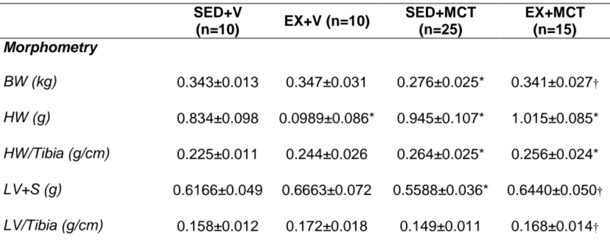
(15) Resumo O exercício físico regular exerce um importante papel na saúde humana global, protegendo, atrasando ou melhorando contra diversas doenças como a obesidade, a diabetes e as doenças cardiovasculares. Sabe-se também que o exercício físico proporciona um fenótipo cardioprotetor permitindo responder melhor contra vários insultos cardíacos, tais como isquemia-reperfusão, infarto do miocárdio, a cardiotoxicidade induzida pela doxorrubicina ou sobrecarga de pressão aguda. A hipertensão arterial pulmonar (HAP) afeta diretamente o ventrículo direito, mas a disfunção do ventrículo esquerdo também já foi descrita recentemente em pacientes com HAP, estando associada com atrofia do ventrículo esquerdo e/ou ativação neuro-humoral. Vendo os potenciais benefícios do exercício físico na fisiologia cardíaca encontrados em vários modelos experimentais, nós projetamos este estudo para analisar os efeitos cardioprotetores hipotéticos do exercício de pré-condicionamento no ventrículo esquerdo (VE) em um modelo animal de hipertensão arterial pulmonar induzida por monocrotalina (MCT). O estudo foi realizado com 115 ratos Wistar machos, submetidos à três intervenções, mostrado aqui em três fases. A primeira fase foi realizada com sessenta ratos, separados aleatoriamente em dois grupos experimentais: sedentário (SED, n=35; manteve-se com o movimento limitado ao espaço da gaiola durante 4 semanas) e exercício (EX n= 25; exercitaram em uma esteira rolante, 5 dias/semana, 60 minutos/dia, a 25 metros/minuto, durante 4 semanas). Depois de terminar durante quatro semanas, alguns animais receberam uma injeção subcutânea de monocrotalina (MCT, 60 mg/kg) e outros o mesmo volume de veículo (V, 1 mL/kg de soro fisiológico), originando os seguintes grupos: i) SED+MCT (n=25), ii) SED+V (n=10), iii) XV.
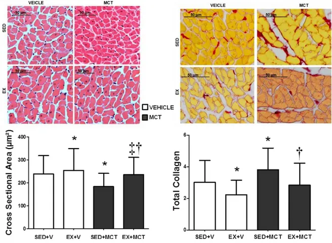
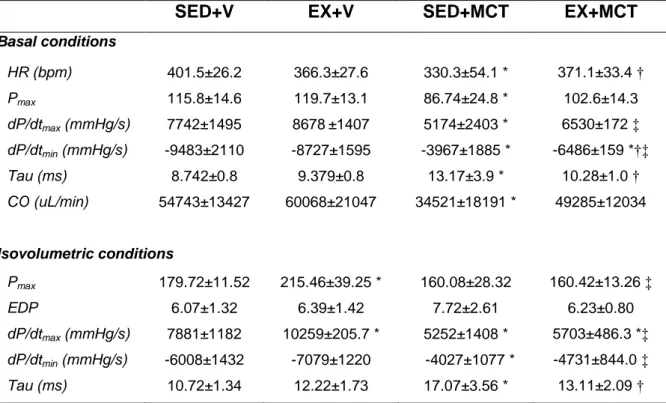
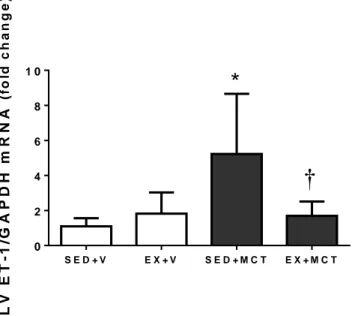
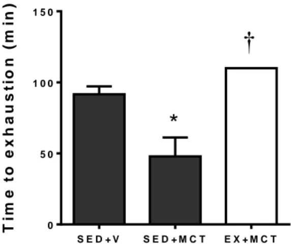
(16) EX+MCT (n=15) e iv) EX+V (n=10). Depois disso, todos os animais mantiveram-se sedentário por mais quatro semanas. Numa segunda fase, foi desenvolvido o estudo de sobrevivência com 40 animais submetidos aos respectivos protocolos experimentais (SED+V, n=5; SED+MCT, n=15; EX+V, n=5; EX+MCT, n=15). A terceira fase foi realizada com 20 animais, onde foi realizado o teste de tolerância ao esforço. No final da primeira fase, os animais foram submetidos a uma avaliação hemodinâmica do VE em condições basais e isovolumétricas, e amostras do VE foram preparadas para análise de microscopia ótica (área da secção transversal de cardiomiócitos e deposição de colagénio) e endotelina (ET-1). Descobrimos que em condições basais, a função sistólica (pressão sistólica de pico e dP/dtmax) e diastólica (dP/dtmin e Tau) foram comprometidos no grupo SED+MCT, mas não no EX+MCT (P<0.05). Sob condições isovolumétricas, SED+MCT mostraram deterioração adicional nos mesmos parâmetros, mas estas alterações foram impedidas no grupo EX+MCT (P <0.05). Esta melhora dos parâmetros hemodinâmicos foi observada juntamente com a prevenção da atrofia dos cardiomiócitos, do aumento da fibrose, e com a normalização de níveis de ET-1 de mRNA (P<0.05). O exercício de pré-condicionamento também melhorou a tolerância ao exercício, exercendo um impactado positivo nos índices de sobrevivência. É importante destacar que as melhorias foram observadas após quatro semanas da última secção de treino, destacando que o fenótipo de proteção promovida pelo exercício físico foi mantido por vários dias.. Palavras chave: Exercício físico; Cardioproteção; Monocrotaline; Atrofia dos cardiomiócitos; Endotelina-1; Taxa de sobrevivência; Tolerância ao exercício.. XVI.
(17) Abstract Regular physical exercise exerts an important role in overall Human health, protecting, delaying or improving against some diseases like obesity, diabetes mellitus and cardiovascular diseases. It is also known that exercise training provides a cardioprotective phenotype allowing an improved response against several cardiac insults such as ischemia-reperfusion, myocardial infarction, cardiotoxicity induced by doxorubicin or acute pressure overload. Pulmonary arterial hypertension (PAH) directly affects the right ventricle but the left ventricle dysfunction was recently described in PAH patients, which is associated with left ventricle (LV) atrophy and/or neurohumoral activation. Seeing that potential benefits of exercise training on cardiac physiology were found in many experimental models, we designed our study to analyze the hypothetical cardioprotective effects of exercise preconditioning on LV in a rat model of PAH induced by monocrotaline (MCT). The study was designed with 115 male Wistar rats, submitted for three interventions, showed here with three phases. The first phase was done with 60 rats were randomly separated in two experimental groups: sedentary (SED; n=35; remained with movement confined to the cage’s space during 4 weeks) and exercise (EX; n=25; exercised on a treadmill, 5 days/week, 60 minutes/day, at 25 meters/minute, during 4 weeks). After ending this 4-week period, some animals from each group received one subcutaneous injection of monocrotaline (MCT; 60 mg/kg) or an equal volume of vehicle (V; 1 mL/kg of saline), originating the following groups: i) SED+MCT (n=25), ii) SED+V (n=10), iii) EX+MCT (n=15) and iv) EX+V (n=10). Afterwards, all animals remained sedentary for additional 4 weeks. Next, animals were submitted to LV hemodynamic evaluation in baseline and isovolumetric. XVII.
(18) conditions, and LV samples were prepared for light microscopy analysis (cardiomyocyte cross sectional area and collagen deposition) and endothelin (ET-1). In a second phase, we developed the survival study with 40 animals submitted to the respective experimental protocols (SED+V, n=5; SED+MCT, n=15; EX+V, n=5; EX+MCT, n=15). The third phase was performed with 15 animals (SED+V, n=5; SED+MCT, n=5; EX+MCT, n=5) for the exercise tolerance test. We found that in baseline conditions, systolic (peak systolic pressure and dP/dtmax) and diastolic function (dP/dtmin and Tau) were compromised in SED+MCT but not in EX+MCT (P<0.05). Under isovolumetric conditions, SED+MCT showed additional deterioration in the same parameters, but these alterations were prevented in EX+MCT (P<0.05). This improved hemodynamic profile was paralleled with prevention of cardiomyocytes atrophy, fibrosis, and with normalization of ET-1 mRNA levels (P<0.05). Exercise preconditioning also enhanced exercise tolerance and positively impacted survival. Of note, these improvements were observed 4 weeks after the cessation of exercise training, highlighting that the protective phenotype promoted by exercise training is maintained for several days.. Key words: Exercise preconditioning; Cardioprotection; Left ventricular dysfunction; Pulmonary arterial hypertension. XVIII.
(19) List of Abbreviations. ADP: deoxyribonucleic acid Akt: protein kinase B Ang-II: angiotensin ATP: adenosine triphosphate CaMK: calcium calmodulin-dependent protein kinase CRP: C-reactive protein DOX: doxorubicin ET-1: endothelin-1 FFR: force-frequency relationships HSP: heat shock protein IGF-1: insulin-like growth factor IL-6: interleukin-6 I-R: ischemia-reperfusion KATP: ATP-sensitive potassium LVD: left ventricle dysfunction LTCC: L-type calcium channels LV: left ventricle MCT: monocrotaline MI: myocardial infarction mitoKATP: mitochondrial inner membrane MHC: myosin heavy chain MnSOD: manganese superoxide dismutase mTOR: mammalian target of rapamycin. XIX.
(20) NCX: sodium/calcium exchanger •NO: nitric oxide PAH: pulmonary arterial hypertension PI3K: phosphoinositide 3-kinase PLN: phospholamban ROS: reactive oxygen species RV: right ventricle RVF: right ventricular failure RyR: ryanodine receptor SERCA2a: sarco/endoplasmic reticulum Ca2+-ATPase sarcKATP: sarcolemmal membrane SOD: superoxide dismutase SR: sarcoplasmic reticulum TNF: tumor necrosis factor Thr17: p-phospholamban. XX.
(21) 1. GENERAL INTRODUCTION. The high physical activity demands imposed to humans and animals in the ancient times have favored the phylogenetic development of a phenotype towards the optimization of aerobic metabolic pathways in order to conserve energy for a potential future food deficiency (4). Moreover, in the ontogenetic point of view, it is known that large periods of high-intensity physical activity induce favorable cardiovascular adaptions to support the metabolic demands of the working skeletal muscles under high exigency conditions (9). Specifically on the heart, the beneficial effect of exercise training seen from animal models indicates an increased oxidative capacity of the myocardium (7), an enhanced cardiomyocyte survival (14), and attenuating left ventricular remodeling (19). Exercise training has been shown as an important preventive measure for cardiovascular diseases, and the benefits are well reported (2, 21). Several human epidemiological studies indicate a significant association of moderate and vigorous exercise training with a reduced incidence of cardiovascular events in healthy individuals (11, 18). Also, animal studies provide evidence of a cardioprotective phenotype induced by exercise training that allows an improved response against several insults such as ischemia-reperfusion (6, 22), myocardial infarction (5, 8), cardiotoxicity induced by doxorubicin (1, 15) or acute pressure overload (20) . Pulmonary arterial hypertension (PAH) is characterized by progressive pulmonary vascular remodeling, imposing an increased overload to the right ventricle (RV). Although initially adapting by developing, among others, RV hypertrophy, it rapidly progresses to ventricular failure and premature death (3, 1.
(22) 10, 13). PAH selectively overloads the RV, but it is known that left ventricle dysfunction (LVD) may also be present in some forms of PAH, leading to a decrease in LV preload, and low cardiac output states (12). Contrarily to RV, the left ventricle (LV) has received less attention in the context of this disease. Mechanisms underlying LVD remain poorly understood but LV atrophy (12) and/or neurohumoral activation (17) have been reported to play a role. Indeed, the chronic ET-1 overactivity in heart disease has been associated with slower relaxation and impaired contractility, favoring the accumulation of fibrosis (16). The benefits of exercise training to improve overall health and protect, delay or improve cardiovascular diseases are well understood, but, the exact mechanisms behind the cardioprotective effects of exercise training have not yet been fully explained, as well as the impact of this on LVD, secondary HAP. Therefore, the objective of this study was to analyze, in a rat model of PAH induced by monocrotaline (MCT), the hypothetical cardioprotective effects of a previous exercise training on LV, trying to identify the underlying mechanisms involved.. 1.1 STRUCTURE OF THE DISSERTATION. This dissertation is presented according the Scandinavian model, being divided in four sections: Section 1: This chapter constitutes the general introduction to the topic, highlighting the relevance of the study and its objectives. Section 2: This chapter, entitled “Acute and chronic mechanisms underlying prior exercise-induced cardioprotection: potential role in left ventricular 2.
(23) dysfunction secondary to pulmonary arterial hypertension” review the mechanisms involved in cardioprotection induced by the exercise training as well as the heart repercussions of the arterial pulmonary hypertension. Section 3: This chapter is an experimental manuscript entitled “Exercise preconditioning. prevents. left. ventricular. dysfunction. and. maladaptive. remodeling secondary to pulmonary arterial hypertension in rats”. It constitutes the experimental part of the dissertation, presenting the material and methods used, the obtained results and their discussion. Section 4: The main conclusions of the dissertation are presented in this chapter. The bibliography references supporting concepts, theories, and/or methods are presented at the end of each chapter.. 3.
(24) REFERENCES. 1. Ascensao A, Magalhaes J, Soares JM, Ferreira R, Neuparth MJ, Marques F, Oliveira PJ, and Duarte JA. Moderate endurance training prevents doxorubicin-induced in vivo mitochondriopathy and reduces the development of cardiac apoptosis. American Journal of Physiology Heart and Circulatory Physiology 289: H722-731, 2005. 2. Berlin JA, and Colditz GA. A meta-analysis of physical activity in the prevention of coronary heart disease. American Journal of Epidemiology 132: 612-628, 1990. 3. Bogaard HJ, Abe K, Noordegraaf AV, and Voelkel NF. The Right Ventricle Under Pressure Cellular and Molecular Mechanisms of Right-Heart Failure in Pulmonary Hypertension. Chest 135: 794-804, 2009. 4. Booth FW, Laye MJ, Lees SJ, Rector RS, and Thyfault JP. Reduced physical activity and risk of chronic disease: the biology behind the consequences. European Journal of Applied Physiology 102: 381-390, 2008. 5. de Waard MC, and Duncker DJ. Prior exercise improves survival, infarct healing, and left ventricular function after myocardial infarction. Journal of Applied Physiology 107: 928936, 2009. 6. Demirel HA, Powers SK, Zergeroglu MA, Shanely RA, Hamilton K, Coombes J, and Naito H. Short-term exercise improves myocardial tolerance to in vivo ischemia-reperfusion in the rat. Journal of Applied Physiology 91: 2205-2212, 2001. 7. Eisele JC, Schaefer IM, Nyengaard JR, Post H, Liebetanz D, Brul A, and Muhlfeld C. Effect of voluntary exercise on number and volume of cardiomyocytes and their mitochondria in the mouse left ventricle. Basic Research Cardioly 103: 12-21, 2008. 8. Freimann S, Scheinowitz M, Yekutieli D, Feinberg MS, Eldar M, and Kessler-Icekson G. Prior exercise training improves the outcome of acute myocardial infarction in the rat - Heart structure, function, and gene expression. Journal of the American College of Cardiology 45: 931-938, 2005. 9. Gielen S, Schuler G, and Adams V. Cardiovascular effects of exercise training: molecular mechanisms. Circulation 122: 1221-1238, 2010. 10. Gomez-Arroyo JG, Farkas L, Alhussaini AA, Farkas D, Kraskauskas D, Voelkel NF, and Bogaard HJ. The monocrotaline model of pulmonary hypertension in perspective. American Journal of Physiology Lung Cellular and Molecular Physiology 302: L363-369, 2012. 11. Hakim AA, Petrovitch H, Burchfiel CM, Ross GW, Rodriguez BL, White LR, Yano K, Curb JD, and Abbott RD. Effects of walking on mortality among nonsmoking retired men. New England Journal of Medicine 338: 94-99, 1998. 12. Hardziyenka M, Campian ME, Reesink HJ, Surie S, Bouma BJ, Groenink M, Klemens CA, Beekman L, Remme CA, and Bresser P. Right ventricular failure following chronic pressure overload is associated with reduction in left ventricular mass: evidence for atrophic remodeling. Journal of the American College of Cardiology 57: 921-928, 2011. 13. Humbert M, Sitbon O, and Simonneau G. Treatment of pulmonary arterial hypertension. New England Journal of Medicine 351: 1425-1436, 2004. 14. Kavazis AN, McClung JM, Hood DA, and Powers SK. Exercise induces a cardiac mitochondrial phenotype that resists apoptotic stimuli. American Journal of Physiology-Heart and Circulatory Physiology 294: H928-H935, 2008. 15. Kavazis AN, Smuder AJ, Min K, Tümer N, and Powers SK. Short-term exercise training protects against doxorubicin-induced cardiac mitochondrial damage independent of HSP72. American Journal of Physiology-Heart and Circulatory Physiology 299: H1515-H1524, 2010. 16. Leask A. TGFbeta, cardiac fibroblasts, and the fibrotic response. Cardiovascular research 74: 207-212, 2007. 4.
(25) 17. Lourenco AP, Roncon-Albuquerque R, Bras-Silva C, Faria B, Wieland J, HenriquesCoelho T, Correia-Pinto J, and Leite-Moreira AF. Myocardial dysfunction and neurohumoral activation without remodeling in left ventricle of monocrotaline-induced pulmonary hypertensive rats. American Journal of Physiology-Heart and Circulatory Physiology 291: H1587-H1594, 2006. 18. Manson JE, Greenland P, LaCroix AZ, Stefanick ML, Mouton CP, Oberman A, Perri MG, Sheps DS, Pettinger MB, and Siscovick DS. Walking compared with vigorous exercise for the prevention of cardiovascular events in women. New England Journal of Medicine 347: 716725, 2002. 19. McMullen JR, Amirahmadi F, Woodcock EA, Schinke-Braun M, Bouwman RD, Hewitt KA, Mollica JP, Zhang L, Zhang Y, Shioi T, Buerger A, Izumo S, Jay PY, and Jennings GL. Protective effects of exercise and phosphoinositide 3-kinase(p110alpha) signaling in dilated and hypertrophic cardiomyopathy. Proceedings of the National Academy of Sciences of the United States of America 104: 612-617, 2007. 20. Moreira-Goncalves D, Henriques-Coelho T, Fonseca H, Ferreira RM, Amado F, LeiteMoreira A, and Duarte JA. Moderate exercise training provides left ventricular tolerance to acute pressure overload. American Journal of Physiology Heart and Circulatory Physiology 300: H1044-1052, 2011. 21. Powell KE, Thompson PD, Caspersen CJ, and Kendrick JS. Physical-Activity and the Incidence of Coronary Heart-Disease. Annual Review of Public Health 8: 253-287, 1987. 22. Taylor RP, Harris MB, and Starnes JW. Acute exercise can improve cardioprotection without increasing heat shock protein content. American Journal of Physiology-Heart and Circulatory Physiology 276: H1098-H1102, 1999.. 5.
(26) 6.
(27) State of The Art. 7.
(28) Acute and chronic mechanisms underlying prior exercise-induced cardioprotection: potential role in left ventricular dysfunction secondary to pulmonary arterial hypertension. ABSTRACT. Growing body of evidence supports the notion that exercise training is capable to provide a protective phenotype. Epidemiological studies suggest a significant association of moderate and vigorous exercise training with a reduced incidence of cardiovascular events and all-cause mortality. Animal studies provide direct evidence that exercise preconditioning confers cardiac protection against cardiac insults such as ischemia-reperfusion, myocardial infarction, doxorubicin cardiotoxicity and acute pressure overload. Moreover, to date, the only practical and sustainable countermeasure capable of promoting protection against cardiac harmful stimuli seems to be the regular practice of endurance exercise. The mechanisms responsible for give the cardioprotector phenotype is still unclear. In this context, this document intents to review the mechanism behind cardioprotection exercise-induced against some pathological stimuli, including. left. ventricular. dysfunction. secondary. to. pulmonary. arterial. hypertension. While cardioprotection afforded by acute exercise has been associated with the increased expression and activity of a few mediators such as ion channels (ex: ATP-sensitive potassium channels), enzymes (ex: manganese superoxide dismutase (MnSOD)) or chaperones (HSP’s), the chronic effect of exercise training seems to induce more profound alterations in the heart. Specifically, cardiac protection conferred by chronic exercise can be 8.
(29) due to morphological remodeling, intrinsic and extrinsic alterations to cardiomyocytes, and improved anti-inflammatory, anti-neurohumoral and antioxidative status. PAH have complex pathophysiology that comprises increased pulmonary vascular resistance and pulmonary vascular remodeling leading to progressive right ventricular failure. Of note, although PAH selectively overloads the right ventricle, left ventricular dysfunction (LVD) also manifests in the course the disease. Potential mechanisms to explain cardiac dysfunction leading to RVF include maladaptive cardiomyocyte remodeling, neurohumoral activation inflammation, and oxidative stress, among others. It was already shown that exercise training protects cardiac function and prevents the activation of maladaptive mechanisms in the presence of chronic and acute LV pressure overload. In addition, exercise training is known to positively modulate oxidative stress, inflammation, neurohumoral activation and endothelial dysfunction in LV heart failure and hypertension, all of which were implicated in the genesis and progression of PAH. This evidence suggests that exercise training may have the potential to confer cardiac protection against LVD secondary to PAH.. Key words: Exercise preconditioning; Cardioprotection; Acute and chronic exercise; Pulmonary arterial hypertension; Right and left ventricular dysfunction. 9.
(30) 1. Introduction Growing body of evidence supports the notion that exercise training is capable to provide a protective phenotype. Two lines of evidence strongly support this concept. First, a wide array of human epidemiological studies suggest a significant association of moderate and vigorous exercise training with a reduced incidence of cardiovascular events and all-cause mortality (94) in persons involved in regular physical exercise. Second, numerous animal studies provide direct evidence that exercise preconditioning confers cardiac protection against cardiac insults such as ischemia-reperfusion (I-R) (47, 61, 88, 106, 108, 121), myocardial infarction (MI) (28, 31, 45, 46), doxorubicin cardiotoxicity (DOX) (5, 70, 76) and acute pressure overload (99). Moreover, to date, the only practical and sustainable countermeasure capable of promoting protection against cardiac harmful stimuli seems to be the regular practice of endurance exercise (107). For instance, and regarding an ischemic event, there are two preconditioning agents, the prior ischemia and adenosine receptor agonist, which were showed to promote delayed preconditioning against subsequent ischemia. However, if continuously administered, these treatments are no longer effective (17). Also, humans rarely know when an ischemic event will occur and so, they will not be able to precondition themselves previously (17). On the other hand, 1 week of exercise training was shown to protect the heart against the deleterious effects of ischemia-reperfusion induced 9 days after the cessation of training (88). Despite the benefits of exercise are widely recognized, the underlying mechanisms remain poorly comprehended. In this sense, diverse experimental models have been used to test the ability of the exercised heart to deal with 10.
(31) different stressors as well as to decipher the molecule(s) responsible for such protective effect. This review is written to try understanding the mechanism behind cardioprotection exercise-induced against some pathological stimuli, including. left. ventricular. dysfunction. secondary. to. pulmonary. arterial. hypertension.. 2. Mechanisms underlying cardiac protection induced by exercise preconditioning The mechanisms underlying the cardioprotective phenotype induced by exercise training are only now starting to be comprehended and seem to include an unknown variety of mediators. Improving our understanding of the molecular basis for exercise-induced cardioprotection will play an important role in developing optimal exercise interventions. When analyzing the available studies dedicated to understand the mechanisms of prior exercise-induced cardiac protection, we felt the need to differentiate between acute and chronic exercise protocols’ effects. While cardioprotection afforded by acute exercise has been associated with the increased expression and activity of a few mediators such as ion channels (ex: ATP-sensitive potassium channels), enzymes (ex: manganese superoxide dismutase (MnSOD)) or chaperones (HSP’s), the chronic effect of exercise training seems to induce more profound alterations in the heart. In the next section of this review, we will summarize the main mediators that have been implicated in prior exercise-induced cardiac protection. We do not intend to individually highlight any of them since we believe that cardiac protection can be explained by multiple factors and can even be stress-specific. Therefore, we start from the view-point that exercise11.
(32) induced cardiac protection is the sum of a myriad of intricate molecular networks, that, in the case of chronic exercise, can act together with functional and structural adaptations.. 2.1- Cardiac protection conferred by acute exercise 2.1.1- Heat shock proteins Heat shock proteins are a group of proteins that play an important role in the maintenance of cellular homeostasis against potentially lethal stimuli (29). They were originally identified on the basis of their induction by hyperthermia but there is a wide range of stimuli that can up-regulate them in different cells, including cardiomyocytes (85, 127). Of note, it seems that members of the 70kDa family (particularly HSP72) are the heat shock proteins most responsible for cardiac protection. Experimental evidences indicate that cardiac overexpression of HSP72 is sufficient to protect the heart against the damaging effects of ischemia (69) and chronic heart failure (120). The mechanism by which HSP72 provides protection is not totally clear. Beside its role in protein synthesis, folding, transport, and degradation, it is though that HSP72 may play a role in augmenting myocardial anti-oxidant capacity as well as preventing apoptosis (85, 103, 107). In this sense, the observation that exercise improved myocardial tolerance to I-R was associated with increased myocardial HSP72 (33, 106), grounded the rational that they could mediate the exercise-induced cardiac protection. However, some experiments demonstrated that the protective effect of exercise on cardiac muscle is independent of HSP’s. In fact, it was shown that exercise training in a cold environment improved myocardial performance after I-R without elevation of myocardial levels of HSP72 (61, 108, 12.
(33) 121), HSP10, HSP40, HSP60, HSP73 or HSP90 (61). Similar data was obtained in response to DOX, where the acute exercise was also shown to provide protection (73, 76, 132, 133) but, independent from myocardial HSP72 levels. (76).. Overall,. these. studies. suggest. that. exercise-induced. cardioprotective phenotype is not dependent on increased myocardial levels of HSP72.. 2.1.2- Anti-oxidants Oxidative stress is caused by an unbalance between the production of reactive oxygen (ROS) or nitrogen species and the antioxidant capacity and repair ability of cell (55) . The overall result of this impairment is cellular damage to macromolecules such as deoxyribonucleic acid (AND), proteins and lipids (4). Cells have developed highly complex antioxidant systems that work all together to protect our body from the noxious effects of oxidative stress. The most efficient enzymatic antioxidants comprise glutathione peroxidase, catalase, and superoxide dismutase (SOD) (55). Exercise seems to increase several key antioxidative enzymes such as MnSOD, glutathione peroxidase, and catalase (52). Of note, it seems that the duration and intensity of exercise protocols are determinant factors since the expression of some of these enzymes may show some variability according to the features of the protocol (55, 107). This ability to augment MnSOD with exercise appears to be maintained in the aged heart Several studies have shown that overexpression or administration of exogenous antioxidants results is cardioprotection, suggesting that it may play an important protective role (44, 111, 124), with a particular role recognized to MnSOD. The strongest evidence that directly link increases in myocardial 13.
(34) antioxidants. and. acute. exercise-induced. cardioprotection. implicates. a. contributory role for MnSOD. In a very elegant study, French and coworkers (47) employed antisense oligonucleotide techniques to silence MnSOD genes and thus prevent exercise-induced increases in myocardial MnSOD activity. With this approach they showed that acute exercise-induced increase in MnSOD is essential to achieve the full protection against IR-induced arrhythmias and infarction (47). However, it should be noted that authors from the same group also reported that protection against myocardial stunning seems to work even in the absence of MnSOD (89), supporting the concept that cardioprotection is multifactorial and, eventually, model specific.. 2.1.3- ATP-dependent potassium channels Located throughout the body in metabolically active tissues, the ATPsensitive. potassium. (KATP). channels. were. first. discovered. in. the. cardiomyocyte sarcolemma, where they are abundantly expressed (75). There is one family of KATP channels in the sarcolemmal membrane (sarcKATP), and another in the mitochondrial inner membrane (mitoKATP) (44). These channels are normally inhibited (closed) by intracellular concentration of adenosine triphosphate (ATP) but certain conditions such as exercise, stress, severe ischemia, and gain-of-function genetic mutations may cause them to open (137). In general terms, KATP activation (opening) reduces calcium entry and preserves energy stores that would otherwise be depleted, therefore providing cardioprotection (17, 137). Several investigations support a protective role for both the sarcKATP and the mitoKATP isoforms through distinct mechanisms (2, 16, 57, 102, 109, 136). Specifically concerning to the exercise-induced 14.
(35) cardioprotective phenotype, their contribution remains relatively uninvestigated but some evidence supports their relevance. Short-term exercise training (1-5 days) increased sarcKATP channel subunit expression in the heart and their blockade resulted in increased infarct size, suggesting that the exerciseacquired resistance to myocardial ischemia-reperfusion injury is dependent on sarcKATP activity (22). Regarding to mitoKATP channel, available data does not support their participation on exercise-induced cardioprotection against IRinduced myocardial infarction (16). However, they seem to play an important role in preventing short-duration IR-mediated arrhythmias (109).. Additional. work is required to clarify the role that sarcKATP and mitoKATP channels play in exercise-induced cardioprotection.. 2.2- Mechanisms underlying cardiac protection induced by chronic exercise Hemodynamic overload due to long-term exercise training typically involves both left and right ventricles, inducing morphological, functional and electrical changes that are globally referred to as the “athletic or athletes” heart (26, 39, 40, 49, 59). Although it is generally assumed that exercise training provides cardiac favorable adaptations, we are only now starting to understand the limits of such adaptation, with some data supporting the notion that even the exercise benefits may be dose-dependent (14). We recognize the importance of this topic but it will not be considered for the purpose of this review. What we would like to highlight is what are the compensatory adaptations that occur with chronic exercise training that translate into improved cardiac function, allowing the heart to respond more efficiently to the daily hemodynamic demands as well 15.
(36) as to more demanding and injurious insults.. 2.2.1- Cardiac growth, cardiomyocyte hypertrophy and hyperplasia Endurance exercise training can induce hypertrophy at the organ as a whole but also at the cellular level. Morphometric studies have shown that this hypertrophy includes proportionate increases of cardiac myocytes and coronary vasculature with no change in the proportion of extracellular collagen (40). Growth of cardiomyocytes is dependent on the initiation of several events in response to an increase in functional load, including activation of signaling pathways, changes in gene expression, increases in the rate of protein synthesis, and the organization of contractile proteins into sarcomeric units (8). Activation of the insulin-like growth factor (IGF)-1/phosphoinositide 3-kinase (PI3K)/protein kinase B (Akt)/ mammalian target of rapamycin (mTOR) signaling pathway is considered a hallmark of adaptive growth of cardiomyocytes, typical from normal postnatal development or exercise training (8, 38, 79, 101). Activation or restoration of this pathway has been associated with enhanced contractile function and improved calcium kinetic (20, 48, 79, 97), enhanced angiogenesis, glucose uptake, proliferation and anti-apoptotic effect (9, 13, 21). Cardiac hyperplasia, i.e. the addition of new cardiomyocytes, is today recognized to occur in the adult heart under different cardiac stressful conditions. Of note, exercise training was shown to activate cardiac stem cells to differentiate into new cardiomyocytes (41, 81, 90, 126) as well as to induce cardiomyocyte proliferation (13). Increased cardiac regeneration ability by prior exercise training may help to explain decreased maladaptive remodeling and improved survival, several weeks after myocardial infarction induction (28, 31, 16.
(37) 45, 46).. 2.2.2- Contractile improvements of cardiomyocyte The ability of the heart to eject blood is highly dependent on myocardial shortening velocity, a propriety largely determined by its myosin heavy chain (MHC) isoforms composition (60). Improvement of cardiac function has been constantly associated with a coordinate increase in alpha- and a decrease in beta-MHC in the rat heart (65, 71, 83). Exercise training generally induces an up-regulation of alpha-MHC in rats (74, 110). Enhancement of contractility has also been associated with enhanced calcium handling. Depolarization of the cardiomyocyte membrane leads to entrance of calcium to the cytosol through the opening of L-type calcium channels (LTCC), triggering further calcium release from the sarcoplasmic reticulum (SR) via ryanodine receptor (RyR). Intracellular calcium then binds to troponin C in the myofilaments and initiates contraction (10, 77, 129). Subsequent relaxation is dependent of calcium detachment from. troponin. C,. which. is. recaptured. into. the. SR. by. sarco/endoplasmic reticulum Ca2+-ATPase (SERCA2a) or extruded from the cell by the sarcolemmal sodium/calcium exchanger (NCX). Exercise was shown to increase the expression and activity of SERCA2a, but not total phospholamban (PLN) (78, 129). This up-regulates the SERCA2a/PLN ratio and therefore allows SERCA2a to increase the rate of calcium uptake. Increased phosphorylation status of PLN at p-phospholamban (Thr17) residue mediated by exercise-induced activation of calcium calmodulin-dependent protein kinase (CaMK) II and by Akt was shown to contribute to increase SERCA activity (42, 80). Akt also seems to regulate LTCC stability, thus 17.
(38) influencing cardiomyocyte calcium entry, handling and contractility (42). Moreover, exercise seems to increase contractility by increasing myofilament responsiveness to calcium (130).. 2.2.3- Increased capillarization Exercise training is associated with adaptations in the coronary microvasculature including increased arteriolar densities and/or diameters. As the heart remodels in response to exercise training, concomitant capillary growth is thought to guarantee that capillary density and perfusion remains normal (40, 86, 128). Exercise training also alters the distribution of coronary vascular resistance so that more capillaries are recruited, resulting in an increase in the permeability-surface area product (40). This may represent an important mechanism to explain why prior exercise training confers protection to IR injury by granting collateral perfusion. Exercise training also increases nitric . oxide ( NO) production and K+ channel activity in coronary resistance vessels,. which may provide a greater intrinsic capacity of local vascular control mechanisms to regulate blood flow to collateral-dependent myocardium and thereby contribute to the improved perfusion observed after exercise training (40).. 2.2.4-. Anti-inflammatory,. anti-neurohumoral. and. anti-oxidative. effect Persons who exercise on a regular basis show a reduction in systemic inflammation (mitogen-stimulated inflammatory cytokine production, skeletal 18.
(39) muscle inflammatory protein content, adipokine production, and serum levels of C-reactive protein (CRP) (54, 118). In addition, it seems that exercise training is able to reduce the local expression of TNF-alpha, IL-1-beta, IL-6, and iNOS in the skeletal muscle of chronic heart failure (HF) patients (51). Regarding neurohumoral activation, exercise training was shown to reduce the circulating levels of angiotensin (Ang-II), aldosterone, vasopressin and natriuretic peptides in HF patients (25, 131). This positive effect was also observed in relation to endothelial function, with regular exercise promoting an increase in nitric oxide bioavailability and number of endothelial progenitor cells (113), as well as decreasing oxidative stress and protein oxidation (131). These overall adaptations may be important in the modulation of maladaptive hypertrophic signaling (7) and explain how exercise training prior to permanent coronary artery ligation protected cardiac function, decreased maladaptive remodeling and improved survival, several weeks after myocardial infarction induction (28, 31, 45, 46).. 3. Pulmonary Arterial Hypertension 3.1- Epidemiology and Pathophysiology Pulmonary arterial hypertension (PAH) is a multi-factorial disease with genetic background and environmental stress as principal components. It is defined as a mean pulmonary artery pressure greater than or equal to 25 mmHg at rest and a mean pulmonary-capillary wedge pressure lesser than or equal to 15 mmHg (53). This disease is rare, with an incidence of approximately 2.4 cases per million diagnosed per year, and a prevalence of approximately 15 cases per million diagnosed per year (68). Two-thirds of PAH patients are 19.
(40) women, with 41-50 years of age at the time of diagnose (122). In the absence of treatment, the survival rate was estimated as low as 2.8 years after diagnosis (135). The pathophysiology of PAH is complex and includes increased pulmonary vascular resistance and pulmonary vascular remodeling leading to progressive right ventricular failure (RVF) (93). Potential mechanisms to explain cardiac dysfunction leading to RVF include maladaptive cardiomyocyte remodeling (11, 23, 104), neurohumoral activation (43, 66), inflammation (18), and oxidative stress (111), among others (10). Of note, although PAH selectively overloads the right ventricle, left ventricular dysfunction (LVD) also manifests in the course the disease (24, 34, 82, 123). In fact, decreased left ventricular systolic and diastolic dysfunction was reported in human and animals settings of chronic pulmonary hypertension, including PAH (3, 15, 23, 27, 36, 82, 95, 125). If the RV response to PAH remains poorly comprehended, even less is known about LVD. In the next section, we will review some of the mechanisms that have been implicated in LVD secondary to PAH.. 3.2- Mechanisms underlying LV dysfunction secondary to PAH A few mechanisms have been proposed to explain LV dysfunction in PAH, including ventricular interdependence and impaired LV filling (123), LV atrophy (63), intrinsic LV myocardial abnormalities (84, 91) and extracellular matrix remodeling (23, 125).. 3.2.1- Ventricular Interdependence. 20.
(41) The phenomenon by which the RV directly influences LV diastolic filling is known as direct ventricular interaction (92, 123). In patients with RV chronic pressure overload, echocardiographic studies have shown altered LV filling dynamics (82, 92). Altered LV filling dynamics seems to be caused by massive RV enlargement as well as flattening and leftward displacement of the interventricular septum (92). Specifically, these structural alterations compress the LV, distorting its cavity geometry (decreased LV end-diastolic volume and abnormal eccentricity index) (92). As a consequence, early diastolic filling is impaired and LV filling is redistributed toward late diastole, impairing LV filling which, according to Frank-Starling’s law, reduces stroke volume (123). Of note, normalization of interventricular septal motion as well as improved venous return to the left atrium normalized LV diastolic and systolic function (95). Finally, it was suggested that LV is affected only in the presence of RV dilation and failure, since mild RV pressure overload in the absence of RV failure does not affect LV structure and function (32, 63).. 3.2.2- LV atrophy LV unloading may cause atrophic remodeling that is associated with diastolic and systolic dysfunction (63, 115). LV atrophic remodeling has been noted in both human and animal settings of chronic pulmonary hypertension and thus may contribute to LV pathophysiology (63, 119). Loss of cardiac mass can be due to either atrophy (a reversible process) or cellular death the cardiomyocytes (115). Currently it remains unknown which of the two processes is responsible for the loss of myocardial mass in the unloaded LV. Although some evidence point to some contribution of cardiomyocyte apoptosis (23), 21.
(42) atrophy is considered the main mechanism (63). Contrarily to the pathways regulating protein synthesis in the heart, protein degradation is poorly studied. It would be interesting to assess the role of the major pathways regulating protein degradation in LV atrophic remodeling secondary to PAH, namely the calcium-dependent calpain system, lysosomal proteolysis and autophagy, and the ubiquitin proteasome system (6).. 3.2.3- Intrinsic LV myocardial abnormalities LV dysfunction in PAH can also be explained, at least partially, to abnormalities intrinsic to cardiomyocytes. For instance, it has been shown that LV muscle strips from MCT-treated rats exhibited negative force-frequency relationships (FFR) (91). The FFR is an important intrinsic regulatory mechanism of cardiac contractility. While the normal myocardium increases developed force with higher frequencies of stimulation showing normal contractile reserve, the failing or dysfunctional myocardium loses this reserve (91). Of note, ET-1 blockade after MCT injection restored the positivity of LV myocardium FFR, suggesting a possible direct detrimental action of ET-1 overexpression in the LV myocardium of PH rats (91). Cardiac ET-1 overexpression has been associated with slower relaxation and impaired contractility through dysfunctional Ca2+ homeostasis and myosin heavy chain (MHC) isoform switch (71, 72). Accordingly, LV from MCT-treated rats was shown to express greater beta (23) and reduced alpha myosin heavy-chain isoform (63), as well as diminished SERCA2a (63).. 3.2.4- Extracellular matrix remodeling 22.
(43) Extracellular matrix composition and fibrillar collagen content were also suggested to be involved in LVD that accompanies severe PH (3, 23, 125). Accumulation of fibrillar collagen (fibrosis) affects cardiac stiffness, promotes arrhythmias and impairs the diffusion of oxygen to cardiomyocytes increasing the susceptibility for heart failure development (35, 64). Collagen is synthesized by myofibroblasts and TGF-beta is its most important activator (87). Neurohumoral factors such as ET-1, Ang II and aldosterone, as well as inflammatory mediators [e.g. interleukin-6 (IL-6), tumor necrosis factor (TNF)alpha] are also involved on activation of myofibroblasts (58). Both neurohumoral and inflammatory mediators (produced locally in LV or released systemically from the RV and lungs) are increased in PAH and therefore may promote a profibrotic environment that can help to explain the increased LV myocardial stiffness (3, 23, 37).. 3.3- Exercise training in PAH For a long time, exercise training was discouraged for PAH patients to avoid provoking PAH symptoms, notably exercise-induced syncope, and due to the risk of sudden cardiac death (34). Recent evidence demonstrates that exercise training is safe, improves exercise tolerance and functional class, provided cardiovascular improvements, reduced lactate production, improved ventilatory reserve, and increased leg blood flow during progressive exercise (30, 96). Our group recently showed that exercise preconditioning protects the RV (98) while others have shown no deleterious effects of exercise when performed after the establishment stable PAH (62, 96). As far as we could address, no study addressed the impact of exercise preconditioning in LVD in 23.
(44) PAH. It was already shown that exercise training protects cardiac function and prevents the activation of maladaptive mechanisms in the presence of chronic (12, 48) and acute (99) LV pressure overload. In addition, exercise training is known to positively modulate oxidative stress, inflammation, neurohumoral activation and endothelial dysfunction in LV heart failure and hypertension (1, 25), all of which were implicated in the genesis and progression of PAH. Based on these data, we anticipate that exercise preconditioning may have a positive impact in LVD secondary to PAH. This is of particular importance if we consider that establishing a clinical diagnosis early in the course of the disease is very hard given the nonspecific nature of the symptoms and the subtleness of the signs on the initial moments of the disease (114). The average time interval between the first symptoms detection and the establishment of the diagnosis was recently estimated to be 27 month (117). In this context, the eventual preventive role of exercise might even assume greater relevance in familial PAH (FPAH), where the disease can develop at an earlier age and assume more severe manifestations in familial members (134). In this case, individuals at risk would eventually benefit from the preconditioning protection recognized to exercise.. 4. Monocrotaline as a model to induce RV and LV dysfunction MCT is a pyrrolizidine alkaloid, derived from Crotalaria spectabilis that can be administrated by intraperitoneal (60 mg/kg), subcutaneous (60 mg/kg), or intravenous injection (1–5 mg/kg). Although the MCT rat model has contributed to a better understanding of vascular remodeling in PH, the underlying basic mechanisms of MCT-induced PH are still unclear. MCT is 24.
(45) converted to its bioactive pyrrolic derivative in the liver by the cytochrome P450 3A (112) that has a half-life of ~3 s in aqueous media and primarily affects the pulmonary arterial bed because lungs are the first major vascular bed after the liver (105). Nonetheless, MCT can injury other sites like the liver or the kidney, which clearly represent an important limitation of this model (19, 116). In the lung, endothelial cells are the site of first damage of MCT. Medial hypertrophy in smaller arteries is present from day 12-14 which is accompanied by a rise in pulmonary artery pressure and both parameters increase progressively with the development of the disease RV hypertrophy developed later than medial thickening and is present only at day 21 postinjection (50). These changes are accompanied by increase in RV systolic and diastolic pressures and ultimately by RV failure (67). MCT model has also been shown to induce LV atrophy, dysfunction, and maladaptive remodeling (23, 63, 91). Summarizing, MCT is a simple model that induce alterations with some similarities with human PAH, such as hemodynamic repercussions, histological changes and high mortality. On the contrary, it diverges from human PAH in the precocious loss of endothelial barrier and in the inflammatory adventitial proliferation (100). However, it is frequently utilized. since it offers technical simplicity,. reproducibility, and low cost compared with other models of PAH, and is one of the most used models to study pharmacologic therapies (56).. 25.
(46) 5. Conclusions It is clear that exercise preconditioning provides a cardioprotective phenotype that confers cardiac protection against several cardiac insults. The mechanisms behind this phenotype are unclear and may differ according to the duration of exercise protocol (acute vs. chronic) and may be stimuli-specific. Little is known about the impact of exercise training in cardiac function in PAH. Given that chronic exercise modulates the main pathologic pathways underlying the disease, it is possible that it can act in multiple targets, preventing both RV and LV dysfunction.. 26.
(47) 6. References. 1. Agarwal D, Haque M, Sriramula S, Mariappan N, Pariaut R, and Francis J. Role of Proinflammatory Cytokines and Redox Homeostasis in Exercise-Induced Delayed Progression of Hypertension in Spontaneously Hypertensive Rats. Hypertension 54: 1393-1400, 2009. 2. Akao M, Ohler A, O’Rourke B, and Marbán E. Mitochondrial ATP-Sensitive Potassium Channels Inhibit Apoptosis Induced by Oxidative Stress in Cardiac Cells. Circulation Research 88: 1267-1275, 2001. 3. Akhavein F, St.-Michel EJ, Seifert E, and Rohlicek CV. Decreased left ventricular function, myocarditis, and coronary arteriolar medial thickening following monocrotaline administration in adult rats. Journal of Applied Physiology 103: 287-295, 2007. 4. Ascensão A, Ferreira R, and Magalhães J. Exercise-induced cardioprotection— biochemical, morphological and functional evidence in whole tissue and isolated mitochondria. International Journal of Cardiology 117: 16-30, 2007. 5. Ascensao A, Magalhaes J, Soares JMC, Ferreira R, Neuparth MJ, Marques F, Oliveira PJ, and Duarte JA. Moderate endurance training prevents doxorubicin-induced in vivo mitochondriopathy and reduces the development of cardiac apoptosis. American Journal of Physiology - Heart and Circulatory Physiology 289: H722-731, 2005. 6. Baskin KK, and Taegtmeyer H. Taking pressure off the heart: the ins and outs of atrophic remodelling. Cardiovascular Research 2011. 7. Benito Ba, and Nattel S. Exercise training as a treatment for heart failure: potential mechanisms and clinical implications. The Journal of Physiology 587: 5011-5013, 2009. 8. Bernardo BC, Weeks KL, Pretorius L, and McMullen JR. Molecular distinction between physiological and pathological cardiac hypertrophy: Experimental findings and therapeutic strategies. Pharmacology & Therapeutics 128: 191-227, 2010. 9. Bersell K, Arab S, Haring B, and K¸hn B. Neuregulin1/ErbB4 Signaling Induces Cardiomyocyte Proliferation and Repair of Heart Injury. Cell 138: 257-270, 2009. 10. Bogaard HJ, Abe K, Vonk Noordegraaf A, and Voelkel NF. The right ventricle under pressure: cellular and molecular mechanisms of right-heart failure in pulmonary hypertension. Chest 135: 794-804, 2009. 11. Bogaard HJ, Natarajan R, Henderson SC, Long CS, Kraskauskas D, Smithson L, Ockaili R, McCord JM, and Voelkel NF. Chronic pulmonary artery pressure elevation is insufficient to explain right heart failure. Circulation 120: 1951-1960, 2009. 12. Boissiere J, Eder V, Machet M-C, Courteix D, and Bonnet P. Moderate exercise training does not worsen left ventricle remodeling and function in untreated severe hypertensive rats. Journal of Applied Physiology 104: 321-327, 2008. 13. Bostrom P, Mann N, Wu J, Quintero PA, Plovie ER, Pan·kov· D, Gupta RK, Xiao C, MacRae CA, Rosenzweig A, and Spiegelman BM. C/EBPβ Controls Exercise-Induced Cardiac Growth and Protects against Pathological Cardiac Remodeling. Cell 143: 1072-1083, 2010. 14. Bostwick JM, and Joyner MJ. The Limits of Acceptable Biological Variation in Elite Athletes: Should Sex Ambiguity Be Treated Differently From Other Advantageous Genetic Traits? Mayo Clinic Proceedings Mayo Fundation 87: 508-513, 2012. 15. Brauchlin AE, Soccal PM, Rochat T, Spiliopoulos A, Nicod LP, and Trindade PT. Severe Left Ventricular Dysfunction Secondary to Primary Pulmonary Hypertension: Bridging Therapy With Bosentan Before Lung Transplantation. The Journal of Heart and Lung Transplantation : the official publication of the international society for heart transplantation 24: 777-780, 2005. 16. Brown DA, Chicco AJ, Jew KN, Johnson MS, Lynch JM, Watson PA, and Moore RL. Cardioprotection afforded by chronic exercise is mediated by the sarcolemmal, and not the 27.
(48) mitochondrial, isoform of the KATP channel in the rat. The Journal of Physiology 569: 913-924, 2005. 17. Brown DA, and Moore RL. Perspectives in innate and acquired cardioprotection: cardioprotection acquired through exercise. Journal of Applied Physiology 103: 1894-1899, 2007. 18. Campian ME, Hardziyenka M, de Bruin K, van Eck-Smit BL, de Bakker JM, Verberne HJ, and Tan HL. Early inflammatory response during the development of right ventricular heart failure in a rat model. European Journal of Heart Failure 12: 653-658, 2010. 19. Carstens LA, and Allen JR. Arterial degeneration and glomerular hyalinization in the kidney of monocrotaline-intoxicated rats. The American Journal of Pathology 60: 75-92, 1970. 20. Ceci M, Ross J, Jr., and Condorelli G. Molecular determinants of the physiological adaptation to stress in the cardiomyocyte: a focus on AKT. Journal of Molecular and Cellular Cardiology 37: 905-912, 2004. 21. Chaanine AH, and Hajjar RJ. AKT signalling in the failing heart. European Journal of Heart Failure 13: 825-829, 2011. 22. Chicco AJ, Johnson MS, Armstrong CJ, Lynch JM, Gardner RT, Fasen GS, Gillenwater CP, and Moore RL. Sex-specific and exercise-acquired cardioprotection is abolished by sarcolemmal KATP channel blockade in the rat heart. American Journal of Physiology - Heart and Circulatory Physiology 292: H2432-H2437, 2007. 23. Correia-Pinto J, Henriques-Coelho T, Roncon-Albuquerque Jr R, Lourenço AP, MeloRocha G, Vasques-Nóvoa F, Gillebert TC, and Leite-Moreira AF. Time course and mechanisms of left ventricular systolic and diastolic dysfunction in monocrotaline-induced pulmonary hypertension. Basic Research in Cardiology 104: 535-545, 2009. 24. Cotrim C, Simoes O, Loureiro MJ, Cordeiro P, Lopes L, Almeida S, Iala M, Miranda R, and Carrageta M. Stress echocardiography in the evaluation of exercise physiology in patients with severe arterial pulmonary hypertension. New methodology. Portuguese Journal of Cardiology : an official journal of the Portuguese Society of Cardiology 24: 1451-1460, 2005. 25. Crimi E, Ignarro LJ, Cacciatore F, and Napoli C. Mechanisms by which exercise training benefits patients with heart failure. Nature Reviews Cardiology 6: 292-300, 2009. 26. D’Andrea A, Riegler L, Morra S, Scarafile R, Salerno G, Cocchia R, Golia E, Martone F, Di Salvo G, Limongelli G, Pacileo G, Bossone E, Calabrò R, and Russo MG. Right Ventricular Morphology and Function in Top-Level Athletes: A Three-Dimensional Echocardiographic Study. Journal of the American Society of Echocardiography 25: 1268-1276, 2012. 27. Davis KL, Mehlhorn U, Laine GA, and Allen SJ. Myocardial edema, left ventricular function, and pulmonary hypertension. Journal of Applied Physiology 78: 132-137, 1995. 28. Dayan A, Feinberg MS, Holbova R, Deshet N, and Scheinowitz M. Swimming Exercise Training Prior to Acute Myocardial Infarction Attenuates Left Ventricular Remodeling and Improves Left Ventricular Function in Rats. Annals of Clinical Laboratory Science 35: 73-78, 2005. 29. De Maio A. Extracellular heat shock proteins, cellular export vesicles, and the Stress Observation System: a form of communication during injury, infection, and cell damage. It is never known how far a controversial finding will go! Dedicated to Ferruccio Ritossa. Cell Stress & Chaperones 16: 235-249, 2011. 30. de Man FS, Handoko ML, Groepenhoff H, van 't Hul AJ, Abbink J, Koppers RJ, Grotjohan HP, Twisk JW, Bogaard HJ, Boonstra A, Postmus PE, Westerhof N, van der Laarse WJ, and Vonk-Noordegraaf A. Effects of exercise training in patients with idiopathic pulmonary arterial hypertension. European Respiratory Journal 34: 669-675, 2009. 31. de Waard MC, and Duncker DJ. Prior exercise improves survival, infarct healing, and left ventricular function after myocardial infarction. Journal of Applied Physiology 107: 928936, 2009. 28.
(49) 32. Dell'Italia LJ. The Forgotten Left Ventricle in Right Ventricular Pressure Overload. Journal American College of Cardiology 57: 929-930, 2011. 33. Demirel HA, Powers SK, Zergeroglu MA, Shanely RA, Hamilton K, Coombes J, and Naito H. Short-term exercise improves myocardial tolerance to in vivo ischemia-reperfusion in the rat. Journal of Applied Physiology 91: 2205-2212, 2001. 34. Desai SA, and Channick RN. Exercise in patients with pulmonary arterial hypertension. Journal of Cardiopulmonary Rehabilitation and Prevention 28: 12-16, 2008. 35. Dhesi P, Tehrani F, Fuess J, and Schwarz ER. How does the heart (not) die? The role of autophagy in cardiomyocyte homeostasis and cell death. Heart failure Reviews 15: 15-21, 2010. 36. Dittrich HC, Chow LC, and Nicod PH. Early improvement in left ventricular diastolic function after relief of chronic right ventricular pressure overload. Circulation 80: 823-830, 1989. 37. Dorfm√ºller P, Perros F, Balabanian K, and Humbert M. Inflammation in pulmonary arterial hypertension. European Respiratory Journal 22: 358-363, 2003. 38. Dorn GW, 2nd, and Force T. Protein kinase cascades in the regulation of cardiac hypertrophy. Journal of Clinical Investigation 115: 527-537, 2005. 39. Drezner JA, Fischbach P, Froelicher V, Marek J, Pelliccia A, Prutkin JM, Schmied CM, Sharma S, Wilson MG, Ackerman MJ, Anderson J, Ashley E, Asplund CA, Baggish AL, Börjesson M, Cannon BC, Corrado D, DiFiori JP, Harmon KG, Heidbuchel H, Owens DS, Paul S, Salerno JC, Stein R, and Vetter VL. Normal electrocardiographic findings: recognising physiological adaptations in athletes. British Journal of Sports Medicine 47: 125-136, 2013. 40. Duncker DJ, and Bache RJ. Regulation of coronary blood flow during exercise. Physiological Reviews 88: 1009-1086, 2008. 41. Ellison GM, Vicinanza C, Mendicino I, Sacco W, Purushothaman S, Indolfi C, Goldspink D, Nadal-Ginard B, and Torella D. Exercise-induced cardiac stem cell activation and ensuing myocyte hyperplasia contribute to left ventricular remodelling. Proceedings of The Physiological Society 11: C17, 2008. 42. Ellison GM, Waring CD, Vicinanza C, and Torella D. Physiological cardiac remodelling in response to endurance exercise training: cellular and molecular mechanisms. Heart 2011. 43. Falcão-Pires I, Gonçalves N, Henriques-Coelho T, Moreira-Gonçalves D, RonconAlbuquerque R, and Leite-Moreira AF. Apelin decreases myocardial injury and improves right ventricular function in monocrotaline-induced pulmonary hypertension. American Journal of Physiology-Heart and Circulatory Physiology 296: H2007-H2014, 2009. 44. Frasier CR, Moore RL, and Brown DA. Exercise-induced cardiac preconditioning: How exercise protects your achy-breaky heart. Journal of Applied Physiology 2011. 45. Freimann S, Kessler-Icekson G, Shahar I, Radom-Aizik S, Yitzhaky A, Eldar M, and Scheinowitz M. Exercise training alters the molecular response to myocardial infarction. Medicine & Science in Sports and Exercise 41: 757-765, 2009. 46. Freimann S, Scheinowitz M, Yekutieli D, Feinberg MS, Eldar M, and Kessler-Icekson G. Prior exercise training improves the outcome of acute myocardial infarction in the rat: Heart structure, function, and gene expression. Journal of the American College of Cardiology 45: 931-938, 2005. 47. French JP, Hamilton KL, Quindry JC, Lee Y, Upchurch PA, and Powers SK. Exerciseinduced protection against myocardial apoptosis and necrosis: MnSOD, calcium-handling proteins, and calpain. Faseb Journal 22: 2862-2871, 2008. 48. Garciarena CD, Pinilla OA, Nolly MB, Laguens RP, Escudero EM, Cingolani HE, and Ennis IL. Endurance training in the spontaneously hypertensive rat: conversion of pathological into physiological cardiac hypertrophy. Hypertension 53: 708-714, 2009.. 29.
(50) 49. George K, Whyte GP, Green DJ, Oxborough D, Shave RE, Gaze D, and Somauroo J. The endurance athletes heart: acute stress and chronic adaptation. British Journal of Sports Medicine 46: i29-i36, 2012. 50. Ghodsi F, and Will JA. Changes in pulmonary structure and function induced by monocrotaline intoxication. American Journal of Physiology 240: H149-155, 1981. 51. Gielen S, Adams V, Mobius-Winkler S, Linke A, Erbs S, Yu J, Kempf W, Schubert A, Schuler G, and Hambrecht R. Anti-inflammatory effects of exercise training in the skeletal muscle of patients with chronic heart failure. Journal of the American College of Cardiology 42: 861-868, 2003. 52. Gielen S, Schuler G, and Adams V. Cardiovascular effects of exercise training: molecular mechanisms. Circulation 122: 1221-1238, 2010. 53. Gladwin MT, and Ghofrani HA. Update on pulmonary hypertension 2009. American Journal of Respiratory and Critical Care Medicine 181: 1020-1026, 2010. 54. Gleeson M. Immune function in sport and exercise. Journal of Applied Physiology 103: 693-699, 2007. 55. Golbidi S, and Laher I. Molecular mechanisms in exercise-induced cardioprotection. Cardiology Research and Practice 2011: 972807, 2011. 56. Gomez-Arroyo JG, Farkas L, Alhussaini AA, Farkas D, Kraskauskas D, Voelkel NF, and Bogaard HJ. The monocrotaline model of pulmonary hypertension in perspective. American Journal of Physiology Lung Cellular and Molecular Physiology 302: L363-369, 2012. 57. Gonca E, and Bozdoğan Ö. Both Mitochondrial KATP Channel Opening and Sarcolemmal KATP Channel Blockage Confer Protection Against Ischemia/Reperfusion-Induced Arrhythmia in Anesthetized Male Rats. Journal of Cardiovascular Pharmacology and Therapeutics 15: 403-411, 2010. 58. Gonzalez A, Ravassa S, Beaumont J, Lopez B, and Diez J. New targets to treat the structural remodeling of the myocardium. Journal of the American College of Cardiology 58: 1833-1843, 2011. 59. Green DJ, Spence A, Rowley N, Thijssen DHJ, and Naylor LH. Vascular adaptation in athletes: is there an ‘athlete's artery'? Experimental Physiology 97: 295-304, 2012. 60. Gupta MP. Factors controlling cardiac myosin-isoform shift during hypertrophy and heart failure. Journal of Molecular and Cellular Cardiology 43: 388-403, 2007. 61. Hamilton KL, Powers SK, Sugiura T, Kim S, Lennon S, Tumer N, and Mehta JL. Shortterm exercise training can improve myocardial tolerance to I/R without elevation in heat shock proteins. American Journal of Physiology - Heart and Circulatory Physiology 281: H1346-H1352, 2001. 62. Handoko ML, de Man FS, Happe CM, Schalij I, Musters RJ, Westerhof N, Postmus PE, Paulus WJ, van der Laarse WJ, and Vonk-Noordegraaf A. Opposite effects of training in rats with stable and progressive pulmonary hypertension. Circulation 120: 42-49, 2009. 63. Hardziyenka M, Campian ME, Reesink HJ, Surie S, Bouma BJ, Groenink M, Klemens CA, Beekman L, Remme CA, Bresser P, and Tan HL. Right ventricular failure following chronic pressure overload is associated with reduction in left ventricular mass evidence for atrophic remodeling. Journal of the American College of Cardiology 57: 921-928, 2011. 64. Harvey PA, and Leinwand LA. The cell biology of disease: cellular mechanisms of cardiomyopathy. The Journal of Cell Biology 194: 355-365, 2011. 65. Hashimoto T, Kambara N, Nohara R, Yazawa M, and Taguchi S. Expression of MHC- β and MCT1 in cardiac muscle after exercise training in myocardial-infarcted rats. Journal of Applied Physiology 97: 843-851, 2004. 66. Henriques-Coelho T, Correia-Pinto J, Roncon-Albuquerque R, Jr., Baptista MJ, Lourenco AP, Oliveira SM, Brandao-Nogueira A, Teles A, Fortunato JM, and Leite-Moreira AF. Endogenous production of ghrelin and beneficial effects of its exogenous administration in 30.
(51) monocrotaline-induced pulmonary hypertension. American Journal of Physiology - Heart and Circulatory Physiology 287: H2885-2890, 2004. 67. Hessel MH, Steendijk P, den Adel B, Schutte CI, and van der Laarse A. Characterization of right ventricular function after monocrotaline-induced pulmonary hypertension in the intact rat. American Journal of Physiology - Heart and Circulatory Physiology 291: H2424-2430, 2006. 68. Humbert M, Sitbon O, Chaouat A, Bertocchi M, Habib G, Gressin V, Yaici A, Weitzenblum E, Cordier J-F, Chabot F, Dromer C, Pison C, Reynaud-Gaubert M, Haloun A, Laurent M, Hachulla E, and Simonneau G. Pulmonary Arterial Hypertension in France: Results from a National Registry. American Journal of Respiratory and Critical Care Medicine 173: 1023-1030, 2006. 69. Hutter JJ, Mestril R, Tam EKW, Sievers RE, Dillmann WH, and Wolfe CL. Overexpression of Heat Shock Protein 72 in Transgenic Mice Decreases Infarct Size In Vivo. Circulation 94: 1408-1411, 1996. 70. Hydock DS, Lien C-Y, Schneider CM, and Hayward R. Exercise Preconditioning Protects against Doxorubicin-Induced Cardiac Dysfunction. Medicine & Science in Sports & Exercise 40: 808-817, 2008. 71. Ichikawa KI, Hidai C, Okuda C, Kimata SI, Matsuoka R, Hosoda S, Quertermous T, and Kawana M. Endogenous endothelin-1 mediates cardiac hypertrophy and switching of myosin heavy chain gene expression in rat ventricular myocardium. Journal of the American College of Cardiology 27: 1286-1291, 1996. 72. Iwanaga Y, Kihara Y, Hasegawa K, Inagaki K, Yoneda T, Kaburagi S, Araki M, and Sasayama S. Cardiac Endothelin-1 Plays a Critical Role in the Functional Deterioration of Left Ventricles During the Transition From Compensatory Hypertrophy to Congestive Heart Failure in Salt-Sensitive Hypertensive Rats. Circulation 98: 2065-2073, 1998. 73. Ji LL, and Mitchell EW. Effects of Adriamycin on heart mitochondrial function in rested and exercised rats. Biochemical Pharmacology 47: 877-885, 1994. 74. Jin H, Yang R, Li W, Lu H, Ryan AM, Ogasawara AK, Van Peborgh J, and Paoni NF. Effects of exercise training on cardiac function, gene expression, and apoptosis in rats. American Journal of Physiology - Heart and Circulatory Physiology 279: H2994-3002, 2000. 75. Kane GC, Liu X-K, Yamada S, Olson TM, and Terzic A. Cardiac KATP channels in health and disease. Journal of Molecular and Cellular Cardiology 38: 937-943, 2005. 76. Kavazis AN, Smuder AJ, Min K, Tümer N, and Powers SK. Short-term exercise training protects against doxorubicin-induced cardiac mitochondrial damage independent of HSP72. American Journal of Physiology - Heart and Circulatory Physiology 299: H1515-H1524, 2010. 77. Kawase Y, and Hajjar RJ. The cardiac sarcoplasmic/endoplasmic reticulum calcium ATPase: a potent target for cardiovascular diseases. Nature Clinical Practice Cardiovascular Medicine 5: 554-565, 2008. 78. Kemi OJ, Ceci M, Condorelli G, Smith GL, and Wisloff U. Myocardial sarcoplasmic reticulum Ca2+ ATPase function is increased by aerobic interval training. European Journal of Cardiovascular Prevention & Rehabilitation 15: 145-148, 2008. 79. Kemi OJ, Ceci M, Wisloff U, Grimaldi S, Gallo P, Smith GL, Condorelli G, and Ellingsen O. Activation or inactivation of cardiac Akt/mTOR signaling diverges physiological from pathological hypertrophy. Journal of Cellular Physiology 214: 316-321, 2008. 80. Kemi OJ, Ellingsen O, Ceci M, Grimaldi S, Smith GL, Condorelli G, and Wisloff U. Aerobic interval training enhances cardiomyocyte contractility and Ca2+ cycling by phosphorylation of CaMKII and Thr-17 of phospholamban. Journal of Molecular and Cellular Cardiology 43: 354-361, 2007.. 31.
(52) 81. Kolwicz SC, MacDonnell SM, Renna BF, Reger PO, Seqqat R, Rafiq K, Kendrick ZV, Houser SR, Sabri A, and Libonati JR. Left ventricular remodeling with exercise in hypertension. American Journal of Physiology - Heart and Circulatory Physiology 297: H1361-1368, 2009. 82. Krayenbuehl HP, Turina J, and Hess O. Left ventricular function in chronic pulmonary hypertension. The American Journal of Cardiology 41: 1150-1158, 1978. 83. Krenz M, and Robbins J. Impact of beta-myosin heavy chain expression on cardiac function during stress. Journal of the American College of Cardiology 44: 2390-2397, 2004. 84. Larsen K-O, Sjaastad I, Svindland A, Krobert KA, Skjønsberg OH, and Christensen G. Alveolar hypoxia induces left ventricular diastolic dysfunction and reduces phosphorylation of phospholamban in mice. American Journal of Physiology - Heart and Circulatory Physiology 291: H507-H516, 2006. 85. Latchman DS. Heat shock proteins and cardiac protection. Cardiovascular Research 51: 637-646, 2001. 86. Laughlin MH, Bowles DK, and Duncker DJ. The coronary circulation in exercise training. American Journal of Physiology - Heart and Circulatory Physiology 302: H10-H23, 2012. 87. Leask A. TGFbeta, cardiac fibroblasts, and the fibrotic response. Cardiovascular Research 74: 207-212, 2007. 88. Lennon SL, Quindry J, Hamilton KL, French J, Staib J, Mehta JL, and Powers SK. Loss of exercise-induced cardioprotection after cessation of exercise. Journal of Applied Physiology 96: 1299-1305, 2004. 89. Lennon SL, Quindry JC, Hamilton KL, French JP, Hughes J, Mehta JL, and Powers SK. Elevated MnSOD is not required for exercise-induced cardioprotection against myocardial stunning. American Journal of Physiology-Heart and Circulatory Physiology 287: H975-H980, 2004. 90. Linke A, Machalica K, Adams V, Woitek F, Erbs S, Sandri M, Hollriegel R, Hambrecht R, and Schuler G. Abstract 1273: Anti-oxidative Effects of Exercise Training Promote the Survival of c-kit+ Cardiac Progenitor Cells and Endogenous Myocardial Repair in Chronic Heart Failure. Circulation 116: II_259-d-260, 2007. 91. Lourenco AP, Roncon-Albuquerque R, Bras-Silva C, Faria B, Wieland J, HenriquesCoelho T, Correia-Pinto J, and Leite-Moreira AF. Myocardial dysfunction and neurohumoral activation without remodeling in left ventricle of monocrotaline-induced pulmonary hypertensive rats. American Journal of Physiology - Heart and Circulatory Physiology 291: H1587-H1594, 2006. 92. Mahmud E, Raisinghani A, Hassankhani A, Sadeghi HM, Strachan GM, Auger W, DeMaria AN, and Blanchard DG. Correlation of left ventricular diastolic filling characteristics with right ventricular overload and pulmonary artery pressure in chronic thromboembolic pulmonary hypertension. Journal of the American College of Cardiology 40: 318-324, 2002. 93. McLaughlin VV, Archer SL, Badesch DB, Barst RJ, Farber HW, Lindner JR, Mathier MA, McGoon MD, Park MH, Rosenson RS, Rubin LJ, Tapson VF, Varga J, Harrington RA, Anderson JL, Bates ER, Bridges CR, Eisenberg MJ, Ferrari VA, Grines CL, Hlatky MA, Jacobs AK, Kaul S, Lichtenberg RC, Lindner JR, Moliterno DJ, Mukherjee D, Pohost GM, Rosenson RS, Schofield RS, Shubrooks SJ, Stein JH, Tracy CM, Weitz HH, Wesley DJ, and Accf/Aha. ACCF/AHA 2009 expert consensus document on pulmonary hypertension: a report of the American College of Cardiology Foundation Task Force on Expert Consensus Documents and the American Heart Association: developed in collaboration with the American College of Chest Physicians, American Thoracic Society, Inc., and the Pulmonary Hypertension Association. Circulation 119: 2250-2294, 2009. 94. Medicine ICWtACoS, Thompson PD, Franklin BA, Balady GJ, Blair SN, Corrado D, Estes NAM, Fulton JE, Gordon NF, Haskell WL, Link MS, Maron BJ, Mittleman MA, Pelliccia A, 32.
Imagem
+2
Documentos relacionados