J of Evolution of Med and Dent Sci/ eISSN- 2278-4802, pISSN- 2278-4748/ Vol.4/ Issue 42/ May 25, 2015 Page 7275
EVALUATION OF RIGHT VENTRICULAR DYSFUNCTION AND PULMONARY
ARTERY HYPERTENSION SECONDARY TO COPD SEVERITY BY
ELECTROCARDIOGRAM AND ECHOCARDIOGRAPHY
Bhupendra Kumar Jain1, Nikhilesh Pasari2, Ashok Bajpai3, Ashwin Songara4HOW TO CITE THIS ARTICLE:
Bhupendra Kumar Jain, Nikhilesh Pasari, Ashok Bajpai, Ashwin Songara. Evaluation of Right Ventricular Dysfunction and Pulmonary Artery Hypertension Secondary to COPD Severity by Electrocardiogram and Echocardiography . Journal of Evolution of Medical and Dental Sciences 2015; Vol. 4, Issue 42, May 25;
Page: 7275-7281, DOI: 10.14260/jemds/2015/1057
ABSTRACT: Patient with COPD carry increased risk of morbidity and mortality due to pulmonary artery hypertension, corpulmonale, cardiac arrhythmias, congestive heart failure and pulmonary embolism. Echocardiography provides a rapid, noninvasive, portable, and accurate method to evaluate the cardiac changes secondary to severe COPD. AIM: To evaluate right ventricular dysfunction and pulmonary artery hypertension secondary to COPD severity as per GOLD guidelines by Electrocardiogram and Echocardiography. MATERIAL AND METHODS: A total 80 of patients of COPD were selected and graded as per GOLD guidelines by spirometry and evaluated by ECG and Echocardiography. RESULT: ECG and Echocardiographic evaluation reveal a direct correlation between cardiac changes and COPD severity. Presence of P pulmonale was seen in 16/34 (47%) in moderate COPD patients, 20/26 (76.9%) in severe COPD patients and 11/12 (91.6%) in very severe COPD patients. The frequencies of Pulmonary artery Hypertension in mild,moderate,severe,and very severe COPD were 1/8 (12.5%), 14/34(41.1%), 18/36 (50%) and 8/12 (66%) respectively. Moderate to severe pulmonary artery hypertension was more commonly seen in COPD Grade 3 and Grade 4 patient as comparison to COPD grade 2 patients. Corpulmonale was observed in 5/12(41.7%) in very severe COPD, 4/26(15.3) in severe COPD and 1/34(2.9%) in moderate COPD patient. CONCLUSION: Prevalence of Pulmonary Artery Hypertension and Corpulmonale has a linear relationship with severity of COPD patients. Severe PAH and corpulmonale is a major complication commonly found in severe and very severe COPD patients. Echocardiography helps in early detection of cardiac changes secondary to severe and very severe COPD cases providing time for early interventions.
KEYWORDS: Chronic obstructive pulmonary disease, Corpulmonale, Electrocardiogram, Echocardiography, pulmonary artery hypertension.
INTRODUCTION: Chronic Obstructive Pulmonary Disease (COPD) kills more than 3 million people every year, making it the 4th largest cause of death in the world.1 Chronic respiratory disease (CRD)
is one of the most common causes of disease burden both globally and in India. CRD includes asthma and chronic obstructive pulmonary disease (COPD) which together may account for an estimated burden of about 100 million individuals in India. Prevalence rates varying from 2% to 22% in men and from 1.2% to 19% in women have been shown in different reports.2 COPD kills half a million
people in India every year,more than those who die due to tuberculosis,malaria or HIV-AIDS. Moreover, these numbers are expected to grow by 160% over the next 2 decades, in contrast to the decline in the number of deaths anticipated due to malaria, TB or HIV-AID.3 Cardiovascular mortality
J of Evolution of Med and Dent Sci/ eISSN- 2278-4802, pISSN- 2278-4748/ Vol.4/ Issue 42/ May 25, 2015 Page 7276 Respiratory and Circulatory Systems are so intimately related that changes in one may cause changes in the other. In severe COPD patients, functional and structural changes of the respiratory system may deeply influence cardiovascular function. This disease process is manifested by progressive airflow limitation, hyperinflation and air trapping, hypoxemia, hypercapnia and elevation in pulmonary vascular pressures. COPD affects pulmonary blood vessels, right ventricle, as well as left ventricle leading to development of pulmonary artery hypertension, corpulmonale, right ventricular dysfunction and may cause left ventricular dysfunction.
Cardiovascular manifestations like Pulmonary arterial hypertension (PAH) and chronic cor-pulmonale are most important complication in severe COPD patient.
ECG can be used for screening of COPD with the common parameters observed may be P pulmonale, Right Ventricular Hypertrophy and Right axis deviation. Echocardiography is noninvasive and portable diagnostic procedure to evaluate Pulmonary Artery Hypertension, Corpulmonale and Right Ventricular dysfunction in severe COPD patients.
AIMS AND OBJECTIVE: To evaluate right ventricular dysfunction and pulmonary artery hypertension secondary to COPD severity as per GOLD guidelines by Electrocardiogram and Echocardiography.
MATERIAL AND METHODS: It is a prospective study of 18 months duration and Data was collected from the OPD and IPD patients attending theDepartment of Respiratory Medicine and Department of Medicine Sri Aurobindo medical college and PG institute indore between June 2013 to November 2014. After permission of departmental ethical committee and informed consent, COPD patient were segregated and diagnosed by symptoms of dyspnoea, chronic cough, chronic sputum production, history of exposure to risk factors and age more than 40 years as per GOLD guidelines. A detailed history and clinical examination was done along with spirometry and radiological tests to confirm the presence of COPD and associated cardiovascular complications were noted. All cases were subjected for CBC, ESR, RBS, Urine Routine/ Microscopy, Lipid Profile, ECG, Spirometry, Chest radiography and Echocardiography.
A standard resting 12 lead ECG was recorded at a paper speed of 25mm/sec in all patients and General electric (GE) E9 MACHINE is used for 2D Echo.
INCLUSION: All the patients were investigated by spirometry and segregated as per GOLD guidelines for COPD with Postbronchodilator forced expiratory volume (FEV1) /forced vital capacity (FVC) ratio
<70% predicted). COPD severity was classified as per GOLD Guidelines: Grade1 (Mild) FEV1 80% of
predicted, Grade 2 (Moderate) 50% FEV1 <80% predicted, Grade 3 (Severe) 30% FEV1 <50%
predicted, and grade 4 (Very severe) FEV1 <30% predicted respectively.
EXCLUSION CRITERIA: Unwilling patients, Non-cooperative patients, Seriously ill patients, known case of Carcinoma, Bronchial asthma, Active TB, Diabetes, Hypertension, collagen vascular disease, patients unable to perform proper spirometry and the presence of congenital, valvular and cardiomyopathy of heart or other cardiac illness.
J of Evolution of Med and Dent Sci/ eISSN- 2278-4802, pISSN- 2278-4748/ Vol.4/ Issue 42/ May 25, 2015 Page 7277 Right ventricle dimension was measured by M-Mode echo and diagnosed as corpulmonale if thickness of anterior wall and septum was >6 mm then it was taken as Right ventricular hypertrophy and Right ventricular dilatation was taken if right ventricular diastolic dimension was >25mm. Right atrial dilatation was taken if right atrial was dilatation >3.6 cm. Right ventricle contractility was also noted and right ventricular systolic dysfunction was said to be present when it was hypokinetic. Tricuspid regurgitant flow was identified by color flow Doppler technique and the maximum jet velocity was measured by continuous wave Doppler without the use of intravenous contrast.
Left ventricular function was also assessed by using the following parameters: EF (ejection fraction) = measure of how much end-diastolic value is ejected from Left Ventricle with each contraction (56%-78%).
STATISTICAL ANALYSIS: The data were analyzed for both groups by using Microsoft Excel 2010 software. Mean±SD was calculated and Fisher exact t-test was applied. P-value of 0.05 was considered as statistically significant.
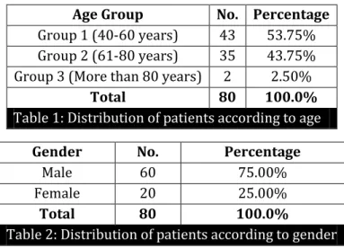
RESULTS: The mean age of the patient was 60±12 year. A total 80 patient of COPD were included and most of patient 43 patient (53.75%) belong to group 1(40-60 year) followed by 35(43.75%) patient in group 2(60-80 year).
Age Group No. Percentage Group 1 (40-60 years) 43 53.75% Group 2 (61-80 years) 35 43.75% Group 3 (More than 80 years) 2 2.50%
Total 80 100.0%
Table 1: Distribution of patients according to age
Gender No. Percentage
Male 60 75.00%
Female 20 25.00%
Total 80 100.0%
Table 2: Distribution of patients according to gender
In our study 60 males and 20 females were included and male: female ratio was 3:1.
COPD Grading No. Percentage Grade-1 Mild (FEV 1 80% of predicted) 8 10.00%
Grade-2Moderate (50% FEV 1< 80% predicted) 34 42.50%
Grade-3 Severe (30% FEV 1< 50% predicted) 26 32.50%
Grade-4Very severe (FEV 1< 30% predicted) 12 15.00%
Total 80 100.0%
Table 3: Distribution of patient according to COPD severity
J of Evolution of Med and Dent Sci/ eISSN- 2278-4802, pISSN- 2278-4748/ Vol.4/ Issue 42/ May 25, 2015 Page 7278
COPD Grading Present Absent P value No. % No. %
Grade-1 (8 Patient) 0 0.00% 8 100%
p <0.001 (Fishers exact t test) Grade-2 (34Patient) 16 47% 18 52.9%
Grade-3 (26 Patient) 20 76.9% 6 23.07% Grade-4 (12 patient) 11 91.6% 1 9.09%
Table 4: Presence of P. Pulmonale According to COPD Grading
In our study there is direct linear correlation between frequency of P pulmonale and frequency of severe COPD Patient. P pulmonale in ECG was found maximum in Grade 4 COPD i.e. (11/12) 91.6% patient followed by Grade 3 COPD i.e. 20/26 (76.9%) patient.
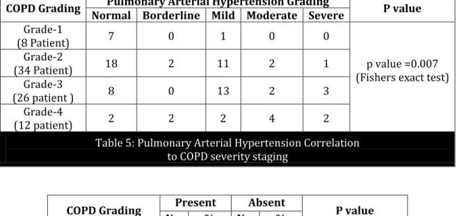
The frequencies of Pulmonary artery Hypertension in mild,moderate,severe,and very severe COPD were 1/8 (12.5%), 14/34 (41.1%), 18/36 (50%) and 8/12 (66%) respectively. In our study moderate to severe pulmonary artery hypertension was more commonly seen in COPD Grade 3 and Grade 4 patient as compared to COPD grade 2 patients.
COPD Grading Pulmonary Arterial Hypertension Grading P value Normal Borderline Mild Moderate Severe
Grade-1
(8 Patient) 7 0 1 0 0
p value =0.007 (Fishers exact test) Grade-2
(34 Patient) 18 2 11 2 1
Grade-3
(26 patient ) 8 0 13 2 3
Grade-4
(12 patient) 2 2 2 4 2
Table 5: Pulmonary Arterial Hypertension Correlation to COPD severity staging
COPD Grading Present Absent P value No. % No. %
Grade-1 (8 patient) 0 0.00% 8 100%
p=0.005 (Fishers exact test) Grade-2 (34 Patient) 1 2.9% 33 97%
Grade-3 (26 Patient) 4 15.3% 22 84.6% Grade-4 (12 Patient) 5 41.6% 7 58.3%
Table 6: Corpulmonale correlation with COPD severity
In our study Corpulmonale secondary to COPD was more common in COPD Grade 3 and Grade 4 patient as comparison to Grade 2 patients.
J of Evolution of Med and Dent Sci/ eISSN- 2278-4802, pISSN- 2278-4748/ Vol.4/ Issue 42/ May 25, 2015 Page 7279 respiratory system and/or hypoxemia. In general, for every 10% decrease in FEV1, all-cause mortality increases by 14%, cardiovascular mortality increases by 28%, and nonfatal coronary event increases by almost 20%.6
2D echocardiography is more sensitive than electrocardiography, radiography and clinical methods in detecting cardiovascular complications like PAH, corpulmonale and R.V. dysfunction in COPD.7
On the basis of these three parameters (dilation, hypertrophy and contractility), an experienced echo cardiographer will be able to make a good qualitative assessment of RV function and will be able to grade it as mild,moderate or severe impairment.8
A good evaluation of pulmonary haemodynamics can be performed with Doppler echocardiography using in association, several measurements,for example: maximal velocity of TR,pulmonary blood flow velocity and new indices of right ventricular dysfunction.9
The triad of decreased RV systolic function, increasing RV sizeand septal bowing form the fundamental basis for the echocardiographic recognition of PAH, with or without demonstration of an increase in Doppler estimated PA systolic pressure.10
Quantitative evaluation of severity of pulmonary hypertension by a pulsed Doppler technique has potential advantages. It can be used to perform repeated examinations without any side effects; it can also be used to follow stages in the development of pulmonary hypertension complicating certain disease processes to evaluate the effects of therapeutic agents and to determine the timing of corrective surgery.11
N K Gupta et al shown that severe PAH is present only in severe or very severe COPD and the incidence of PAH is directly proportional to severity of disease.12 In our study also moderate to severe
pulmonary artery hypertension is more commonly seen in severe to were severe COPD as comparison to moderate COPD patient.
In a study Vikramb Vikhe et al corpulmonale was present in 70% of patient and more commonly seen in severe COPD patient.7 In our study also corpulmonale was more common in very
severe COPD patient (41.6%) and severe COPD patient (15.3%) as comparison to moderate COPD patient (2.9%).
Despite of its benefits, echocardiography in COPD is not without inherent drawbacks. The substernal location of the right ventricle itself and also the difficulties posed by the over inflation of lungs, which reduces the window available for examination, leads to problems in obtaining a good echocardiographic study. But most studies report that adequate examination can be obtained in more than70% of the patients.7
J of Evolution of Med and Dent Sci/ eISSN- 2278-4802, pISSN- 2278-4748/ Vol.4/ Issue 42/ May 25, 2015 Page 7280
REFERENCES:
1. Salvi S. COPD: The neglected epidemic. Textbook of Pulmonary and Critical Care Med 2011, Ed: Jaypee Publications, Vol. 2; 971-974.
2. Jindal SK. Indian Study on Epidemiology of Asthma, Respiratory Symptoms and Chronic Bronchitis (INSEARCH) A Multi Centre Study (2006-2009) - Final Report. New Delhi: Indian Council of Medical Research; 2010.
3. WHO 2008 Oct, www.who.int/ healthinfo /global burden disease/projections/en/index.html. The Global Burden of Disease. Accessed on 22nd Dec 2011.
4. BV Murali Mohan, Tiyas Sen, R Ranganatha. Systemic Manifestations of COPD. SUPPLEMENT TO JAPI, FEBRUARY 2012; VOL. 60 p 44-47.
5. Chemla D, Castelain V, Humbert M, Simonneau JLHG, Lecarpentier Y, Hervé P. New Formula for Predicting Mean Pulmonary Artery Pressure Using Systolic Pulmonary Artery Pressure. Chest2004; vol126: 1313–17.
6. Sin DD,Man SF. Chronic obstructive pulmonary disease as a risk factor for cardiovascular morbidity and mortality. Proc Am Thorac Soc. 2005; 2 (1): 8-11.
7. Vikramb Vikhe, Prakash S Shende, Rahul S Patil, Krunal K Tamakuwala, Amol S Patil, Ankur P Gupta. Cardiovascular complications in chronic obstructive pulmonary disease with reference to 2d echocardiography findings. National journal of medical research 2013, volume 3; Issue 4: 385-388.
8. Luke S. Howard, Julia Grapsa, David Dawson, Michael Bellamy, John B. Chambers, Navroz D. Masani, Petros Nihoyannopoulos and J. Simon R. Gibbs. Echocardiographic assessment of pulmonary hypertension: standard operating procedure. European Respiratory Review, Vol. 21; NUMBER 125: 239-248.
9. A. Chaouat, R. Naeije and E. Weitzenblum. Pulmonary hypertension in COPD. Eoropean Respiratory Journal 2008, Vol 32; NUMBER 5: 1371-1385.
10.Justin D. Roberts and Paul R. Forfia. Diagnosis and assessment of pulmonary vascular disease by Doppler echocardiography. Pulmonary Circulation 2011, Vol. 1; No 2: 160-181.
11.Akira Kitabatake, Michitoshiinoue, Masato Asao, Tohrumasuyama, Jun Tanouchi, Toshio Morita, Masayoshi Mishima, Masaaki Uematsu, Takashi Shtmazu, Masatsuguhori and Hiroshi abe. Noninvasive evaluation of pulmonary hypertension by a pulsed doppler technique. Circulation 1983, vol. 68; No. 2: p302-309.
J of Evolution of Med and Dent Sci/ eISSN- 2278-4802, pISSN- 2278-4748/ Vol.4/ Issue 42/ May 25, 2015 Page 7281 sa
AUTHORS:
1. Bhupendra Kumar Jain 2. Nikhilesh Pasari 3. Ashok Bajpai 4. Ashwin Songara
PARTICULARS OF CONTRIBUTORS:
1. Assistant Professor, Department of Respiratory Medicine, Sri Aurobindo Medical College & PG Institute.
2. Senior Resident, Department of Respiratory Medicine, Sri Aurobindo Medical College & PG Institute.
3. Professor, Department of Respiratory Medicine, Sri Aurobindo Medical College & PG Institute.
FINANCIAL OR OTHER
COMPETING INTERESTS: None
4. Junior Resident, Department of Respiratory Medicine, Sri Aurobindo Medical College & PG Institute.
NAME ADDRESS EMAIL ID OF THE CORRESPONDING AUTHOR:
Dr. Bhupendra Kumar Jain, Assistant Professor,
Sri Aurobindo Medical College &
PG Institute, Indore-Ujjain State Highway, Near MR 10 Crossing, Indore.
E-mail: [email protected]