Diana Marisa Marques dos Santos
Degree in Biochemistry
Improving a bacterial pyranose 2-oxidase from
Arthrobacter siccitolerans through directed evolution
Dissertation to obtain the Master Degree in Biochemistry for Health
Supervisor: Lígia O. Martins, PhD, ITQB-UNL
i
Diana Marisa Marques dos Santos
Degree in Biochemistry
Improving a bacterial pyranose 2-oxidase from
Arthrobacter siccitolerans through directed evolution
Dissertation to obtain the Master Degree in Biochemistry for Health
Supervisor: Lígia O. Martins, PhD, ITQB-UNL
Júri:
Presidente: Prof. Doutor Pedro Matias Arguente: Prof. Doutora Manuela M. Pereira
Vogal: Doutora Margarida Archer
Instituto de Tecnologia Química e Biológica António Xavier
iii
Improving a bacterial pyranose 2-oxidase from
Arthrobacter siccitolerans through directed evolution
Copyright Diana Santos, ITQB, UNL
O Instituto de Tecnologia Química e Biológica António Xavier e a Universidade Nova de Lisboa têm o direito, perpétuo e sem limites geográficos, de arquivar e publicar esta dissertação através de exemplares impressos reproduzidos em papel ou de forma digital, ou por qualquer outro meio conhecido ou que venha a ser inventado, e de a divulgar através de repositórios científicos e de admitir a sua cópia e distribuição com objetivos educacionais ou de investigação, não comerciais, desde que seja dado credito ao autor e editor.
v
Acknowledgments
Nesta fase final da minha dissertação de mestrado, gostaria de demonstrar a imensa gratidão com todas as pessoas que me apoiaram e que contribuíram ao longo do último ano para a minha formação como pessoa e investigadora. Não poderia deixar de agradecer a todo o ITQB, especialmente ao laboratório MET por me fornecerem todas as condições e equipamentos sem os quais não poderia ter elaborado esta dissertação.
Vou ficar eternamente grata à minha orientadora: professora Lígia Martins por todo o apoio e transmissão de conhecimento que me facultou! Um obrigada nunca vai ser suficiente por nunca me deixar desanimar quando as coisas não corriam da forma desejada, por me fazer acreditar na ciência, por ter sempre total disponibilidade para me ajudar mesmo quando o tempo era escasso e por me encorajar a lutar pelos meus objetivos todos os dias com otimismo. Terei sempre uma grande admiração pela grande cientista que é mas também pela excelente pessoa que demonstra todos os dias ser. Sem dúvida, um exemplo a seguir!
Não poderia deixar de agradecer à Dra. Vânia Brissos que tanto me ajudou ao longo de toda a minha dissertação de mestrado. Sempre com total disponibilidade para me esclarecer todas as dúvidas e fornecer valiosos conselhos que a vasta experiência lhe ensinou e que com todo o gosto me transmitiu. Obrigada pelo tempo, todo o conhecimento e ajuda que disponibilizou e que tanto me foram úteis. Quero ainda agradecer à Dra. Sónia Mendes, pela energia positiva que sem dúvida contagia as pessoas ao redor e pela ajuda que sempre me foi disponibilizada. Aos membros do MET que ao longo deste ano estiveram presentes no laboratório (Patrícia, Ana, Manuel, Lorenzo e Diogo) um obrigada pelos bons momentos passados e por tornarem por vezes momentos de stress em momentos divertidos.
Gostaria ainda de agradecer à Dra. Mónica Serrano pela ajuda e disponibilidade que demonstrou ter quando necessitei de utilizar a técnica de microscopia de fluorescência e ao professor Willem van Berkel e Dr. Adrie Westphal (Univ. Wageningen, Holanda) pelo modelo da proteína que construíram. Aos meus pais, por serem o meu refúgio quando atravesso momentos menos bons, pelo trabalho constante que desenvolvem todos os dias para me proporcionarem a oportunidade de tirar um curso superior, de seguir sempre os meus sonhos e concretizar os meus objetivos. Nunca haverá palavras suficientes para agradecer todo o apoio incondicional e motivação que me transmitiram não só durante esta fase mas sim durante toda a minha vida.
Ao meu namorado e acima de tudo melhor amigo Daniel, obrigada por todo o apoio durante esta fase, por nunca me deixar desmotivar e pela compreensão quando nem sempre lhe conseguia dar a atenção devida. Da mesma forma, um obrigada a todos os meus amigos por sempre me apoiarem, acreditarem em mim e compreenderem quando nem sempre podia despender o tempo devido com eles.
vii
Resumo
Piranose 2-oxidases (P2Ox) são flavoproteínas que catalisam a oxidação do carbono 2 de aldopiranoses, com redução concomitante de O2 a H2O2 e, que apresentam, um grande interesse
biotecnológico. Recentemente, foi caracterizada pela primeira vez, uma P2Ox bacteriana, AsP2Ox de Arthrobacter siccitolerans. As bactérias apresentam maior rapidez de crescimento, em comparação com os fungos, e existe grande disponibilidade de ferramentas de biologia molecular em sistemas bacterianos. No entanto, verificou-se que a eficiência catalítica de AsP2Ox era seis-ordens de magnitude inferior à exibida pelas enzimas fúngicas. Neste trabalho, a evolução dirigida foi utilizada na engenharia da enzima AsP2Ox, para melhoramento da sua eficiência catalítica. Otimizaram-se protocolos de mutagénese, seleção e crescimento de células hospedeiras, lise celular e ensaios enzimáticos. A aplicação de uma estratégia validada de mutagénese e “screening” permitiu a seleção de um variante, 2C9, com atividade enzimática superior à observada na enzima nativa, a partir de uma biblioteca de 25 000 clones. Após purificação, verificou-se que o seu nível de produção era 6 vezes mais baixo relativamente à enzima nativa e mostrava ser inativado pela luz. Este variante contém duas mutações não sinónimas (A35T e F300V) e uma sinónima (Q343Q). Com o objetivo de avaliar o efeito das mutações nas propriedades do variante, foram construídos os mutantes “single” (A35T e F300V) e o mutante duplo 2C9* (A35T/F300V), sem a mutação sinónima. Concluiu-se que a mutação Q343Q era a única responsável pela foto-inativação da enzima, e, em conjunto com a mutação F300V, contribuía para os níveis de produção mais baixos. Verificou-se ainda que a mutação A35T era neutra, mas indispensável para, combinada com a mutação F300V, numa interação epistática, resultar numa eficiência catalítica 3 vezes superior no variante 2C9*, relativamente à enzima nativa. Assim sendo, o mutante 2C9* foi selecionado como parente para uma segunda geração de evolução dirigida.
Palavras-chave: flavoproteínas, enzimas formadoras de peróxido, evolução dirigida, eficiência catalítica, biosensores
ix
Abstract
Pyranose 2-oxidases (P2Ox) are flavoproteins that catalyze the oxidation of several aldopyranoses to yield the corresponding 2-keto-aldoses with concomitant reduction of O2 to H2O2 and are enzymes that
show many biotechnological applications. Recently, a bacterial P2Ox from Arthrobacter siccitolerans (AsP2Ox) was characterized for the first time since bacteria grow faster as compared to fungi and have well-established genetic and molecular biological tools allowing for higher enzyme production yields. Directed evolution has proven to be a powerful approach to improve enzyme efficiency and robustness required for biotechnological applications. Therefore, in this work the optimization and validation of critical steps of directed evolution was performed, namely mutagenesis protocols, cell growth, lysis and high-throughput enzymatic assays. One round of evolution through error prone PCR was performed and a total of 25 000 clones were screened to find variants with improved activity for D-glucose and dioxygen. One hit variant, 2C9, was identified, showing higher activity than wild-type and containing two non-synonymous (A35T and F300V) and one non-synonymous mutation (Q343Q). The variant enzyme was produced at a larger scale, purified and characterized. It was observed that 2C9 was produced at 6-fold lower yields as compared to wild-type and showed photo-inactivation behaviour. Therefore single mutants (A35T and F300V) and double 2C9* without the silent mutation (A35T/F300V) were constructed and characterized to unveil the role of mutations from the catalytic and structural viewpoints. The introduction of the synonymous mutation Q343Q resulted in the photo-inactivation of the enzyme and was co-responsible, along with the F300V mutation, to the variant lower production yields. Mutation A35T was neutral but in combination with the functional F300V mutation, an epistatic effect became evident, resulting in the 3-fold higher catalytic efficiency exhibited by 2C9* as compared with the wild-type enzyme. Therefore, 2C9* was selected as a parent for the second generation of directed evolution.
Keywords: flavoproteins, hydrogen peroxide forming enzymes, directed evolution, catalytic efficiency, biosensors
xi
Table of contents
List of Figures ... xiii
List of tables ... xvii
List of Abbreviations ... xix
1. Introduction ... 1
1.1. Fungal pyranose oxidases: General properties and catalysis... 1
1.2. Pyranose 2-oxidase from Arthrobacter siccitolerans (AsP2Ox) ... 2
1.3. Structural characterization of P2Ox ... 3
1.4. Applications of P2Oxs ... 5
1.5. Protein engineering ... 6
1.5.1 Methods for introducing mutations in a target gene ... 6
1.5.2 Methods of analysis and isolation of variants from a library of mutants ... 8
1.6 Context of the project ... 9
2. Material and Methods ... 11
2.1 Bacterial strains, plasmids and media ... 11
2.2 Preparation of E. coli electrocompetent cells ... 11
2.3 Transformation of E. coli cells ... 11
2.4 Heterologous expression of target genes... 12
2.4.1 Cell growth of E. coli strains overexpressing recombinant genes ... 12
2.4.1.1 Overproduction of AsP2Ox at different temperatures ... 12
2.4.1.2 Co-production of AsP2Ox in the presence of chaperones ... 12
2.4.1.3 Co-production of AsP2Ox with Hyper ... 13
2.4.2 Cell disruption ... 13
2.4.3 Determination of protein concentration ... 14
2.4.4 SDS-PAGE analysis ... 14
2.4.5 Enzymatic assays ... 14
2.5 Fluorescence Microscopy ... 14
2.6 Directed evolution ... 15
xii
2.6.2 Overexpression of AsP2Ox and variants in 96-well plates ... 15
2.6.3 Cell disruption in 96 well plates ... 16
2.6.4 Spectrophotometric high-throughput activity screening using D-glucose and 1,4-BQ as substrates ... 16
2.6.5 ‘Activity-on-plate’ high-throughput screening using D-glucose and O2 as substrates ... 16
2.6.6 Activity re-screening using glucose and O2 as substrates ... 17
2.7 Site-directed mutagenesis ... 17
2.8 Production and purification of wild-type and recombinant AsP2Ox variants... 18
2.9 Spectroscopic analysis of FAD-ligation ... 19
2.10 Kinetic analysis ... 19
3. Results and Discussion ... 21
3.1 Development of an in vivo activity screening for AsP2Ox ... 21
3.2. Optimization of growth conditions to increase the solubility of recombinant AsP2Ox in E. coli cells ... 24
3.3 Directed Evolution of AsP2Ox ... 27
3.3.1 Validation of high-throughput screenings in 96-well plates ... 27
3.3.2 Selection of the best conditions to overexpress asP2Ox in E.coli ... 27
3.3.3 Selection of the cell disruption method in 96-well plates ... 28
3.3.4 Generation of AsP2Ox mutant libraries using epPCR ... 29
3.3.5 High-throughput activity screening of the mutant library using 1,4-BQ as electron acceptor ... 30
3.3.4. “Activity-on-plate” high-throughput activity screening using O2 as electron acceptor ... 31
3.4. Kinetic and biochemical characterization of the hit 2C9 variant ... 35
3.4.1 Structural analysis of mutations that improved the activity of 2C9 ... 35
3.4.2 Spectroscopic analysis and identification of FAD ligation ... 35
3.5 Site-directed mutagenesis ... 37
4. Conclusions ... 43
xiii
List of Figures
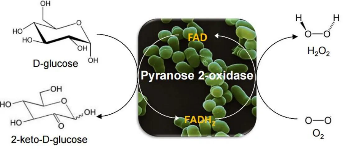
Figure 1.1 Schematic reaction catalyzed by P2Ox: first the transfer of two electrons from D-glucose to
the FAD cofactor occurs followed by O2 reduction resulting in the formation of 2-keto-D-glucose and
H2O2. ... 2
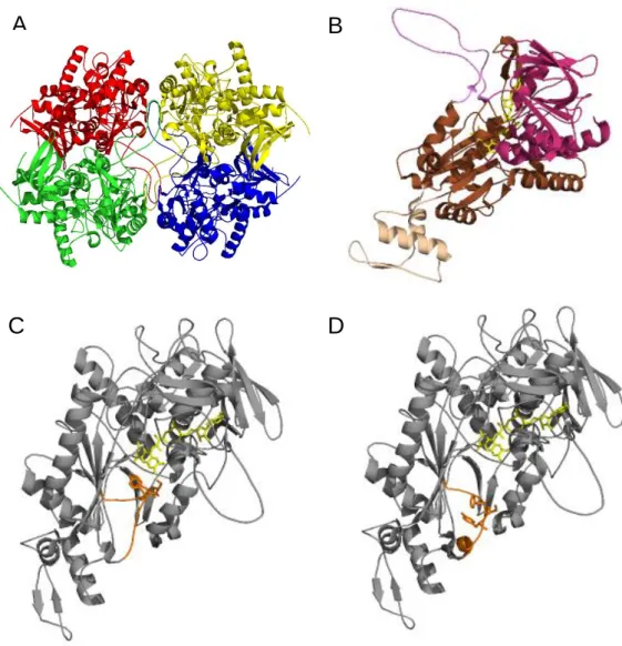
Figure 1.2 Crystal structure of P2Ox from T. multicolor19. (A) Homotetramer structure and subunit
assembly. (B) Structure of P2Ox subunit. In purple is represented the oligomerization arm and in beige the head domain. The Rossman domain is represented in pink and the substrate binding domain in brown. The FAD cofactor is shown as a yellow stick representation. Subunit structure with the substrate-recognition loop represented in orange in a closed (C) and open (D) conformation. The gating segment (Phe454, Ser455 and Tyr456) is shown as an orange and FAD cofactor as a yellow stick representation in
(C) and (D). (A), (B), (C) structures corresponds to PDB code 1TT0 and (D) to PDB code 2IGO. ... 4
Figure 1.3 Overview of directed evolution of proteins with all steps needed to achieve a desired property:
generation of diversity, the choice of a good method of screening/selection, identification of a variant with an improved property and repetition of the cycle until a variant with the desired property is found. 7
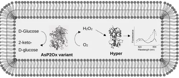
Figure 3.1 Hyper-based screening for in vivo detection of enzymatic H2O2 production. The objective was
to identify better variants of AsP2Ox through a higher production of H2O2 that would result in an
increased excitation spectrum of Hyper at 500 nm and a decrease at 420 nm. The reduced spectrum is shown as a solid line and the oxidized spectrum as a dashed line. Adapted from62………21
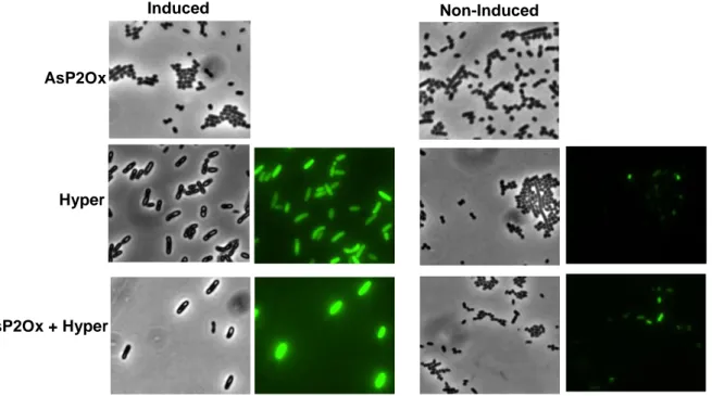
Figure 3.2 Images of induced and non-induced cell cultures obtained by bright-field and fluorescence
microscopy producing AsP2Ox, Hyper and co-producing both proteins in the BL21 star strain. When only AsP2Ox was produced, no fluorescence was detected as expected. ... 22
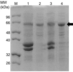
Figure 3.3 SDS-PAGE of BL21 star crude extracts. (M) Marker, non-induced (1) and induced (2) cell
extracts overproducing AsP2Ox, induced (3) and non-induced (4) cell extracts overproducing Hyper and, induced (5) and non-induced (6) cell extracts overproducing both AsP2Ox and Hyper proteins. The arrows show the bands that correspond to the AsP2Ox protein (64 kDa) and to the Hyper protein (66 kDa). ... 22
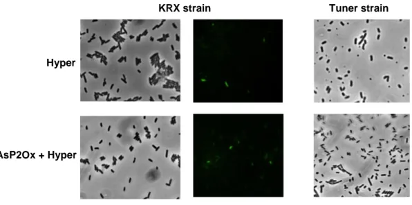
Figure 3.4 Images of cell cultures obtained by bright-field and fluorescence microscopy producing Hyper
and co-producing both proteins (AsP2Ox and Hyper) in KRX and Tuner strains. No fluorescence was detected when Tuner strain was used as expression strain. ... 23
Figure 3.5 SDS-PAGE of KRX and Tuner crude extracts. (M)-Marker, (1) KRX cell extracts
overproducing Hyper, (2) KRX cell extracts overproducing Hyper and AsP2Ox, (3) Tuner cell extracts overproducing Hyper and (4) Tuner cell extracts overproducing AsP2Ox and Hyper. AsP2Ox has a molecular weight of 64 kDa and Hyper of 66 kDa approximately. ... 23
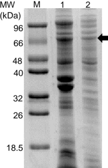
Figure 3.6 SDS-PAGE performed with the insoluble and soluble fractions of recombinant E. coli cell
extracts of cells overproducing AsP2Ox. (M) marker, (1) insoluble fraction and (2) soluble fraction. The arrow shows the band that corresponds to the AsP2Ox protein (64 kDa). ... 24
xiv
Figure 3.7 SDS-PAGE of insoluble and soluble fractions of cells grown at different temperatures afterIPTG induction. (M) marker, insoluble (1) and soluble fraction (2) of crude extracts of cells incubated at 10ºC after IPTG induction and insoluble (3) and soluble fractions (4) of crude extracts of cells incubated at 25ºC after IPTG induction. The arrow indicates the band that corresponds to AsP2Ox (~64 kDa). 25
Figure 3.8 SDS-PAGE of insoluble and soluble fractions of induced and non-induced cells
overproducing AsP2Ox and chaperones. (M) marker, soluble fractions of crude extracts where genes coding for chaperones were induced (1) and non-induced (2), insoluble fractions of crude extracts of cells where genes coding for chaperones were induced (3) and non-induced (4). Black arrows correspond to chaperone proteins: GrpE, DnaJ, GroEL and DnaK that present molecular weights of 22, 40, 60 and 70 kDa respectively. ... 26
Figure 3.9 Landscape correspondent to six variant libraries using different MnCl2 concentrations: 0.01
mM (∆), 0.10 mM (○), 0.20 mM (♦), 0.30 mM (●), 0.40 mM (▲) and 0.50 mM (x). Activity of clones relative to the wild-type is plotted in descending order. ... 29
Figure 3.10 Directed evolution landscape corresponding to the first generation of mutants. (A) Activity
relative to wild-type of 2052 clones screened in the first generation. (B) Re-screening of the best 66 variants found in the first generation. Enzymatic assays were performed at 37⁰C, in 100 mM sodium phosphate buffer at pH 6.5 in the presence of 10 mM D-glucose and 1 mM 1,4-benzoquinone. ... 30
Figure 3.11 Overview of ‘activity-on-plate’ screening used for the directed evolution of AsP2Ox enzyme.
... 32
Figure 3.12 ‘Activity-on-plate’ high-throughput assay (with 10 U HRP and 10 mM D-glucose) using BL21
star cells transformed with wild-type AsP2Ox plasmid and grown in LA media in the presence of 10 μM IPTG. (A) Non-induced cells incubated in the presence of 20 mM ABTS. Cells overexpressing asP2Ox gene in the presence of (B) 20 mM, (C) 10 mM, (D) 5 mM and (E) 1 mM of ABTS, respectively. ... 32
Figure 3.13 ‘Activity-on-plate’ high-throughput assay (10 U HRP, 1 mM ABTS and 10 mM D-glucose)
using BL21 star cells expressing (A) wild-type asP2Ox gene and (B-D) mutant library constructed using epPCR in the presence of 0.01 mM MnCl2. Colonies within circles represent variants with improved
activity visible by the darker colour. ... 33
Figure 3.14 Rescreening using the same conditions than previously used of the best 153 variants
through ‘activity-on-plate’. The seven clones showing a faster and stronger purple colour than wild-type were chosen for a new rescreening. Colonies within circles represent variants with improved activity and inside rectangles are represented colonies overexpressing wild-type asP2Ox. ... 33
Figure 3.15 Rescreening of the best seven variants through ‘activity-on-plate’. The wild-type AsP2Ox
and variants have shown different growth rates and consequently very weak or inexistence activity.. 34
Figure 3.16 Enzymatic activity using crude extracts of wild-type AsP2Ox and 2C9 in the presence of
100 mM D-glucose, 10 U HRP and 1 mM ABTS in 100 mM sodium phosphate buffer at pH 6.5. In the control reaction no crude extract was added... 34
xv
Figure 3.17 Model structure of wild-type AsP2Ox where the FAD cofactor is represented in yellow sticks.A35 is in red, F300 in green and Q343 in blue colour. The distances from A35, F300V and Q343 to the FAD cofactor are represented in blue in Ångström (Å). ... 35
Figure 3.18 SDS-PAGE of BL21 star crude extracts overproducing wild-type and 2C9 purified proteins.
(M) Marker, (1) crude extracts of cells overproducing wild-type AsP2Ox protein, (2) AsP2Ox after purification using a metal affinity chromatography, (3) crude extracts of cells overproducing 2C9 variant protein and (4) 2C9 after purification using a metal affinity chromatography. The arrow indicates the band that corresponds to AsP2Ox proteins (~64 kDa)... 36
Figure 3.19 (A) UV-vis spectra of AsP2Ox as isolated (solid line) and of 2C9 fractions after purification
(dashed and dotted lines) (B) UV-vis spectra of purified 2C9 as isolated (solid line) and after treatment with 0.4% (w/v) SDS or 5%TCA followed by heating and centrifugation (dotted line). ... 37
Figure 3.20 SDS-PAGE of BL21 star crude extracts overproducing wild-type and mutant proteins. (M)
Marker, crude extracts overproducing (1) wild-type, (2) 2C9, (3) 2C9*, (4) A35T and (5) F300V proteins. The arrow indicates the band that corresponds to AsP2Ox proteins (~64 kDa). ... 38
Figure 3.21 UV-vis spectra of (A) 2C9*, (B) A35T and (C) F300V as isolated (solid line) and after
treatment with 0.4% (w/v) SDS (dotted line). ... 39
Figure 3.22 Apparent steady-state kinetic analysis of (A) wild-type, (B) 2C9*, (C) A35T and (D) F300V
proteins for D-glucose using O2 as electron acceptor. Reactions were performed in 100 mM sodium
phosphate buffer at pH 6.5, using O2 saturated solutions at 37°C, and the ABTS-peroxidase assay. The
xvii
List of tables
Table 2.1 Plasmid harbouring the wild-type asP2Ox gene and plasmids containing genes for the
different chaperones that were used in the co-expression using the BL21 star strain. ... 13
Table 2.2 Primers used in the site-directed mutagenesis for the construction of different mutants.
Fwd-primer forward; Rev-Fwd-primer reverse. The nucleotides in bold and underlined correspond to the base substitutions. ... 18
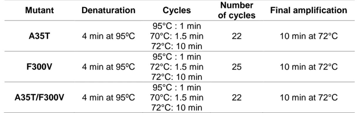
Table 2.3 Conditions used in the PCR reactions for the construction of different mutant enzymes using
site-directed mutagenesis... 18
Table 3.1 Final OD600nm of cell cultures co-producing AsP2Ox and different chaperones and protein
concentration in cell crude extracts. 26
Table 3.2 Final OD600nm of cell cultures of BL21 star and KRX strains grown in different media, protein
concentration and specific activity in crude cell extracts. Enzymatic assays were performed at 37⁰C, in 100 mM sodium phosphate buffer at pH 6.5 in the presence of 10 mM D-glucose and 1 mM 1,4-benzoquinone. ... 27
Table 3.3 Protein concentration and specific activity in crude extracts after using different disruption
methods. Cells were disrupted using enzymatic (lysozyme 2 mg/mL), physical (freeze/thaw using Liquid Nitrogen (LN)) or freeze/thaw through incubation at -80⁰C) and chemical (B-PER® lysis solution)
methods. ... 28
Table 3.4 Average of base substitutions in the asP2Ox gene after epPCR performed in the presence of
different concentrations of manganese chloride. Four to five clones were randomly selected from libraries generated after epPCR at different MnCl2 concentrations, plasmids were extracted, transformed
on DH5-α cloning strain, purified and sent to sequencing. ... 31
Table 3.5 Total protein concentration achieved from 1L of culture of Bl21 star cells producing wild-type,
2C9, 2C9*, A35T and F300V proteins determined after purification using a metal affinity chromatography and specific activity achieved after enzymatic assays at 37⁰C, in 100 mM sodium phosphate buffer at pH 6.5 in the presence of 1 M D-glucose, 1 mM ABTS and 10 U HRP. ... 38
Table 3.6 Apparent steady-state kinetic parameters of AsP2Ox (wild-type and variants) determined
using D-glucose and O2. Enzymatic assays were performed at 37⁰C, in 100 mM sodium phosphate
xix
List of Abbreviations
1,4-BQ - 1,4-benzoquinone
1,5-AG - 1,5-anhydro-D-glucitol
ABTS - 2,2’-azino-bis (3- ethylbenzothiazoline-6-sulfonic acid)
BLAST - Basic Local Alignment Search Tool
B-PER - Bacterial Protein Extraction Reagent
APS - Ammonium persulfate
AsP2Ox - Pyranose 2-oxidase from Arthrobacter siccitolerans
BSA - Bovine Serum Albumin
DCPIP - 2,6-Dichlorophenolindophenol
epPCR - Error prone polymerase chain reaction
FACS - Fluorescence-activated cell sorting
FAD - Flavin adenine dinucleotide
GMC- Glucose-methanol-choline
GOx - Glucose oxidase
HRP - Horseradish peroxidase
IPTG - Isopropyl β-D-1-thiogalactopyranoside kDa - Kilodalton
LB – Luria-Bertani
OD600nm - Optical density at 600 nm
P2Ox - Pyranose 2-oxidase
PCR - Polymerase chain reaction
RT - Room temperature
SDS-PAGE - Sodium dodecyl sulfate polyacrylamide gel electrophoresis
SOB - Super Optimal Broth
SDM – Site-directed mutagenesis SSM - Site-saturation mutagenesis
TB - Terrific Broth
xx
TEMED - N, N, N’, N’-TetramethylethylenediamineTris – Tris (hydroxymethyl)aminomethane UV-Vis - Ultraviolet-Visible
1
1. Introduction
1.1. Fungal pyranose oxidases: General properties and catalysis
Pyranose oxidases (P2Ox, pyranose: oxygen oxidoreductase; EC 1.1.3.10; synonym, glucose 2-oxidase) belong to the glucose-methanol-choline (GMC) oxidoreductase superfamily of enzymes1. This
family includes enzymes such as glucose oxidase, choline oxidase, cholesterol oxidase, cellobiose dehydrogenase, aryl-alcohol oxidase, pyridoxine 4-oxidase, among others1. P2Oxs are flavin adenine
dinucleotide (FAD)-dependent enzymes, exhibiting the typical UV–Visible spectrum of the FAD cofactor with absorption maxima at 360 and 455 nm2.
P2Oxs catalyze the regiospecific oxidation of sugar molecules at an alcohol moiety (-CH-OH) to the corresponding aldehyde with concomitant reduction of molecular oxygen to hydrogen peroxide3,4.
Several aldopyranoses and disaccharides such as D-glucose, D-galactose, D-mannose, D-arabinose, D-fructose, L-sorbose, D-xylose, D-fucose, maltose and trehalose are oxidized at the C2 position to yield the corresponding 2-keto-aldoses5,6. These enzymes can also use quinones as electron acceptors
in replacement to O2 and show a higher efficiency for substituted benzoquinones, structural analogues
of benzoquinone (e.g. DCPIP), metal ions such as ferricyanide and ferrocenium, and radicals (e.g. the cation radical ABTS+)7. The specific activity of P2Ox for the different sugar substrates varies
considerably, but D-glucose has been identified as the favourite substrate4. Oxidation at the C3 position,
as a side reaction, was also observed with 2-deoxy-D-glucose, 2-keto-D-glucose and methyl β-D-glucosides, indicating that when the C2 hydroxyl group is compromised, P2Oxs have the ability to oxidize sugar substrates at a different position, demonstrating an alternative regioselectivity8,9,10.
The first P2Ox enzyme was isolated from the fungus Polyporus obtusus, followed by its characterization in several other P2Oxs, mainly from wood-degrading, white- and brown-rot, basidiomycetes11. In these
microorganisms, P2Oxs are located in the hyphal periplasmic space and are released to the extracellular space in the later stages of the fungal cycle development11. Fungal enzymes have glucose,
D-galactose and D-xylose as preferred substrates most likely due to their abundance in lignocellulose material originated from wood rot. Their physiological role has been associated with the generation of H2O2 that is an essential substrate for lignin-degrading peroxidases5,12. Furthermore, it was shown that
in some species of white-rot fungi, P2Oxs are involved in secondary metabolic pathways that lead to the production of the antibiotic cortalcerone, protecting the microorganism from bacterial attack5. As
quinones act as alternative electron acceptors to O2, it has been claimed that P2Oxs can also get
involved in the reduction of quinones during the process of ligninolysis11.
The reaction catalyzed by P2Oxs can be divided in two half-reactions: a reductive half-reaction, where the transfer of two electrons from a sugar substrate to the oxidized FAD occurs leading to the formation of a 2-keto-sugar and reduced FADH2, and an oxidative half reaction, where two electrons from the
reduced FADH2, are transferred to molecular oxygen, with concomitant formation of H2O23,13 (figure1.1).
The first evidence for the formation of an intermediate in a flavoprotein oxidase was detected in P2Ox, the C-4a-hydroperoxyflavin intermediate, observed during the oxidative half reaction, when O2 acts as
2
reduction, the mechanism of the reaction catalyzed by P2Oxs was classified as a ping-pong bi-bi type, characteristic of flavoprotein oxidoreductases1. Typically, P2Oxs activity is measured through H2O2production in a coupled spectrophotometric assay using a commercial peroxidase and a chromogen substrate, or using an O2 electrode by measuring the rate of O2 consumption2.
Figure 1.1 Schematic reaction catalyzed by P2Ox: first the transfer of two electrons from D-glucose to the FAD
cofactor occurs followed by O2 reduction resulting in the formation of 2-keto-D-glucose and H2O2.
1.2. Pyranose 2-oxidase from Arthrobacter siccitolerans (AsP2Ox)
P2Oxs from fungal origin have been extensively studied and characterized. Consequently, in our lab it was settled as an objective to isolate and characterize a P2Ox enzyme from bacterial origin: Arthrobacter siccitolerans (AsP2Ox). In these microorganisms, recombinant proteins are easier and cheaper to produce, since they reach higher cell densities in shorter periods of time as compared with fungi, and there are plenty of genetic and molecular tools available14. A bioinformatics analysis was
performed using Basic Local Alignment Search Tool (BLAST) in all bacterial genomes available using as template the sequence of the fungal P2Ox from Trametes multicolor. The top 100 Blast sequence hits from bacterial genomes showed an identity between 24 and 39% to the template and 85 out of 100 sequences identified belong to members of the Actinobacteria3 phyla, one of the most dominant in the
bacteria domain, present in varied ecosystems such as alkaline saline soils, marine sponges, deep sea sediments, hot springs, gut and medicinal plants. Actinobacteria displays an important biological role in biogeochemical cycles, bioremediation, bioweathering and plant growth promotion15,16. Furthermore,
they produce a large number of pharmaceutically bioactive compounds such as antibiotics, antitumor agents, anti-inflammatory compounds, enzyme inhibitors and a wide variety of industrial and clinically important enzymes16. The most represented putative P2Ox sequences (62%) are from species of the
Streptomyces (14), Arthrobacter (17) and Microbacterium (25) genera. The A. siccitolerans genome
revealed an ORF encoding a 519 amino acid polypeptide that showed 26% homology with the P2Ox template sharing 13 of the 15 strictly conserved residues present in GMC family in the: (i) FAD-binding domain, (ii) flavin attachment loop and (iii) substrate binding domain that was putatively assigned as a
3
pyranose 2-oxidase (AsP2Ox)3,17,18. The gene was cloned and the enzyme was overproduced inEscherichia coli. After purification, AsP2Ox showed a yellow colour and the typical flavoprotein visible absorption spectrum with two peaks at 390 and 460 nm, with a flavin-protein molar ratio of approximately 1:13. Unlike most known fungal enzymes, in AsP2Ox, the FAD molecule is not covalently bonded to the
protein and the enzyme is monomeric with a molecular mass of 64 kDa, while fungal counterparts are tetrameric enzymes. AsP2Ox exhibited an optimal temperature at 37⁰C, an optimum pH of 6.5 and oxidizes D-glucose at the highest efficiency, using additionally D-galactose, D-xylose, L-arabinose and D-ribose as electron donors, coupling their oxidation to the reduction of both dioxygen and 1,4-benzoquinone (1,4-BQ), the preferred electron acceptor3.It shows a Km value 2-orders of magnitude
higher and a kcat between 2 to 3 orders of magnitude lower for D-glucose and dioxygen when compared
with P2Ox characterized from fungal origins resulting in a 6 to 7-orders of magnitude reduced catalytic efficiency (kcat/Km)11,12.
1.3. Structural characterization of P2Ox
Only three crystal structures were reported to date for P2Oxs from fungal origin: T. multicolor19,
Peniophora sp.20 and Phanerochaete chrysosporium10. All structures are homotetrameric proteins, in
some cases glycosylated, constituted by a dimer of dimers with molecular masses in the range of 280-320 kDa2 (figure 1.2-A). FAD is covalently linked to the monomers by a histidyl linkage21. As shown in
figure 1.2-B, each subunit of the homotetramer can be seen as a central, peanut-shaped elongated ‘body’ with an ‘arm’ projected to the outside that is responsible for the contact with neighbouring subunits and a ‘head’ (αββα motif) that possibly acts as a immobilizer of the enzyme in the fungal cell-wall19. The
FAD-subdomain presents a Rossman fold (βαβ mononucleotide-binding motif) and the substrate-binding subdomain is composed by a central six-stranded β sheet packed between the active site and three α helices19.
The substrate has to pass through a large, internal cavity that contains water, before accessing the active site of each subunit19. The amino acid residues with a role in the catalytic reaction of T. multicolor,
and that are conserved in other fungal P2Oxs, are His548, Asn593, Gln448, His450 and Val546. With the support of Asn593, the His548 amino acid residue most probably acts as a base and deprotonates the substrate at the 2-OH group with a subsequent hydride transference to the flavin N(5) atom19. In
AsP2Ox, the main catalytic amino acid residues (His440, Asn484 and Gln340) are conserved, however catalytic residue His450 in T. multicolor P2Ox appears as Met342 in AsP2Ox and Val546 in T. multicolor P2Ox as Ser438 in AsP2Ox, which can have implications in the catalytic reaction.
The active site of T. multicolor P2Ox is gated by a dynamic, highly conserved loop (residues 450-461) that presents several residues that efficiently bind both electron-donor (sugar) and electron-acceptor substrates (oxygen or quinone derivates) and are therefore determinant for the substrate binding and catalysis19,22. Through the analyses of two P2Ox crystal structures, one in a sugar free form
(acetate-bonded, figure 1.2-C) in native P2Ox and the other in a sugar-bonded form (2-keto-D-glucose-(acetate-bonded, figure 1.2-D) in the mutant H167A enzyme, it is possible to observe at least two loop conformations that were assigned to the two half-reactions of the enzyme19.
4
Figure 1.2 Crystal structure of P2Ox from T. multicolor19. (A) Homotetramer structure and subunit assembly. (B)
Structure of P2Ox subunit. In purple is represented the oligomerization arm and in beige the head domain. The Rossman domain is represented in pink and the substrate binding domain in brown. The FAD cofactor is shown as a yellow stick representation. Subunit structure with the substrate-recognition loop represented in orange in a closed (C) and open (D) conformation. The gating segment (Phe454, Ser455 and Tyr456) is shown as an orange and FAD
cofactor as a yellow stick representation in (C) and (D). (A), (B), (C) structures corresponds to PDB code 1TT0 and (D) to PDB code 2IGO.
When the P2Ox enzyme is in complex with small ligands (e.g. acetate), the loop is in a fully closed conformation with the active site less accessible, providing a more hydrophobic environment, appropriate for the reaction of the reduced enzyme with electron-acceptors such as dioxygen, and for the accommodation and stabilization of the C(4a)-hydroperoxyflavin intermediate, during the oxidative half-reaction (figure 1.2-C)19,23,24. The structure of the H167A mutant shows that the loop is in an open
conformation, suitable to accommodate the sugar substrates in the reductive half-reaction (figure 1.2-D)9,22. The observed conformational transition is apparently due to the 454FSY456 motif in the
substrate-recognition loop, which, in the oxidative half-reaction, comes closer to the active site, blocking access of larger substrates, such as sugars22. It was demonstrated that these residues are intolerant to
substitution, but in AsP2Ox only the serine residue is conserved, 346ASP348, with structural and functional
B
A
5
consequences that are yet to be understood. Among members of the GMC family of enzymes with three-dimensional structures available, only P2Oxs present a covalently bonded FAD which is recognized to facilitate the substrate oxidation and help to increase the redox potential of the enzyme19,25. Thesequence motif associated with flavinylation of the P2Ox is 165STHW16819 and in AsP2Ox, the
corresponding motif has two alanine residues that are not conserved: 125AAHW128. As previously
mentioned, AsP2Ox does not present a covalently bonded FAD cofactor, which can relate to the alteration of this motif despite its absence apparently not affects the redox potential of the enzyme (-50 mV) that is slightly higher that the fungal counterpart with known redox potential (-105 mV)1.
1.4. Applications of P2Oxs
In the last decades there has been an increased interest in the use of P2Oxs as a biocatalyst for the construction of biosensors or biofuel cells by biotechnological industries. P2Ox has the potential to substitute the widely used glucose oxidase (GOx) as an analytical reagent in diagnostic kits. The main advantages of P2Ox in relation to GOx are the high range of substrates and the lack of an anomer preference (GOx only oxidizes the β-anomer of D-glucose), which leads to an improvement in the response time of the reaction2,26.
P2Oxs can be used in biosensors and biofuel cells through the utilization of redox mediators such as osmium, redox polymers, ferrocenes or benzoquinone that have the ability of transfer electrons from the enzyme to the electrode11. The number of diabetic patients is increasing nowadays and it is possible to
employ P2Oxs in biosensors constructed for the determination of 1,5-anhydro-D-glucitol (1,5-AG), a natural analogue of D-glucose present in human cerebrospinal fluid and in serum2,27. It is known that a
decrease of this compound in serum is related with hyperglycemia or renal dysfunction and therefore P2Ox can be used as a marker of glycemic control in diabetes mellitus2,28. A commercial biosensor with
P2Ox (GlycoMark) based at H2O2 measurement is currently in the market and other detection methods
are being tested using, for example, electrochemical impedance spectroscopy29,27. Recently, P2Ox was
immobilized and stabilized in carbon nanotubes, to improve the glucose sensitivity of biosensors and to increase the power density of biofuel cells. When tested in the determination of sugars in soft drinks, P2Ox showed a higher sensitivity compared to the GOx enzyme30,31.
Another potential application of P2Ox is in carbohydrate chemistry for the development of efficient routes that convert bulk carbohydrates into more valuable products and materials32,33. This application relies
on the large number of products with industrial interest that can be produced using 2-keto-D-glucose as intermediate: D-fructose, mannitol, 2,3-di-keto-4-deoxy-D-glucose, that are part of the pathway for the production of rare sugars, fine chemicals and drugs32. One example of a drug produced from
2-keto-D-glucose is the β-pyrone antibiotic cortalcerone34. This antibiotic is rare in nature and can be produced
using two enzymatic steps: D-glucose is first oxidized to 2-keto-D-glucose by P2Ox which is then dehydrated to cortalcerone by pyranosone dehydratase35,36. In addition to 2-D-glucose, other
keto-sugars can be produced using P2Ox, including 2-keto-galactose, a precursor of the rare sugar D-tagatose, a low-calorie and noncariogenic sweetener, and 2-deoxy-3-keto-D-glucose, a precursor for the production of vitamins B1 and B632,37.
6
Anaerobic conditions are sometimes extremely important for electroanalytical procedures but they are often difficult to achieve38. Therefore, P2Ox can be used as oxygen scavenger to create anaerobicconditions and consequently increase the compound lifetime through the prevention of oxygen-based reactions39. With this objective, P2Ox was tested in an oxygen removal system and it was demonstrated
that this enzyme is as good as the common scavenging systems but with the advantages of allowing for a long-term pH stability and higher versatility in buffer conditions in terms of ionic strength39,38.
1.5. Protein engineering
The majority of organisms’ native enzymes are not suitable for application in cost-effective industrial bioprocesses due to their poor stability, relatively low reaction rates, product inhibition or limited substrate conversion yields40. Protein engineering allows to design enzymes to new or desirable
tailor-made properties, appropriate for industrial applications, within short time frames and with the help of a range of strategies that goes from rational computational design to entirely random mutagenic libraries41.
It is based on the use of recombinant DNA to change protein amino acid sequences42 and there are
three main approaches that can be followed for enzyme engineering: rational design, semi-rational
design and directed evolution.
1.5.1 Methods for introducing mutations in a target gene
The process of rational design is the most classical method and deals with preconceived amino acid sequence changes into a target gene through the use of a site-directed mutagenesis (SDM)42. This
approach is very useful when the structure of a protein is well known, either from a model or a crystal structure, and when it is possible to identify a region of the protein critical for its catalysis or stability43.
Rational mutagenesis is thus useful to test hypotheses about the structural and functional roles of specific amino acid residues in an enzyme44. The ‘whole plasmid single round PCR’ from Stratagene
(‘QuickChange SDM kit’) is the most commonly approach used to replace specific amino acids into a target gene. In this strategy, two oligonucleotide primers containing the desired mutation(s) to be replaced are constructed (complementary to the opposite strands of a double-stranded DNA plasmid template) and their extension occurs in a PCR reaction through the action of DNA polymerases44. Both
strands of the template are replicated without the displacement of the primers and a mutated plasmid is obtained with breaks that do not overlap42. Afterwards, a selective digestion is performed for the
elimination of methylated, non-mutated parent DNA template, and a circular, nicked vector containing the mutant gene is obtained and transformed into competent cells where the nicked vector is repaired by the cell machinery44. Finally, mutant proteins are overproduced, purified and characterised, allowing
to assess their functional performance45,46.
The main advantage of directed evolution, a ‘Darwinian selection’ that mimics the natural evolution, is the non-requirement of previous knowledge on structure-function relationships47. It relies only on the
principle of mutation and selection, and in a first step diversity is created in the parent gene using random mutagenesis or recombination through DNA shuffling, to generate a library of mutant variants46,48. In a
second step, appropriated methods are used to identify library members that present improved phenotypes through a strategy of selection or screening49. The improved variant is used as parent for
7
the next generation of mutagenesis and screening, and iterative rounds of evolution follow until the desired properties are reached46 (figure 1.3). Directed evolution has successfully been used to improvecharacteristics such as substrate specificity, organic solvent resistance, thermostability and up- or down-shifted optimum pH of a large number of enzymes50. Random mutagenesis is commonly achieved using
E. coli mutator strains, chemical mutagenesis or error-prone polymerase chain reaction (epPCR) for the construction of libraries of enzyme variants47,49. The most popular method is epPCR which is performed
under conditions that conduct to a low fidelity performance of DNA polymerases during DNA synthesis. Usually, a low mutation rate is preferred to ensure a high probability to find beneficial mutations, since most random mutations are either neutral or deleterious46. With the aim of controlling the rate of
mutation, it is possible to change (1) the number of PCR cycles, (2) the concentration of DNA polymerase, (3) the concentration of MgCl2, (4) the concentration of dNTPs or the use of mutagenic
dNTP analogues and, (5) the concentration of MnCl2 in the reaction mixture51,52. The drawbacks of this
strategy are the high tendency of A → G and T → C nucleotide transitions which lead to a large GC content and the inherent bias introduced by polymerases that limits the type of mutations that can be achieved46,52. These inconvenient can be reduced through the use of unbalanced dNTP concentrations
and appropriate mixtures of DNA polymerases49. The beneficial mutations achieved by this strategy can
be subsequently combined through recombination, namely DNA shuffling53. Recombination allows the
mixture of homologous genes encoding for variants of the same gene or of genes coding for structurally similar proteins of different origin40,47. In DNA shuffling, selected genes suffer a controlled fragmentation
(~50 to 150 base pairs) with DNaseI and are subsequently combined in an in vitro PCR reaction without the need of adding primers since the fragments themselves align and cross-prime to each other42.
Finally, the small amount of full-length gene present in the reassembly reaction is amplified through the use of appropriate flanking primers in a standard PCR reaction46. Among the resulting genes, the most
promising ones are selected by screening and isolated54. The advantages of this approach is the
possibility to accumulate beneficial mutations that can be additive and to eliminate deleterious mutations, which is not possible through the use of non-recombinative strategies as epPCR55.
Figure 1.3 Overview of directed evolution of proteins with all steps needed to achieve a desired property: generation
of diversity, the choice of a good method of screening/selection, identification of a variant with an improved property and repetition of the cycle until a variant with the desired property is found.
Semi-rational design is a strategy based on the previous knowledge of structure-function relationships i.e. of amino acid residues that are important for catalysis, and is useful when a dramatic alteration of the specificity or regioselectivity of the enzyme is required47. Through site-saturation mutagenesis
8
(SSM), it is possible to substitute an amino acid residue for all the other nineteen amino acids available at once. Synthetic oligonucleotides with a randomized codon flanked by wild-type sequences (mutagenic primers) are used to produce every possible amino acid single mutant (‘smart libraries’)40.Due to technical similarities, all methodologies used for SDM can also be applied in SSM with differences in the nature of mutagenic primers; in SSM these are degenerated, i.e. a mixture of oligonucleotides that are identical to the parental DNA (allowing the annealing) except for specific targeted positions that permit creating the diversity of amplified products56. After a faster and simpler
screening, since a smaller library of variants is generated, as compared with random mutagenesis, the best amino acid residue for a given position is chosen considering the property of interest56,57,58. There
is also the possibility to randomize several codons simultaneously40.
In addition to above mentioned traditional methods of protein engineering, there is a tool that is becoming increasingly important: computational protein design that is based on the combination of a force field and a search algorithm to identify the amino acid sequence most compatible with a three-dimensional conformation of a given protein42. The algorithm has the capability of change at selected
positions, the original amino acid to all other nineteen possible amino acids that result in new conformations with different energies42. Finally, the conformations that present lower energies are the
most favourable and are retained.
1.5.2 Methods of analysis and isolation of variants from a library of mutants
In directed evolution, selection/screening is the most crucial and challenging step and it needs to be sufficiently sensitive and fast enough to find improved variants from large mutant libraries. These can generally be divided in to selection and screening or in vivo versus in vitro methodologies. The differences between these strategies are based on how the genotype (genetic information encoding for a protein) and the phenotype (the function of the translated protein) are linked.
In the selection strategies there is no need to analyse each library mutant individually due to the presence of a direct link between cell survival or growth and the improved or acquired enzyme function49.
Selection methods directly eliminate undesired enzyme variants without the requirement of special instruments49 and their advantages are the possibility of analysing a much larger library of mutants in a
reduced time scale50. The main disadvantages of the in vivo systems relate to the fact that some proteins
are toxic for the cells and thus only cellular-harmless enzymes or enzymes that synthetize essential nutrients for cell growth can be engineered, and to the limited genetic diversity that is commonly present due to low transformation efficiencies40,48.
In screening methods, a gene is associated with a certain phenotype (enzymatic activity for a certain substrate, for example) and each mutant protein is evaluated individually for the desired property50. The
most simple screening is performed in agar plates after growing the library of mutants in the presence of inducer followed by lysis and reaction with enzyme substrates resulting in the formation of a visual sign (fluorescence or colour) that allows for the identification of clones producing improved enzymes40.
Another common screening method is based in cultivation of the library, disruption and enzyme assays in microtiter plates40. Screening methods based on agar or microtiter plates assays only allow to explore
9
size59. More recently, fluorescence-activated cell sorting (FACS) has appeared as a high sensitivitytechnique of high-throughput screening that allows to analyse a large number of variants (108 per day)
of a library, with single cell resolution60. In a FACS sorter, cells enter in single droplet form and are
illuminated with the use of a focused laser beam. Then, fluorescence of individual cells is measured and a charge is applied to the cells of interest that with the application of an electrostatic field leads to its deflection into a collection tube59. The use of fusion reporter proteins provides a very useful tool for
single-cell fluorescence analysis and can be used in directed evolution approaches to eliminate inactive variants that present destabilizing mutations59. It is possible to couple the activity of a target enzyme to
the co-overproduction of a fluorescent protein such as green fluorescent protein (GFP) allowing the set-up of a high-throughput screening of enzyme activities coset-upled to a FACS system41. As the sequence
space of proteins is very large, it is almost impossible to explore all mutations that could occur in a protein, which can be considered the major drawback of the directed evolution strategy43.
1.6 Context of the project
The research focus of the Microbial and Enzyme Technology Lab at ITQB has been to explore enzymes with potential to be used in the areas of health, environmental and industrial biotechnology. The set-up of biocatalytic processes for best performance depends on the development and combination of multidisciplinary research strategies at the biocatalyst level: i) selection of an enzyme with industrial interest, ii) biochemical characterization for providing structural, catalytic and stability fingerprints, iii) engineering to improve their performance and robustness and iv) application to the synthesis, degradation or modification of compounds of interest. The research activities are at the interface of protein science and protein technology, and have focused on the isolation, characterization and engineering of useful bacterial oxidoreductases (laccases, metalloxidases, azoreductases and more recently DyP type peroxidases).
The major aim of this work was to set-up a directed evolution approach to improve the catalytic efficiency of AsP2Ox for D-glucose and dioxygen, with the final purpose of replacing GOx in a large range of industrial and analytic applications. Firstly, we tested the co-production in E. coli cells of recombinant AsP2Ox with the Hyper protein (from hydrogen peroxide), a specific fluorescent probe that detect H2O2
inside living cells61, in order to develop a fast and reliable in vivo AsP2Ox activity screening method that
would allow to discriminate between clones that contain active and inactive AsP2Ox based on the fluorescence of recombinant whole cells62. Secondly, several strategies were followed to increase the
amounts of soluble AsP2Ox in E. coli cells and facilitate the detection of AsP2Ox oxidase activity in cell crude extracts: overproduction of AsP2Ox in various E.coli strains, cultivation of cells at different temperatures and culture media, and co-production of AsP2Ox and different chaperones in recombinant cells. Thirdly, the best conditions for mutagenesis and screening of AsP2Ox mutants were identified after optimization and validation of directed evolution protocols at the level of cell growth, lysis and enzymatic assays, in 96 well-plates and in Petri dishes. Finally, a library of 25 000 variants was created by epPCR and screened using a colorimetric high-throughput ‘activity-on-plate’ assay to detect improved variants of AsP2Ox in the oxidation of D-glucose in the presence of oxygen. A hit 2C9 variant containing 3 mutations was identified and through the complementary use of site-directed mutagenesis
10
and biochemical characterization of enzyme variants it was possible to unveil the critical role of acquired mutations from the catalytic and structural viewpoints and select the best candidate for a second generation of evolution.11
2. Material and Methods
2.1 Bacterial strains, plasmids and media
E. coli strain DH5α (Novagen) was used for routine propagation and amplification of plasmid constructs.
E. coli Tuner (DE3, Novagen), KRX (Promega) and BL21 star (DE3, Novagen) strains were used to express the genes coding for wild-type AsP2Ox and mutants cloned in pET-15b plasmid (Novagen), the plasmids containing the chaperone genes tested (table 2.1) and the Hyper gene cloned in pET-28b (Novagen). In BL21 star and Tuner strains the target genes are under the control of the T7 promoter, induced by isopropyl β-D-1-thiogalactopyranoside (IPTG), and in the KRX strain the genes are under the control of rhaPBAD promoter, induced by rhamnose. Luria-Bertani medium (LB) and Terrific Broth medium (TB) were used for the maintenance and growth of E. coli strains, supplemented with appropriated antibiotics. LB medium contains (per liter): 10 g of tryptone, 5 g of yeast extract and 10 g of NaCl. TB medium contains the following components (per liter): 12 g of tryptone, 24 g of yeast extract, 4 mL of glycerol (86%), 2.3 g of KH2PO4 and 12.5 g of K2HPO4. Super Optimal Growth medium (SOB)
was used for the growth of electrocompetent cells. SOB medium contains (per liter): 20 g of tryptone, 5 g of yeast extract, 0.584 g of NaCl and 0.186 g of KCl. All culture media were sterilized in an autoclave at 121ºC and stored at room temperature until use.
2.2 Preparation of E. coli electrocompetent cells
An LB agar plate was streaked out with a frozen stock of E. coli cells and incubated overnight at 37⁰C. A single colony was picked to inoculate 20 mL of SOB medium and the culture was incubated overnight at 37⁰C, 180 rpm. Growth in 500 mL of SOB medium was started at an optical density at 600 nm (OD600nm) of 0.05 and incubated at 37⁰C, 180 rpm. After 2.5 h (OD600nm ≃ 0.8), cells were transferred to
ice-cold centrifuge bottles and spun down (5,000 rpm, 15 min at 4⁰C). The supernatant was discarded, and the cell pellets were washed with 250 mL of a sterile ice-cold 10% glycerol solution. The cells were centrifuged, re-washed in 250 mL of the same solution, centrifuged for a third time and re-suspended in 500 μL of the same solution. Aliquots of 150 μL were frozen in liquid nitrogen and stored at -80⁰C. For the co-expression of two recombinant genes in the same E. coli strain, first the E.coli cells were transformed with the plasmid harbouring the gene encoding for one of the proteins and with the resulting colonies, electrocompetent cells were prepared as previously described. Afterwards, the second plasmid was transformed into these electrocompetent cells so that the co-expression of genes coding for both recombinant proteins is possible.
2.3 Transformation of E. coli cells
One microliter of purified plasmids or five microliters of ligation mixtures were added to an aliquot of 150 μL electrocompetent cells (previously thawed on ice) mixed and placed on ice for 5 min. This mixture was transferred to a sterile and ice-cold electroporation cuvette, which was placed in the Xcell ShockPod chamber (Gene Pulser XcellTM, Biorad) and pulsed using set conditions of C = 25 μF, PC = 200 Ω, V = 2.5 kV. Immediately afterwards, 1 mL of LB medium was added; the suspension was transferred to an Eppendorf and incubated at 37⁰C for 1 h, 180 rpm. The cells were diluted (to obtain single colonies)
12
and were spread to an LB agar plate supplemented with appropriate antibiotic and incubated at 37⁰C overnight.2.4 Heterologous expression of target genes
2.4.1 Cell growth of E. coli strains overexpressing recombinant genes
2.4.1.1 Overproduction of AsP2Ox at different temperatures
The plasmid pSM-1 (containing the wild-type asP2Ox gene) was transformed into E. coli BL21 star cells and afterwards the obtained single colonies were used to inoculate 10 mL of LB medium supplemented with 100 µg/mL ampicillin and the cells were grown overnight at 37°C, 180 rpm. Fresh cultures were transferred to two Erlenmeyer flasks containing 20 mL of LB medium supplemented with 100 µg/mL ampicillin, in order to start the growth with an OD600nm 0.05. Cultures were incubated at 37°C, 180 rpm
and when OD600nm≃ 0.6, 100 µM IPTG was added to the cultures and the temperature was decreased
to 25°C in one culture and to 10°C in the other culture. In the following day, cells were collected by centrifugation (8,000 rpm, 10 min at 4°C).
2.4.1.2 Co-production of AsP2Ox in the presence of chaperones
From a fresh agar plate, individual E. coli colonies with pSM-1 plasmid and individual colonies presenting both pSM-1 and one of each plasmid present in table 2.1 were picked and transferred to 96-well plates (Sarstedt) containing 200 μL of LB medium supplemented with 100 μg/mL ampicillin (cells containing the pSM-1 plasmid) and with 100 μg/mL ampicillin and 20 μg/mL chloramphenicol (cells containing the additional plasmids). In order to avoid evaporation only the interior wells were used while the peripheral wells were filled with water and plates were sealed with a foil. Cultures were grown for 24 h at 37⁰C, 750 rpm in a Titramax 1000 shaker (Heidolph). Twenty microliters were used to inoculate 180 μL of TB medium supplemented with antibiotics in 96-well plates that were incubated at 37⁰C, 750 rpm. After 2.5 h of incubation, induction of genes coding for the chaperones (table 2.1) was performed by adding 0.5 mg/mL L-arabinose and/or 5 ng/mL tetracycline, and after more 1 h of incubation, 100 µM IPTG was added to all cultures for the induction of the gene coding for AsP2Ox. The growth proceeded at RT for a 24 h period after which cells were harvested by centrifugation (4,000 rpm, 30 min at 4⁰C).
For scaling-up, individual E. coli colonies harbouring both pSM-1 and pG-KJE8 plasmids from a fresh agar plate were used to inoculate 20 mL of LB medium supplemented with 100 µg/mL ampicillin and 20 μg/mL chloramphenicol and growth proceeded overnight at 37°C, 180 rpm. Cultures were transferred to two Erlenmeyer flasks containing 100 mL of LB medium supplemented with 100 µg/mL ampicillin and 20 μg/mL chloramphenicol, in order to start growth with an initial OD600nm 0.05. Cultures were
incubated at 37°C, 180 rpm and when OD600nm≃ 0.4, 0.5 mg/mL L-arabinose and 5 ng/mL tetracycline
were added and the temperature was decreased to 25°C. When OD600nm≃ 0.6, 100 µM IPTG was added
13
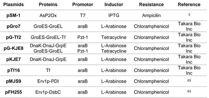
Table 2.1 Plasmid harbouring the wild-type asP2Ox gene and plasmids containing genes for the different
chaperones that were used in the co-expression using the BL21 star strain.
2.4.1.3Co-production of AsP2Ox with Hyper
The plasmid pSM-1 containing the asP2Ox gene was transformed into E.coli BL21 star, Tuner and KRX strains that were previously transformed with the Hyper-containing plasmid. Single colonies were used to inoculate 10 mL of LB medium supplemented with 100 µg/mL ampicillin (for cells with pSM-1 plasmid only), with 10 µg/mL kanamycin (for cells with Hyper-containing plasmid only) and 100 µg/mL ampicillin plus 10 μg/mL kanamycin (for cells harbouring both the pSM-1 and Hyper-containing plasmids). Cells were grown overnight at 37°C, 180 rpm. These cultures were used to inoculate 20 mL of LB medium supplemented with appropriated antibiotics at a starting OD600nm = 0.05. Cultures were incubated at
37°C, 180 rpm, and when OD600nm≃ 0.6, 100 µM IPTG was added to recombinant E. coli BL21 star and
Tuner strains, and 0.2 % rhamnose to recombinant E. coli KRX and the temperature was decreased to 25°C.
2.4.2 Cell disruption
For cultures grown in 96-well plates, a chemical cell disruption method was employed: cell pellets were suspended in 100 μL of 40% Bacterial Protein Extraction Reagent (B-PER®) lysis solution (Thermo
Scientific). After this, plates were centrifuged (4,000 rpm, 30 min at 4⁰C) and the supernatant collected (crude extracts).
For cultures grown at larger scales, the cell pellets were suspended in 20 mM Tris-HCl buffer at pH 7.6 containing 5 mM MgCl2, 10 μg/mL DNase I and 5 μg/mL of a mixture of protease inhibitors (antipain and leupeptin). The cell suspensions were disrupted in a French press (Thermo IEC) operating at 900 psi in a process repeated 3 times, allowing for the complete cellular rupture. The lysates were then centrifuged (13,000 rpm, 1 h at 4°C) to separate the non-soluble fraction (i.e. the pellet corresponding to cell debris) from the soluble fraction (crude cell extracts in the supernatant).
Plasmids Proteins Promotor Inductor Resistance Reference
pSM-1 AsP2Ox T7 IPTG Ampicilin 3
pGro7 GroES-GroEL araB L-Arabinose Chloramphenicol Takara Bio
Inc
pG-Tf2 GroES-GroEL-Tf Pzt-1 Tetracycline Chloramphenicol Takara Bio Inc pG-KJE8 DnaK-DnaJ-GrpE GroES-GroEL araB Pzt-1 L-Arabinose Tetracycline Chloramphenicol Takara Bio Inc
pKJE7 DnaK-DnaJ-GrpE araB L-Arabinose Chloramphenicol Takara Bio Inc
pTf16 Tf araB L-Arabinose Chloramphenicol Takara Bio
Inc
pMJS9 Erv1p-PDI araB L-Arabinose Chloramphenicol 63
14
2.4.3 Determination of protein concentration
Protein concentration was determined using the Bradford assay with bovine serum albumin (BSA) as standard64. A calibration curve was performed with known concentrations of BSA: 0 - 0.6 mg/mL and
20 μL of sample was added to 1 mL or 180 μl (96-well-plate) of Bradford reagent (100 mg of Comassie Blue G dissolved in 45% of ethanol, 100 mL orthophosphoric acid and 750 mL of distilled water, for 1 L of solution), mixed and 10 min later, the absorbance was measured at 595 nm in a spectrophotometer Novaspec III (Amersham Biosciences) or in a Synergy 2 (BioTek) micro plate reader. The protein measurements were performed in triplicate.
2.4.4 SDS-PAGE analysis
Sodium dodecyl sulfate polyacrylamide gel electrophoresis (SDS-PAGE), a method for separating proteins based on the difference of their molecular weight, was utilized to monitor the recombinant protein production as well as the level of protein purification. The running gel was prepared as follows: 12.5% (v/v) acrylamide, “Lower Tris buffer” (375 mM Tris-HCl, 0.1% SDS, pH 8.8), 0.1% (w/v) SDS, 0.1% (w/v) ammonium persulfate (APS), 0.06% (v/v) N, N, N’, N’-tetramethylethylenediamine (TEMED). For the stacking gel: 5% (v/v) acrylamide, “Upper Tris buffer” (125 mM Tris-HCl, 0.1% SDS, pH 6.8), 0.1% (w/v) SDS, 0.1% (w/v) APS, 0.1% (v/v) TEMED. The electrophoresis was performed at 220 V during 45 min in the “Electrophoresis Buffer” (192 mM glycine, 25 mM Tris, 0.1% SDS, pH 8.3). Before the application of the protein samples (10 μg of total protein in each well for crude extracts and 1 μg of purified protein) these were mixed with 2 × “Sample Preparation Solution” (2% SDS, 6.255 mM TrisHCl pH 6.8, 5% 2-mercaptoethanol, 0.5 mM DTT, 5% glycerol, and 0.025% bromophenol blue) and denatured at 99°C for 10 min. Gels were stained with Coomassie® blue staining solution (50% ethanol, 10% acetic acid, 0.05% (v/v) Coomassie brilliant blue R-250) with a slow agitation for homogeneous staining. Destaining was performed by using a solution of 10% ethanol and 10% acetic acid.
2.4.5 Enzymatic assays
The activity of AsP2Ox was measured using a horseradish peroxidase (HRP) coupled assay in which the hydrogen peroxide generated by AsP2Ox reaction is used as substrate by the HRP enzyme (Panreac AppliChem) to oxidise two molecules of 2,2'-Azinobis(3-ethylbenzthiazoline-6-sulfonic acid) (ABTS) generating the radical form of ABTS (green) that could be easily monitored at 420 nm (ε = 36,000 M-1 cm-1)65. Reactions were performed at 37ºC in a Nicolet Evolution 300 spectrophotometer from
Thermo Industries (Madison). The initial activity in crude cell extracts was measured by adding the enzymatic mixture of 100 mM sodium phosphate buffer at pH 6.5 containing 100 mM of D-glucose, 1 mM ABTS and 10 U HRP. One Unit of P2Ox activity was defined as the amount of enzyme that is necessary for the consumption of 2 μmol of ABTS per min, which equals the consumption of 1 μmol of O2 per min.
2.5 Fluorescence Microscopy
Cells co-producing heterologous Hyper and AsP2Ox proteins were grown overnight and in the following day 500 μl of each culture was centrifuged (6,000 rpm, 2 min). The cell pellet was suspended in