Faculdade de Medicina da Universidade de Lisboa
Topical bacteriophage therapy
of the infected diabetic foot
Doutoramento em Medicina
Especialidade de Medicina Interna
João João Mendes
2014
Faculdade de Medicina da Universidade de Lisboa
Topical bacteriophage therapy
of the infected diabetic foot
Tese orientada pelo Professor Doutor José Melo Cristino da Faculdade de Medicina da Universidade de Lisboa, e co-orientada pela Professora Doutora Patrícia Cavaco Silva do Instituto Superior de Ciências da Saúde Egas Moniz e pelo Professor Doutor Andrzej Górski da The Medical University of Warsaw / Institute of Immunology and Experimental Therapy, Polish Academy of Sciences, Wroclaw
Doutoramento em Medicina
Especialidade de Medicina Interna
Todas as afirmações efetuadas no presente documento são da exclusiva responsabilidade do seu autor, não cabendo qualquer responsabilidade à Faculdade de Medicina da Universidade de Lisboa pelos conteúdos nele apresentados
João João Mendes
2014
Conselho Científico da Faculdade de Medicina da Universidade de Lisboa em reunião
de 22 de julho de 2014
Table of Contents
Abstract ...ix
Resumo ...xi
Acknowledgements ...xv
List of Abbreviations ... xvii
List of Figures ... xix
List of Tables... xxi
List of Publications ... xxiii
Section A ... 1
Aims and Thesis Outline ... 3
General Introduction ... 9
Chronic wounds ... 9
Definitions and epidemiology ... 9
Healing pathophysiology ... 9
Bacterial infection ... 9
Wound infection management principles... 11
Diabetic foot infections ... 14
Definitions and epidemiology ... 14
Pathophysiology... 14 Assessment ... 16 Treatment ... 18 Bacteriophages ... 20 Introduction ... 20 Bacteriophage taxonomy ... 20 Bacteriophage lifecycle ... 20 Bacteriophage-biofilm interactions ... 22
Bacterial resistance to bacteriophages ... 23
Biological basis of bacteriophage therapy safety ... 25
Bacteriophage therapy ... 26
Introduction ... 26
Historical perspective ... 26
Bacteriophage therapy design ... 27
Human applications ... 33
Regulatory issues ... 35
Advantages and disadvantages of bacteriophage therapy relative to chemical antibiotics ... 36
Bacteriophage applications for the treatment of diabetic foot infections ... 37
Section B ... 51
Chapter 1: Epidemiological survey of diabetic foot infections ... 53
Specific framework ... 53
References ... 55
Clinical and bacteriological survey of diabetic foot infections in Lisbon. ... 57
Chapter 2: In vitro design of the bacteriophage cocktail ... 73
Specific framework ... 73
References ... 75
In vitro design of a novel lytic bacteriophage cocktail with therapeutic potential against organisms causing diabetic foot infections ... 77
Chapter 3: Rat model of diabetic wound infection ... 95
Specific framework ... 95
References ... 97
A rat model of diabetic wound Infection for the evaluation of topical antimicrobial therapies. ... 99
Chapter 4: In vivo topical bacteriophage therapy of infected diabetic wounds ... 119
Specific framework ... 119
References ... 124
Wound healing potential of topical bacteriophage therapy on diabetic cutaneous wounds. ... 125
Section C ... 145
General discussion ... 147
Future prospects ... 153
References ... 155
Abstract
Diabetes mellitus is a global epidemic, with an estimated 171 million diabetic patients worldwide. Diabetic wound infections, particularly diabetic foot infections (DFI), are a major source of morbidity and their treatment consumes a significant amount of medical economic resources. In addition to debridement, the current first-line treatment for DFIs is antibiotic therapy. However, an increasing number of antibiotic-resistant DFI cases require alternative treatments. One potential alternative is bacteriophage therapy (BT). The purpose of this study is to develop and evaluate a topically-delivered bacteriophage treatment protocol (including formulation and dosage regimen) with potential efficacy in diabetic wound infections.
The principles of wound infection management are discussed, and a comprehensive overview of DFI assessment and management is provided. The principles and current applications of BT are also examined, with particular regard to the potential for the inclusion of topical BT in the DFI therapeutic strategy.
In order to provide a thorough understanding of the epidemiology of DFI, a transversal observational multicenter study was conducted. Forty-nine hospitalized and ambulatory patients were enrolled, and 147 microbial isolates, comprising 43 different species, were cultured. This survey enabled the identification, characterization, and clinical correlation of the main bacteria involved, thus allowing the choice of the bacterial targets (Staphylococcus aureus, Pseudomonas aeruginosa and Acinetobacter baumannii). These targets were chosen not only because of their frequency but also because of their association with multi-drug resistance.
Following the epidemiological study, five previously morphologically and genetically characterized bacteria-specific lytic bacteriophages (S. aureus F44/10 and F125/10, P. aeruginosa F770/05 and F510/08, and A. baumannii F1245/05 bacteriophages) were tested against these strains. The bacteriophage spot test procedure was used, and their combination was shown to have broad host ranges for the different target bacterial species. Subsequently, the in vitro activity against planktonic cells and established biofilms was studied. Target bacteria during planktonic growth were challenged with their specific bacteriophages, which revealed effective, early killing at 4 hours, but this was followed by bacterial regrowth to pre-treatment levels within 24 hours. However, by using metabolic activity as a measure of cell viability within established biofilms, significant cell impairment following bacteriophage exposure was found when repeated treatment (every 4 hours) was applied. The greatest effects occurred in both planktonic and biofilm cells, showing a bacteriophage/bacterium input multiplicity (IM) of 10. These complementary studies on both planktonic cells and established biofilms allowed us to define a high IM (≥10) and a treatment protocol of multiple doses (every 4 hours for 24 hours).
When in vitro testing showed the feasibility of the project and indicated the optimal dosage regimen, the opportunity to study the bacteriophage solutions led to the development of an animal model of diabetic wound infection, which was suitable for the evaluation of topical antimicrobial therapies. The Galiano et al. murine splinted, excisional wound-healing model was used as a template and further refined and adapted using male chemically induced diabetic Wistar rats. After every step of the procedure was optimized, a model was established, which accurately reproduced not only the healing of infected wounds in humans but also the current treatment standard of care, including sharp debridement.
This led to the subsequent in vivo testing of the antimicrobial activity and wound-healing capability of the topically delivered bacteriophage protocol against wounds with S. aureus, P. aeruginosa, and
A. baumannii infections, using single strains of the bacteria, the aforementioned animal model, and a previously optimized pig wound infection model. In conjunction with sharp debridement, the bacteriophage treatment effectively decreased bacterial colony counts and improved planimetric and histological parameters in S. aureus and P. aeruginosa infections, but was not as effective against A. baumannii. Although the improvements were more significant in the rodent model than in the porcine model, our results suggested that topically administered bacteriophage treatment when applied in conjunction with wound debridement may be effective in resolving chronic wound infections. Incidentally, the results of the in vitro and in vivo studies were not only complementary but also yielded similar microbiological results, further highlighting the consistency and efficacy of the protocol across the tested scenarios.
However, this project is not without limitations. Only one strain each of the bacteria was used in the in vivo testing, which meant that the effectiveness of the treatment was limited to these strains and cannot be universally assumed to be effective against related, similar, or other untested strains. Moreover, although the animal models are appropriate in terms of their biological similarity to humans, they fail to account for the full complexity of human DFIs, which could influence the effectiveness of the protocol. Furthermore, only one type of BT protocol was tested. It was demonstrably effective, but it may not be the most efficacious form of BT protocol. Additionally, bacteriophage resistance was not examined or tested, so even if the BT protocol is effective, this study cannot predict outcomes for all scenarios in which the BT protocol may be used.
Nevertheless, collectively these studies show that a high IM (≥10) and multiple dose (every 4 hours for 24 hours), topical BT protocol may be an effective therapeutic approach to diabetic wounds infected with different pathogens, particularly those identified as antibiotic-resistant. These results represent the first step in the development of a fully regulated human clinical trial that explores the potential of BT in diabetic wound infections. The ultimate goal of this study is to transform BT into a viable, everyday strategy for treating DFIs.
Resumo
A diabetes mellitus configura-se hoje como uma epidemia global, com uma estimativa mundial de cerca de 171 milhões de diabéticos. As infeções de feridas em diabéticos, particularmente as infeções do pé diabético, são uma importante fonte de morbidade, e representam uma significativa fonte de consumo de recursos médicos e económicos. A par do desbridamento mecânico, o atual tratamento de primeira linha para as infeções do pé diabético é a terapêutica antibiótica. No entanto, são cada vez mais as infeções por bactérias resistentes aos antibióticos, o que incita ao desenvolvimento de terapêuticas alternativas. Uma alternativa potencial é a terapêutica bacteriofágica. O objetivo deste estudo foi o desenvolvimento e avaliação de um protocolo de terapêutica bacteriofágica tópica (incluindo a formulação e posologia) com potencial eficácia no tratamento de infeções de feridas de diabéticos.
São abordados os princípios gerais da terapêutica das infeções da pele e tecidos moles, com especial referência às infeções do pé diabético. Também os princípios gerais e as aplicações atuais e potenciais da terapêutica bacteriofágica são detalhados, destacando-se o potencial para a sua inclusão na estratégia terapêutica nas infeções do pé diabético.
A fim de proporcionar uma visão global da epidemiologia das infeções do pé diabético, conduziu-se um estudo observacional, multicêntrico e transversal. Foram recrutados 49 doentes de ambulatório e em hospitalização, tendo-se isolado 147 microrganismos, pertencendo a 43 diferentes espécies. Este estudo permitiu a identificação, caracterização e correlação clínica das principais bactérias responsáveis pelas infeções do pé diabético, permitindo, assim, a escolha dos alvos bacterianos (Staphylococcus aureus, Pseudomonas aeruginosa e Acinetobacter baumannii). Estes microrganismos foram escolhidos não só pela sua frequência, mas também pela sua associação à resistência aos antibióticos.
Na sequência do estudo epidemiológico, foram testados nestas estirpes cinco bacteriófagos líticos bactéria-específicos previamente caracterizados morfológica e geneticamente (bacteriófagos S. aureus F44/10 e F125/10, P. aeruginosa F770/05 e F510/08 e A. baumannii F1245/05), utilizando o procedimento de spot test para bacteriófagos. As combinações de bacteriófagos bactéria-específicos demostraram ter amplos host range para as diferentes espécies de bactérias-alvo. Posteriormente, a atividade destas combinações sobre células planctónicas e em biofilme maduro foi estudada in vitro. As bactérias-alvo em crescimento planctónico foram infetadas com diferentes combinações dos seus bacteriófagos específicos, obtendo-se eficácia inicial (nas primeiras 4 horas), porém, com recrescimento bacteriano para os níveis pré-tratamento em 24 horas. No entanto, em células de biofilmes maduros – usando a atividade metabólica como uma medida da viabilidade celular – obteve-se uma importante limitação da viabilidade após exposição repetida (a cada 4 horas) às diferentes combinações dos bacteriófagos específicos. Os maiores efeitos, tanto sobre células planctónicas como sobre as células em biofilme, ocorreu com uma input multiplicity de 10. Estes estudos complementares em células planctónicas e em biofilme maduro permitiram definir um protocolo terapêutico utilizando uma input multiplicity elevada (≥ 10) e múltiplas doses (a cada 4 horas durante 24 horas).
Uma vez que os testes in vitro mostraram a viabilidade do projeto e indicaram a posologia ideal, a oportunidade de estudar as soluções de bacteriófagos levou ao desenvolvimento de um modelo animal de infeção da ferida diabética adequado para a avaliação das terapêuticas tópicas antimicrobianas. O modelo murino excisional de cicatrização de feridas com utilização de tala de Galiano et al. foi adaptado e aperfeiçoado, utilizando ratos Wistar machos com diabetes mellitus
induzida quimicamente. Depois de otimizar cada etapa do processo, foi estabelecido um modelo que não só reproduz com precisão o processo de cicatrização de feridas infetadas em seres humanos, mas que reproduz igualmente o atual protocolo de tratamento destas feridas, incluindo o desbridamento mecânico.
Isto levou ao subsequente ensaio in vivo – no modelo animal acima mencionado, bem como num outro modelo porcino de infeção de ferida previamente otimizado – da atividade antimicrobiana e da capacidade de cicatrização de feridas do protocolo de terapêutica bacteriofágica tópica em feridas infetadas por estirpes individuais de S. aureus, P. aeruginosa e A. baumannii. A terapêutica bacteri-ofágica tópica, em conjunto com o desbridamento mecânico, resultou efetivamente na diminuição da contagem de colónias bacterianas bem como na redução da área de ferida e melhoria dos parâ-metros histológicos em feridas infetadas por S. aureus e P. aeruginosa, não tendo sido demonstrada tanta eficácia nas infetadas por A. baumannii. Estes resultados, embora mais expressivos no modelo de roedor do que no modelo porcino, sugerem que a terapêutica bacteriofágica tópica, quando aplicada em conjunto com o desbridamento mecânico, pode ser eficaz na resolução de infeções de feridas crónicas. Aliás, os resultados dos ensaios in vitro e in vivo não só são complementares, como também apresentam resultados microbiológicos semelhantes, destacando a consistência e eficácia do protocolo entre os cenários testados.
No entanto, este projeto não é isento de limitações. Apenas uma estirpe de cada uma das bactérias foi usada no teste in vivo, o que significa que a evidência da eficácia terapêutica é limitada a estas estirpes, não podendo ser assumida como eficácia universal, nomeadamente em estirpes não testadas. Ademais, e independentemente da similaridade biológica dos modelos animais com os humanos, eles não conseguem dar conta de toda a complexidade das infeções do pé diabético que pode influenciar a eficácia do protocolo. Além disso, apenas um tipo de protocolo de terapêutica bacteriofágica foi testado. Apesar de ter demonstrado eficácia, pode não ser o protocolo mais eficaz. Por fim, também a resistência aos bacteriófagos não foi avaliada, o que significa que, mesmo que o protocolo seja eficaz, o estudo não pode prever os resultados para todos os cenários em que o protocolo possa ser utilizado.
Coletivamente, estes estudos demostram que um protocolo de terapêutica bacteriofágica tópica usando uma input multiplicity alta (≥ 10) e doses múltiplas (a cada 4 horas durante 24 horas) pode ser uma abordagem terapêutica eficaz para a terapêutica de feridas diabéticas infetadas por diferentes agentes patogénicos, especialmente aqueles associados a resistência aos antibióticos. Estes resultados representam um primeiro passo no desenvolvimento de um ensaio clínico controlado e regulamentado para a avaliação do potencial da terapêutica bacteriofágica nas infeções de feridas de diabéticos. O objetivo final deste estudo é transformar a terapêutica bacteriofágica numa alternativa viável para o tratamento das infeções do pé diabético.
who never stopped believing – to my parents,
who laid the foundations of the man I am – to my grandparents,
who gave me the love and time that made everything else possible – to Maria Maria,
who taught me that life is not just Medicine – to Natália, João, and António,
Acknowledgements
The path towards completing this thesis has involved several years of work, and many people have contributed to the ideas presented and the knowledge gained. The author recognizes his debt to those who helped and positively influenced him along the way. It is, however, not possible to list them all here.
First, I want to thank my primary supervisor, Professor Melo Cristino, who gave me the latitude to explore the topic in my own way while constantly providing encouragement, advice, and guidance. I also wish to express my gratitude to my co-supervisors, Professor Patrícia Cavaco Silva and Professor Andrzej Górski, for their continuous encouragement and invaluable suggestions during this work.
I gratefully acknowledge the funding sources that made the work for my doctoral thesis possible. I would like to thank Professor Miguel Garcia, who enabled this research work at Technophage— Investigação e Desenvolvimento em Biotecnologia, S.A. and, together with Tecnifar—Indústria Técnica Farmacêutica, S.A., provided funding for this research. My work was also supported by a Fundação para a Ciência e a Tecnologia PhD studentship grant for medical residents (SFRH/ SINTD/60025/2009).
I thank my colleagues at TechnoPhage, S.A. for providing technical support, especially the stimulating team exercises aimed at developing solutions to the identified problems and for the sleepless nights we worked together. I am deeply indebted to my colleague and co-author, Clara Leandro, for her extremely valuable support and insights, without which this thesis would not have been possible. I would also like to thank Carlos São José and Madalena Pimentel for offering thorough and excellent feedback on the papers and on an earlier version of this thesis.
The epidemiological study was made possible only by the commitment of all professionals in various health facilities. I would therefore like to thank all those involved at Associação Protetora dos Diabéticos de Portugal and Centro Hospitalar de Lisboa Central EPE. I am especially indebted to José Neves, whose constant, positive influence and implicit encouragement sustained me throughout the work for this PhD.
The animal studies were also possible only through the total engagement of the Instituto de Medicina Molecular Animal Facility and the Faculdade de Medicina da Universidade de Lisboa Experimental Surgery staff, as well as the technical support provided by the Instituto de Medicina Molecular BioImaging Unit and Histology Service staff. I especially thank Dolores Bonaparte, whose experience in animal research was essential for the development of the project.
All the laboratory work for the epidemiological study and the biofilm studies was done at the Faculdade de Medicina Veterinária da Universidade Técnica de Lisboa. This was made possible by the unwavering support of Professor Cristina Lobo Vilela, whose positive attitude and enthusiasm were and continue to be a true inspiration.
I would also like to thank Dr.ª Alexandrina Quintino and Professor António Sousa Guerreiro, Heads of the Internal Medicine Department of the Centro Hospitalar de Lisboa Central, and Rita Barata Moura, my clinical supervisor, for their constant encouragement and for giving me enough time to complete the thesis.
Finally, I want to thank my family. My beloved wife Natália and our sons João and António are a continuous and powerful source of inspiration and energy. I extend my special gratitude to my grandparents, parents, and sister for their never-ending support.
List of Abbreviations
AB – alamarBlue
Abi – abortive Infection system BT – bacteriophage therapy CFU – colony-forming units
CRISPR – Clustered Regularly Interspaced Short Palindromic Repeats CRTB – clinically relevant tissue burden
DFI – diabetic foot infection DFU – diabetic foot ulcer DG – dermal gap
DM – diabetes mellitus DNA – deoxyribonucleic acid EG – epithelial gap
ESBL – extended-spectrum β-lactamase ds – double-stranded
EMA – European Medicine Agency EPS – extracellular polymeric substance FDA – Food and Drug Administration GT – granulation tissue
HCP – health care provider HDI – hair-density index
ICTV – International Committee for Taxonomy of Viruses IM – input multiplicity
i.p. – intraperitoneal i.v. – intravenous
MDR – multi-drug resistant
MRCN – methicillin-resistant coagulase-negative Staphylococcus spp. other than Staphylococcus epidermidis
MRSA – methicillin-resistant Staphylococcus aureus MRSE – methicillin-resistant Staphylococcus epidermidis P.H.A.G.E. – Phages for Human Applications Group Europe PDR – pan-drug resistant
RM – Restriction-Modification SDI – skin-damage index ss – single-stranded
Sie – superinfection exclusion system TAT – topical antimicrobial therapy TSA – tryptone soy agar
TcPO2 – transcutaneous oxygen pressure US – United States
List of Figures
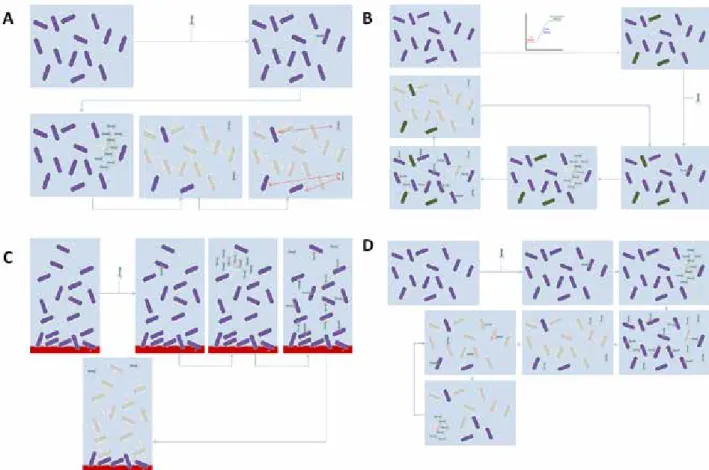
Figure A0-1 – General outline of Section B of the thesis. ... 7
Figure A1-1 – Qualitative aspects of acute and chronic wounds. ... 10
Figure A1-2 – Wound infection continuum. ... 11
Figure A1-3 – Common wound infection management principles. ... 13
Figure A1-4 – Diabetic foot infection pathophysiology. ... 15
Figure A1-5 – Management of diabetic foot infections. ... 19
Figure A1-6 – Common steps of the bacteriophage lytic replication process properly correlated with graphical representation of the one-step-growth curve. ... 21
Figure A1-7 – Bacteriophage-biofilm interactions. ... 23
Figure A1-8 – Theories suggested to explain bacteria-bacteriophage coexistence and coevolution. ... 24
Figure A1-9 – Timeline depicting the comparative evolution of bacteriophage therapy, discovery of antibiotics and the Staphylococcus aureus resistance profile, as an illustrative example of the bacterial resistance evolution. ... 27
Figure A1-10 – Bacteriophage therapy concepts: prêt-à-porter or sur-mesure. ... 28
Figure A1-11 – Bacteriophage kinetics. ... 30
Figure B1.0-1 – Study outline of epidemiological survey of diabetic foot infections. ... 54
Figure B1.1-1 – A box plot representing the ulcer duration data stratified by the microbial isolate. ... 65
Figure B2.0-1 – In vitro design of a lytic bacteriophage cocktail with therapeutic potential against organisms causing diabetic foot infections. ... 74
Figure B2.1-1 – Morphological and genomic characteristics of the bacteriophages used for bacteriophage therapy. ... 82
Figure B2.1-2 – Time-kill curves of the target bacteria during planktonic growth when challenged with their specific bacteriophages (alone or in combination). ... 85
Figure B2.1-3 – Analysis of the bacteriophages’ activity against bacterial biofilms. ... 87
Figure B3.0-1 – Wound kinetics as quantified by planimetry and histology. ... 96
Figure B3.2-1 – Schematic representation of diabetic rodent models of excisional wound healing. ... 101
Figure B3.2-2 – Illustration of specific techniques. ... 103
Figure B3.2-3 – Final study design: optimized rodent wound infection model. ... 106
Figure B3.2-5 – Results of the final wound-infection model study. ... 110 Figure B3.2-6 – Quantitative histologic evaluation of epithelial gap, dermal gap,
and granulation tissue area in the negative control and both infected
(Staphylococcus aureus or Pseudomonas aeruginosa) groups on days 9 and 11. ... 111 Figure B4.1-1 – Topical bacteriophage therapy protocol in the rodent
diabetic chronic wound infection model. ... 120 Figure B4.1-2 – Topical bacteriophage therapy protocol in the pig chronic
wound infection model. ... 122 Figure B4.2-1 – Schematic depicting bacteriophage cocktail preparation. ... 127 Figure B4.2-2 – Average swab colony count in infected rats;
and wound closure kinetics in rats ... 132 Figure B4.2-3 – Histological wound analysis in rats. ... 134 Figure B4.2-4 – Average swab colony count in infected pigs;
and wound closure kinetics in pigs. ... 136 Figure B4.2-5 – Histological wound analysis in pigs. ... 138
List of Tables
Table A1-1 – PEDIS classification system. ... 17 Table B1.1-1 – Clinical and microbiological characteristics
of diabetic foot infections stratified by the sample collection method. ... 62 Table B1.1-2 – Distribution of the diabetic foot infection isolates. ... 64 Table B1.1-3 – Distribution of the diabetic foot infection isolates
in relation to current antibiotic therapy. ... 66 Table B1.1-4 – Relationship between multi-drug resistant organisms
and recent (≤3 months) or current antibiotic therapy. ... 67 Table B2.1-1 – Susceptibility of wound bacterial isolates
to candidate bacteriophages for bacteriophage therapy. ... 83 Table B3.2-1 – Evaluation of 3 strategies for preventing unintentional critical colonization. ... 109 Table B3.2-2 – Bacterial colony counts from tissue or swab culture
List of Publications
This doctoral thesis is based on the following articles, which, as first author, I published in peer-reviewed journals:
Paper 1:
Mendes JJ, Marques-Costa A, Vilela C, Neves J, Candeias N, Cavaco-Silva P, Melo-Cristino J. Clinical and bacteriological survey of diabetic foot infections in Lisbon.
Diabetes Res Clin Pract. 2012 Jan;95(1):153-61. doi: 10.1016/j.diabres.2011.10.001. Epub 2011 Oct 21.
PMID: 22019426 [PubMed – indexed for MEDLINE]
JJM conceived the study and developed the data collection tools. JJM, AMC, JN and NC collected the data. CV contributed to the conception and design of the study and conducted the microbiological analysis. PCS and JMC helped with the writing of the manuscript. JJM analyzed the overall data and prepared the manuscript.
Paper 2:
Mendes JJ, Leandro C, Mottola C, Barbosa R, Silva FA, Oliveira M, Vilela CL, Melo-Cristino J, Górski A, Pimentel M, São-José C, Cavaco-Silva P, Garcia M.
In vitro design of a novel lytic bacteriophage cocktail with therapeutic potential against organisms causing diabetic foot infections.
J Med Microbiol. 2014 May 28. pii: jmm.0.071753-0. doi: 10.1099/jmm.0.071753-0. [Epub ahead of print]
PMID: 24869663 [PubMed – as supplied by publisher]
JJM and CL conceived and designed the overall project. CSJ, MP, and MG planned the experiments on bacteriophage isolation and characterization. MO and CLV planned the experiments on established biofilms. CL, RB, and FAS conducted the bacteriophage isolation, amplification, purification, and DNA extraction. CL and RB additionally performed the bioinformatics analysis and assessed the bacteriophages’ host range and activity against planktonic cells. CM conducted the experiments on established biofilms. CSJ, MO, CLV, PCS, JMC, and AG contributed reagents, materials, and analysis tools and helped with the writing of the manuscript. CSJ, MP, and MG analyzed the data related to the bacteriophages’ morphological and genomic characterization. JJM analyzed the overall data and prepared the manuscript.
Paper 3:
Mendes JJ,Leandro C, Bonaparte D, Pinto A.
A rat model of diabetic wound Infection for the evaluation of topical antimicrobial therapies. Comp Med. 2012 Feb;62(1):37-48.
PMID: 22330650 [PubMed - indexed for MEDLINE]
JJM conceived and designed the overall project. JJM, CL and DB conducted the animal studies. AP conducted the histologic analysis. JJM analyzed the overall data and prepared the manuscript.
Paper 4:
Mendes JJ,Leandro C, Corte-Real S, Barbosa R, Cavaco-Silva P, Melo-Cristino J, Górski A, Garcia M. Wound healing potential of topical bacteriophage therapy on diabetic cutaneous wounds.
Wound Repair Regen. 2013 Jul-Aug;21(4):595-603. doi: 10.1111/wrr.12056. Epub 2013 Jun 11.
PMID: 23755910 [PubMed - indexed for MEDLINE]
JJM and CL conceived and designed the overall project. JJM, CL and RB conducted the experi-ments. PCS, JMC, and AG contributed reagents, materials, and analysis tools and helped with the writing of the manuscript. JJM analyzed the overall data and prepared the manuscript.
Parts of the introduction are also based on the following publication: Mendes JJ, Neves J.
Diabetic foot infections: Current diagnosis and treatment. The Journal of Diabetic Foot Complications. 2012;4(2): 26-45. JJM prepared the manuscript. JN reviewed the manuscript.
Aims and Thesis Outline
Aims
The main objective is the development of a topically delivered bacteriophage treatment protocol (including formulation and dosage regimen) with potential efficacy in diabetic wound infections.
The secondary objectives are the following:
– conduct an epidemiological survey of diabetic foot infections in Lisbon, allowing the identification and characterization of the main bacteria involved and their correlation with clinical data;
– investigate, in vitro, the antimicrobial activity of bacteriophage solutions against planktonic cells and established biofilms;
– develop an animal model of diabetic wound infection suitable for the evaluation of topical antimicrobial therapies, namely topical bacteriophage therapy; and
– investigate, in vivo, the antimicrobial activity and wound-healing capability of bacteriophage solutions delivered topically to wounds in an animal model of diabetic chronic wound infection.
Thesis outline
This thesis is divided into three sections (A, B, and C).
Section A includes the aims and outline of the thesis as well as the general introduction, which
describes the context of the research, providing a theoretical foundation for the thesis. The General Introduction presents background information on chronic wounds, specifically diabetic foot infections, and provides the current framework for their diagnosis and treatment. In addition, it gives a general outline of bacteriophage biology and bacteriophage therapy, especially concerning topical therapy.
Section B, whose general outline is presented in Figure A0-1, is divided into four chapters. Chapter 1 presents the epidemiological survey of diabetic foot infections in Lisbon, allowing the identification, characterization and clinical correlation of the main bacteria involved, and thus justifying the chosen bacterial targets. Chapter 2 presents a detailed characterization of the bacteriophages used with regard to their spectrum of activity, and their genetic and morphological structure. It also describes their activity against planktonic cells and established biofilms, justifying the posology and dosage regimen used in the animal studies. Chapter 3 presents the optimization of a new wound infection model in chemically induced diabetic Wistar rats, which is suitable for the evaluation of topical antimicrobial therapies, namely bacteriophage therapy. Chapter 4 presents the study of the antimicrobial and wound-healing capability of bacteriophage solutions delivered topically to wounds in two (rodent and porcine) animal models of diabetic chronic wound infection. Each chapter is introduced by a short text that frames it according to the general outline of the thesis.
Section C includes chapters on the general discussion and future research. The chapter on
General discussion summarizes and discusses the main findings, as well as the implications and methodological limitations of the results obtained. The chapter on Future prospects presents future research directions that could provide the next steps along the path to a practical and widely applicable topical bacteriophage therapy. In addition, it also presents potential applications and future research directions outside the area of bacteriophage therapy.
Figur e A0-1 – Gener al outline of Section B of the thesis. Each num ber ed shaded ar ea corr esponds to a chap ter . F or additional de tails please re fer to the t ex t.
General Introduction
Chronic wounds
Definitions and epidemiology
Chronic wounds, such as diabetic foot ulcers (DFUs), chronic venous leg ulcers, and pressure ulcers, are defined as wounds that have failed to proceed through an orderly and timely reparative process to produce anatomic and functional integrity over a period of 3 months (1). It has been estimated that around 1% of the population is affected by non-healing wounds, and the most affected population is over the age of 65 (2). Therefore, future aging trends in the world’s population are likely to lead to a significant increase in the incidence of these wounds over the next 20 years. This is of particular relevance because chronic wounds are a major source of pain and disability in patients, and their treatment involves considerable economic cost (3).
Healing pathophysiology
Human skin is a complex and uniquely constructed organ with vital functions. Wounds occur when the integrity of the skin tissue is disrupted. In the acute wound setting, when this protective barrier is broken, the physiologic process of healing is immediately set in motion. In this classic model (4), wound healing is divided into three sequential but overlapping phases: (1) the inflammatory phase; (2) the proliferative phase (re-epithelialization, granulation and neo-angiogenesis); and (3) the remodeling phase (extracellular matrix remodeling). This complex, orchestrated biochemical cascade is characterized by signature events, cells, and their molecular regulators. For a complete review of the various cellular and inflammatory pathways involved in acute wound healing please refer to Schreml et al. (5).
Although most acute wounds heal in an uncomplicated fashion, following the aforementioned orderly and timely repair process, a large number of non-healing wounds fail to establish a sustained anatomic and functional result (6). Despite the differences in origin, non-healing wounds display common clinical features, include the following: the presence of necrotic tissue, lack of adequate blood supply, and excess exudate (7). Continuous bacterial clinical or sub-clinical infections (8) limit the cytokine-mediated switch to the later granulation tissue formation phase, resulting in prolonged inflammation and increased neutrophil infiltration with consequent protease activity. A persistent inflammatory phase is commonly witnessed by histopathology, which is associated with a delay in the formation of mature granulation tissue and failure of re-epithelialization (9).
Bacterial infection
As previously noted, clinical or subclinical infection is considered a common reason for impaired wound healing (10). Recent studies have emphasized both qualitative and quantitative aspects of wound microbiology, as well as the host’s immune response, as critical determinants of wound outcome.
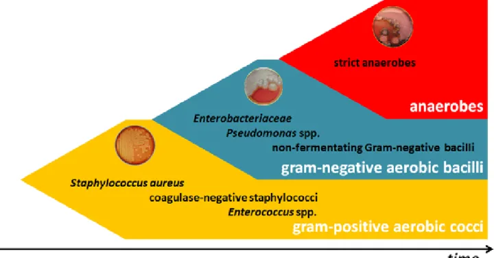
In relation to qualitative aspects, although important differences exist in the microbiology of various chronic wounds, some common concepts can be presented (Figure A1-1). Gram-positive cocci, namely Staphylococcus aureus, are the first microorganisms to colonize1 and acutely infect2
breaks in the skin. Chronic wounds develop a complex polymicrobial microbiology, including aerobic gram-negative rods and anaerobes. Nonfermenting gram-negative bacilli are found in many patients 1 Presence of multiplying bacteria in a wound but not causing tissue damage
with chronic or previously treated infections, and Pseudomonas aeruginosa is specifically associated with wounds treated with wet dressings (11). Anaerobes are rarely the sole pathogen, but they often participate in a mixed infection with aerobes, especially in cases of deep tissue infection (12). These mixed infections provide an optimal opportunity for microbial synergy, which increases the net pathogenic effect and hence the severity of infection (13). Thus, the presence of specific pathogens is less important than are the composition of the polymicrobial wound microbiota and the presence of additional potentiating factors, namely bacterial biofilms. Bacterial biofilms are formed when planktonic phenotype bacteria attach to the wound surface and colonize into highly organized structures composed of extracellular polymeric substance (EPS) produced by the microorganisms and the host’s surrounding tissues (14). Within the biofilm, both the protective outer coating and the altered bacterial phenotype contribute to the enhanced resistance of microorganisms to the host response, as well as to various antibiotic treatments. The described formation and behavior of the entire biofilm community is directed by signaling molecules that are produced when microorganisms reach a critical number—critical colonization3. This phenomenon is termed “quorum sensing,” and
it has been shown to be a key regulator of the expression of virulence factors as well as a modulator of host immunity (15).
Figure A1-1 – Qualitative aspects of acute and chronic wounds. Gram-positive cocci, namely S. aureus, are the first microorganisms to colonize and acutely infect breaks in the skin. Chronic wounds develop a complex polymicrobial microbiota, including aerobic gram-negative rods and anaerobes.
Assuming that the qualitative microbiology remains constant, the probability of wound infection increases as the microbial load increases to a critical level, when infection or a failure to heal is considered almost inevitable (13). Breidenbach et al. (16) established this critical level in complex extremity wounds, as a bacterial tissue count >105 of colony-forming units (CFU)/g. However, there
are exceptions to this rule of thumb because various organisms have different intrinsic virulence 3 Presence of multiplying bacteria in a wound adversely affecting wound healing while not causing classical clinical sings of infection
potentials. A good example is ß-hemolytic streptococci, which are able to induce tissue damage at 102 CFU per gram of tissue, while greater counts of less pathogenic organisms may be of little clinical
significance (17).
Another critical factor is the efficacy of the host’s immune response in dealing with wound microbiota. Infection is facilitated by local potentiating factors, such as tissue necrosis and hypoxia (caused by poor local perfusion accentuated by the hypermetabolic state and microbial cellular metabolism), which impair the immune cell activity in the wound environment (18).
All these complex interactions have been systematized in the wound infection continuum (Figure A1-2). This concept describes the effects of increasing bacteria quantity and diversity in wound tissue and their relationship to the quality of the host’s immune response (19).
Figure A1-2 – Wound infection continuum. Wound infection can be defined as the process by which organisms bind to tissue, multiply, and then invade tissue and elicit an immune response. It can be illustrated as an infection continuum or shown as an equation. The quantity of microbes representing the states of colonization, critical colonization, and infection are unique and related to the quality of the host immune response. At a certain quantity, these organisms may begin quorum sensing or communicating chemically, triggering the expression of virulence factors, namely the production of biofilm.
Wound infection management principles
Correctly identifying the etiology of a chronic wound, as well as the local and systemic factors that may contribute to poor wound healing, is essential to an adequate therapy and key in successful
wound management (1). However, regardless of the specific type of wound, general wound management principles exist for all chronic wounds (20) (Figure A1-3).
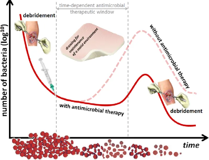
The initial and most important step in the management of any chronic wound, infected or not, is to remove the necrotic material and debris until normal tissue appears. This is called debridement, which reveals the healthy tissue required for wound healing while the wound bed is cleansed of bacterial biofilms (21). Although there are several modalities of debridement, sharp debridement is generally regarded as fast and effective and, as elegantly demonstrated by Wolcott et al. (22), also opens a time-dependent antimicrobial therapeutic window. A serial debridement strategy further enhances this effect, by enabling the frequent disruption of the biofilm and increasing its vulnerability to treatment (23). This strategy is not only theoretical but also has been recognized as effective in a retrospective analysis of patients in a randomized controlled trial of growth factor therapy in DFUs (24).
Antimicrobial therapy may further enhance the reduction of the number of bacteria in chronic wounds (25). Although systemic infection is treated with systemic antibiotics (13), these do not effectively decrease bacterial levels in granulating wounds where, theoretically, topically applied antimicrobials (topical antibiotics or antiseptics) could be more effective (26). Irrespective of the definitions of antibiotics and antiseptics, for which a lack of consensus within the literature exists, these may be divided into two categories: antimicrobial solutions used to irrigate wounds and antimicrobial preparations designed for longer periods of contact time. The former usually have only a brief contact time with the wound surface and include hypochlorites and substances, such as potassium permanganate. The latter are normally developed as creams, ointments, and impregnated dressings, including most topical antibiotics (e.g. fusicidic acid) and silver-based products (e.g. silver sulphadiazine). Some products (available in different forms) fall into both categories: povidone iodine, chlorhexidine and hydrogen peroxide (27). Controversy has long surrounded the use of topical antimicrobial agents because of the lack of adequate proof of their efficacy, reports of cytotoxicity, and risk of antibiotic-resistance induction, which depends on the particular formulation, concentration of active ingredient, and duration of exposure (28). Most efficacy studies are suboptimal and have varied designs that are not easily comparable (28). Moreover, most have not considered or have even excluded debridement (27). However, a systematic review of controlled trials (29) showed that several topical substances hastened healing and induced a few improved outcomes. A recent Cochrane systematic review (30) concluded that evidence supported the use of a topical antiseptic in a specific chronic wound type. Regardless of the decision to initiate topical antimicrobial therapy, general consensus exists about its discontinuation when bacterial balance has been achieved because protracted courses of antibiotics may inhibit wound healing and promote the development of resistant organisms (20).
After preparation of the wound bed by using debridement and antimicrobial agents, a moist environment, which has been accepted as the best topical environment for open wounds (20), should be maintained. If, despite all efforts of optimization, the wound fails to heal in a timely fashion, surgical closure is generally recommended.
Figure A1-3 – Common wound infection management principles. The initial and most important step in the management of any chronic wound is debridement, which opens a time-dependent antimicrobial therapeutic window. Antimicrobial therapy (topical antibiotics or antiseptics) may further enhance the reduction of the number of bacteria in chronic wounds.
Diabetic foot infections
Definitions and epidemiology
The world is facing a major epidemic of diabetes mellitus (DM). There are an estimated 171 million diabetic patients worldwide, and this number is expected to double by the year 2030 (31). All DM patients are at risk of developing a diabetic foot ulcer (DFU), which is a full-thickness wound below the ankle, irrespective of duration. Based on current studies, the annual population-based incidence is 1 to 4% with a prevalence of 4 to 10%, and the estimated lifetime risk is 25% (32). According to a study published by the Eurodiale study group (33), approximately 58% of DFU patients will become clinically infected. Patients with DM frequently require minor or major amputations of the lower limbs (15 to 27%), and in more than 50% of cases, infection is the preponderant factor (34). Major amputation is associated with significant morbidity and mortality (ranging from 13 to 40% at 1 year to 39 to 80% at 5 years (32)), in addition to immense social, psychological and financial consequences (35). The treatment of diabetic foot infections (DFI) accounts for up to one-quarter of all diabetic admissions in both Europe and the United States (US), making it the single most common reason for DM-related hospital admission (36). The solution to this predictable progression includes the development of structured screening tools to identify those at risk and the implementation of standardized education and prevention protocols. However, as stated by Lavery et al. (37), even with the best preventive standard of care, 9% of patients with DM will still develop a DFI, with the consequent risk of amputation.
Pathophysiology
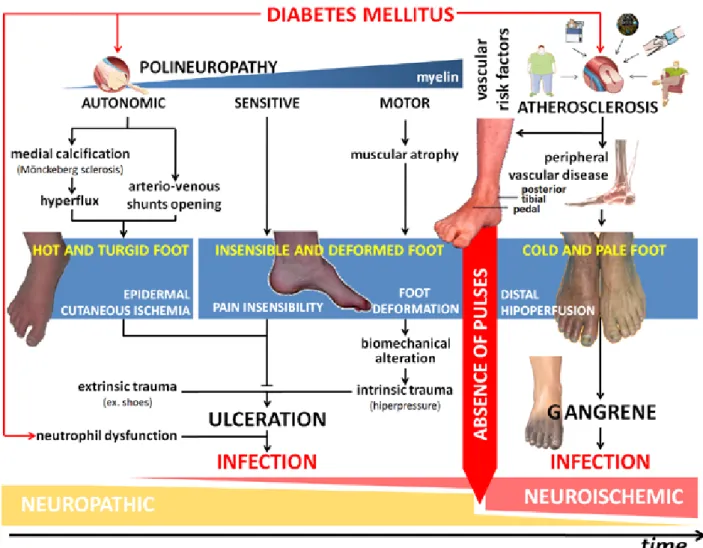
A prior DFU is an almost obligatory prerequisite for DFIs, even though, in some cases, the wound may have closed before DFI presentation (37). DFUs have a multifactorial nature, as numerous observational studies have indicated. It is well established that insulin deficiency (absolute or relative) is the basis of the biochemical abnormalities that lead to the organic complications of DM and the biological deficits of tissue healing and regeneration (38). DFUs result from a complex interaction of two major risk factors: neuropathy—both symmetric and bilateral, with varying degrees of alterations in autonomic, sensory and motor functions —plays the main role, while peripheral vascular disease resulting from atherosclerosis plays a secondary role (Figure A1-4). Approximately 50 to 60% of all DFUs can be classified as neuropathic. Signs or symptoms of vascular compromise are observed in 40 to 50% of all patients. The vast majority of patients have neuroischemic ulcers, and only a minority of patients have purely ischemic ulcers (39). However ulceration of the diabetic foot, either neuropathic or ischemic, does not occur spontaneously; it follows some form of extrinsic (e.g., low-pressure trauma from ill-fitting shoes) or intrinsic trauma (e.g., from the atrophy induced by motor neuropathy of the foot’s intrinsic muscles) (40).
Once the protective layer of skin is broken, deep tissues are exposed to bacterial colonization. In DFUs, in addition to the considerations discussed in the previous chapter, infection is facilitated by ischemia, the particular anatomy of the foot (i.e., it is divided into several compartments, which explains the rapid spread of infection), and intrinsic immunological deficits, especially in terms of neutrophil dysfunction (41).
Figure A1-4 – Diabetic foot infection pathophysiology. Diabetic foot ulcer results from the complex interaction of a number of risk factors. Neuropathy (with alterations in motor, sensation, and autonomic functions) plays the central role and causes ulcerations because of trauma or excessive pressure in a deformed foot without protective sensibility. Once the protective layer of skin is broken, deep tissues are exposed to bacterial colonization. Infection is facilitated by diabetes mellitus-related immunological deficits, especially in terms of neutrophils, and it rapidly progresses to the deep tissues.
Bacterial infection
The microbiologic features of DFIs develop according to the already described microbiological principles of chronic wounds, with some specificities. Acute infections in patients who have not recently received antimicrobials are often monomicrobial (almost always with an aerobic gram-positive cocci), whereas chronic infections are often polymicrobial (including gram-gram-positive and gram-negative aerobes and anaerobes (18)). However, the impaired host’s defenses around necrotic tissue allow low-virulence bacteria, such as coagulase-negative staphylococci and Corynebacterium species, to assume a pathogenic role (11).
The intrinsic pathophysiological characteristics of DFIs, along with long periods of hospitalization, complex surgical procedures, and prolonged broad-spectrum antibiotic therapy, are also predisposed to infection with antibiotic-resistant organisms (e.g., methicillin-resistant S. aureus [MRSA] or vancomycin-resistant enterococci [VRE]) (18, 42).
Assessment
Recognizing important risk factors and making a logical treatment-oriented assessment of DFIs requires a consistent and thorough diagnostic approach. Such evaluation involves the careful assimilation of global medical foot and wound history, a systemized and detailed physical examination, and the results of complementary diagnostic procedures. Various systems have been proposed to classify DFUs, but none has gained widespread acceptance. The International Working Group of the Diabetic Foot developed the PEDIS classification system (43), which consists of internationally applicable guidelines that can reliably predict the outcome of diabetic foot management (44). The PEDIS system classifies all DFUs into the subcategories of five main parameters (perfusion, extent/size, depth/tissue loss, infection and sensation), according to strict criteria (Table A1-1). Although it was not developed as a guide for daily management or to predict the outcome of an individual patient, it considers all the potentially useful information obtained from the patient’s clinical history, foot examination, and diagnostic exams. Consequently, the use of this systematic examination ensures that important aspects are not overlooked.
The assessment of DFIs based on the PEDIS classification has been reviewed elsewhere (45). In this chapter, we will consider only the evaluation of infection. The diagnosis of infection is clinical, based on the presence of symptoms and signs of inflammation (39), and it must always be confirmed and classified according to the depth of involvement. In the PEDIS grading system, three parameters are particularly relevant to clinical management and outcome: the involvement of skin only (Grade 2); the involvement of deeper structures (Grade 3); and the patient’s systemic inflammatory response (Grade 4). To categorize the patient definitively into one of these groups, different diagnostic procedures are indicated.
All patients should have a complete blood count with differential, erythrocyte sedimentation rate and c-reactive protein testing. However, caution must be exercised when interpreting laboratory tests because no marker is sufficiently sensitive or specific to confirm the diagnosis of DFI, and tests are often misleading, even in the case of severe lesions (46). In these patients, the most sensible sign of infection is often recalcitrant hyperglycemia, despite regular anti-hyperglycemic regimens.
Another problem is determining the presence of osteomyelitis. The International Working Group on the Diabetic Foot has proposed consensus criteria (47) for diagnosing diabetic foot osteomyelitis; however, these remain to be validated by a properly designed trial. A positive probe-to-bone test (i.e., when a sterile metal probe reveals a hard and gritty surface compatible with bone) in the presence of DFI appears to have a relatively variable positive predictive value, while a negative test in a low-risk patient markedly decreases the likelihood of osteomyelitis (48). This simple technique may be complemented by imaging studies (e.g., plain radiographs and magnetic resonance imaging). However, the gold standard criterion for diagnosing osteomyelitis is a characteristic histopathology (acute or chronic inflammatory cells or necrosis) associated with a positive culture from a bone specimen ideally obtained at the time of surgical debridement or by fluoroscopic- or computed tomography-guided percutaneous biopsy (47).
Table A1-1 – PEDIS classification system. The International Working Group of the Diabetic Foot’s PEDIS system classifies all foot ulcers in subcategories of five main categories (perfusion, extent/ size, depth/tissue loss, infection and sensation), according to strict criteria. PaCO2: partial pressure of carbon dioxide in the arterial blood; TcpO2: transcutaneous oxygen pressure
Categories Grades Description
Perfusion
grade 1
No symptoms or signs of peripheral artery disease in the affected foot in combination with
– palpable dorsal pedal and posterior tibial artery or – ankle-brachial index 0.9 to 1.10 or
– toe-brachial index >0.6 or
– TcpO2 >60 mmHg
grade 2
Symptoms or signs of peripheral artery disease but not of critical limb ischemia: – presence of intermittent claudication or
– ankle-brachial index < 0.9, but with ankle pressure >50 mmHg or – toe-brachial index<0.6, but systolic toe blood pressure >30 mmHg or
– TcpO2 30-60 mmHg or
– other abnormalities on non-invasive testing, compatible with peripheral artery disease but with critical limb ischemia
grade 3
Critical limb ischemia:
– systolic ankle blood pressure <50 mmHg or – systolic toe blood pressure <30 mmHg or
– TcpO2 < 30 mmHg
Extent Wound size after debridement (measured in square centimeters)
Depth
grade 1 Superficial full-thickness ulcer, not penetrating deeper than the dermis
grade 2 Deep ulcer, penetrating to subcutaneous structures (fascia, muscle or tendon)
grade 3 Deep ulcer, penetrating any of the subsequent layers of the foot (bone or joint)
Infection
grade 1 No symptoms or signs of infection
grade 2
Infection involving the skin and the subcutaneous tissue only at least two of the following items must be present:
– local swelling or induration
– erythema >0.5 to 2 cm around the ulcer – local tenderness or pain local warmth
– purulent discharge (thick, opaque to white or sanguineous secretion)
grade 3 Infection involving structures deeper than skin and subcutaneous tissues orerythema >2 cm plus one of the items described above
grade 4
Foot infection accompanied by signs of systemic inflammatory response syndrome: – temperature >38 ºC or <36 ºC
– heart rate >90 beats/min
– respiratory rate >20 breaths/min (or PaCO2 <32 mmHg)
– white blood cell count >12.000 or <4.000 cells/mm3
(or 10% band forms)
Sensation
grade 1 No loss of protective sensation
grade 2
Loss of protective sensation
absence of perception of the one of the following tests:
– absent pressure sensation, determined with a 10-g monofilament on two out of three sites on the plantar side of the foot or
– absent vibration sensation, determined with a 128-Hz tuning fork tested on the hallux
In the absence of suspected osteomyelitis, bacteriological sampling, which must be done after mechanical debridement and cleansing of the wound with gauze soaked in sterile physiological saline, is indicated if infection is clinically suspected. The best sampling technique remains a matter of debate. While tissue biopsy and fluid aspirate are considered the gold standard for diagnosing wound infection (13), such invasive tests are infrequently performed for superficial wounds or in many practice settings, such as outpatient clinics, because of concerns about enlarging the ulcer or inducing pain (13, 49). Superficial swabbing of the wound is discouraged, but swabbing the base of the ulcer is acceptable if it is the only possible option (50). Independent of the sampling method, specimens must be placed in transport medium and be sent to the microbiology laboratory as quickly as possible. Assuming that at present, there are no completely reliable microbiological methods to distinguish between pathogenic and nonpathogenic microorganisms, microbiologists and clinicians must collaborate closely to interpret the results, taking into account the sampling conditions, transport time and conditions, and the type of bacteria isolated.
Treatment
When a DFI patient presents to the care team, a multidisciplinary management strategy should be rapidly implemented (Figure A1-5) because evidence suggests that this reduces the incidence of major amputation. The multidisciplinary team should include the following: a diabetologist, a surgeon with relevant expertise in managing DFI, a tissue viability nurse and ideally a podiatrist, as well as access to other specialist services (e.g., vascular surgeons and orthopedists) (50).
The literature includes excellent, complete reviews on the current treatment strategies of DFIs (18, 45), which is beyond the scope of this thesis. In this chapter, we will consider only infection control. Drainage and surgical debridement are two different but complementary surgical procedures that are essential in infection control. Drainage is the incision of an area of tissue phlegmon or abscess, and should be the first-line treatment for all DFIs, if these are present. Debridement, following the principles already described, should follow and be performed as soon as possible.
Randomized clinical trials have shown that systemic antibiotics selected according to the severity of infection are clinically valuable in DFIs (18, 51); as in the majority of infectious diseases, they must be provided as early as possible. However, as authoritative guidelines emphasize (18, 50) and a recent systematic review confirms (52), no particular antimicrobial regimen has been shown to be superior to others in DFI treatment. The initial empirical antibiotic therapy in DFIs should aim to cover the most common pathogens, based on the known local epidemiology of DFIs. Moreover, the therapy and should subsequently be refined according to clinical response and microbiological results (11). The optimal duration of antibiotic therapy has not been clearly established, but it could be 1 to 2 weeks for simple forms of infection, and 2 to 4 weeks for moderate to severe forms of skin and soft tissue infections (18). The application of topical antibiotics, although not currently advisable for most clinically infected chronic wounds, may be considered for a properly managed wound with subclinical infection that is failing to heal, or to help in the removal of biofilms that have been implicated in persistent infections (28).
Figure A1-5 – Management of diabetic foot infections. The multidisciplinary team must consider draining invasive infections, debriding necrosis, and promptly starting empirical antibiotic therapy, complemented by appropriate vascular reconstruction. Complete and permanent off-loading of the wound should follow. Accumulating evidence indicates that negative pressure wound therapy (NPWT) should be included in the treatment pathway. Assuming appropriate attention to all these steps, a wound that fails to improve should prompt the clinician to consider alternative and adjunctive therapies. Control of vascular risk factors and a biomechanically sound surgical reconstruction, with or without amputation, must be considered in the final common pathway of the treatment plan in order to minimize the risk of recurrent ulceration.
Once the infection has been controlled, revascularization must be immediately considered. The main objective in patients with DFIs is to obtain sufficient perfusion to control the infection and save the limb, in which the temporary improvement of perfusion obtained with endovascular therapy may be sufficient to promote healing and prevent amputation (53).
After the wound has healed, the control of vascular risk factors should be addressed. In the presence of deformity, a biomechanically sound foot reconstruction must be completed to prevent the recurrence of foot ulceration. On the other hand, if the wound fails to improve despite repeated surgical interventions, alternative and/or innovative therapies should be considered (e.g., growth factors and hyperbaric oxygen therapy). However, if all these treatments fail or are not considered, amputation remains the only option in cases of severe infection, especially in the neuroischemic foot. Major (leg or thigh) amputations should be exceptional, occurring only in cases of uncontrolled life-threatening infection.
Bacteriophages
Introduction
Bacteriophages are viruses that consist of a genome contained within a protein coat and specifically infect bacteria. They are the most abundant entities on earth (the estimates range from 1030 to 1032
(54) ), and they play key roles in regulating the microbial balance in every ecosystem where this has been explored (55).
Bacteriophages were discovered independently by two microbiologists: in 1915 by the British Felix Twort (56) and in 1917 by the French-Canadian Felix d’Hérelle (57). Although Twort did not pursue his discovery, d’Hérelle systematically investigated the nature of bacteriophages (58) and explored their ability to function as therapeutic agents (59).
Bacteriophage taxonomy
As viruses that infect bacteria, bacteriophages are genotypic and phenotypically different from viruses that infect archaea (archaeovirus) and eukarya (eukaryovirus). The name “bacteriovirus” was recently proposed as scientifically appropriate (60). The classification of bacteriophage was assigned to the International Committee for Taxonomy of Viruses (ICTV), which recognizes 14 families of bacteriophages (61). Eleven of these families are not grouped in a superior taxonomical category, while the other three are included in the order Caudovirales. This comprises the vast majority of known bacteriophages (96 %) and its members have in common deoxyribonucleic acid (DNA) genomes and a complex morphology with a capsid of regular symmetry (the head) and a DNA injection apparatus of helicoidal symmetry (the tail). The morphology of the tail defines the three families of the order: Myoviridae (with a long, contractile tail), Siphoviridae (with a long, non-contractile tail), and Podoviridae (with a short tail).
The ICTV classification is based mainly on morphological analysis, nucleic acid type, and host organism. This classification has been greatly criticized during the past few years because it is dependent on electron microscopic images and does not take into account the rapidly increasing amount of genomic and proteomic data (62). Furthermore, innumerable bacteriophages whose genomes have been completely sequenced—especially prophages of lysogenic bacteria and bacteriophages of non-cultivable hosts—for which no electron microscopic images are available. Because bacteriophage genomes are highly mosaic, it is now becoming clear that a strictly hierarchical taxonomy cannot represent the complex relationships between viral species. Thus, there is increasing consensus that bacteriophage classification should be based on genomic data (63).
Bacteriophage lifecycle
Lysis of the host cell by bacteriophages is a complex process consisting of a cascade of events involving several structural and regulatory genes. Moreover, not all bacteriophages replicate in a similar way, and there are significant differences in their replication cycles between strictly lytic4
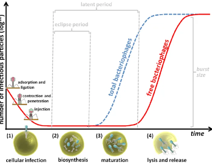
and temperate bacteriophages. However, for a specific group of bacteriophages, Caudovirales, morphogenesis is so similar that a standard process has been suggested (64). Bacteriophages do not have their own metabolism, and require the metabolism, energy resources, and materials of their hosts to replicate. Common steps in the replication process of bacteriophages can be properly correlated with the graphical representation of the one-step growth curve, which translates the experimental interaction between the bacteriophage and its host over time (Figure A1-6).
4 Strictly lytic (often described as virulent) bacteriophages are both lytic and incapable of displaying a lysogenic cycle. Throughout the text the term
Figure A1-6 – Common steps of the bacteriophage lytic replication process properly correlated with graphical representation of the one-step-growth curve (a myovirus is used here as example). Once a bacteriophage encounters a target bacterium the process of bacteriophage replication takes place: (1) the bacteriophage adsorbs and ligates to bacterial surface receptors, then the sheath contracts and the hollow tail tube is thought to penetrate through the cell cytoplasmic membrane, injecting bacteriophage nucleic acid in the bacterial cytoplasm; (2) the genetic material of the bacteriophage takes up the biosynthetic machinery of the host and, during the eclipse period, mRNA expression occurs resulting in directed macromolecular biosynthesis; (3) during maturation, the previously synthesized bacteriophage structural proteins are assembled, and bacteriophage particles accumulate inside the cell; (4) at the end of the latent period, the accumulation of lytic proteins results in cell lysis and release of bacteriophages. The burst size corresponds to the average number of progeny bacteriophage particles produced per infected bacterium.
Cellular infection
Bacteriophages must encounter target bacteria before cellular infection occurs. This process of extracellular search occurs through diffusion and other means of movement (65, 66), which is followed by virion adsorption to the host bacteria. Adsorption is the process by which specific bacteriophage receptor binding proteins (tail fibers in Myoviridae), through the effects of diffusion and Brownian motion5, come into contact with specific and chemically complementary locations
(bacterial receptors) on the bacterial surface. Generally, after an initial weak and reversible interaction, 5 Random movement of microscopic particles suspended in a fluid resulting from the impact of molecules of the surrounding medium