Hajer Radhouani
Genomic and pr
oteomic study of antimicr
obial resistance in Escherichia coli and Enterococcus spp. recover ed fr om wild animals 2013
University of Trás-os-Montes and Alto Douro
School of Life and Environmental Sciences
Genomic and proteomic study of antimicrobial
resistance in Escherichia coli and Enterococcus spp.
recovered from wild animals
PhD Thesis in
Technologic, Comparative and Molecular Genetics
Hajer Radhouani
Prof. Dr. Gilberto Paulo Peixoto Igrejas
Prof. Dr. Patrícia Alexandra Curado Quintas Dinis Poeta
Prof. Dr. Carmen Torres Manrique
Jury composition:
Prof Dr. Luís Herculano Melo de Carvalho Prof Dr. John Elmerdahl Olsen
Prof Dr. Luísa Vieira Peixe Prof Dr. António Carlos Matias Correia Prof Dr. Joana Cecília Valente Rodrigues Azeredo
Prof Dr. Maria de Fátima Silva Lopes Prof Dr. Carmen Torres Manrique
Prof Dr. Patrícia Alexandra Curado Quintas Dinis Poeta Prof Dr. Gilberto Paulo Peixoto Igrejas
This thesis was specifically prepared to obtain the PhD degree in Technologic, Comparative and Molecular Genetics.
Financial support was provided by FCT (Fundação para a Ciência e a Tecnologia) and POPH - QREN / FSE (Programa Operacional Potencial Humano - Quadro de Referência Estratégico Nacional / Fundo Social Europeu) through a doctoral degree grant with reference SFRH/BD/60846/2009.
The presented doctrines are the exclusive responsibility of the author.
The partial reproduction of this thesis is authorized only for research purposes, by written declaration of the person concerned, who commits to do so.
University of Trás-os-Montes and Alto Douro, ___/___/_____
|
A
CKNOWLEDGEMENTS
Here I am about to write my acknowledgements; it would take several pages to thank all those who participated directly or indirectly in the achievement of this PhD so I would like to briefly express my sincere thanks:
Institutionally, to the Rector magnificus of University of Trás-os-Montes and Alto Douro, Professor Carlos Alberto Sequeira, the Department of Genetics and Biotechnology, the Institute for Biotechnology and Bioengineering, the Center of Genomics and Biotechnology, the Department of Veterinary Sciences, and the Centre of Animal and Veterinary Research, for your willingness to provide the best conditions for the development and realization of this work.
To Fundação para a Ciência e a Tecnologia (FCT) and Programa Operacional Potencial Humano/Fundo Social Europeu (POPH/FSE) for financial support.
To Professor Gilberto Igrejas, as my advisor, I would like to express my special appreciation and thanks. You have been a tremendous mentor for me and I appreciate all your contributions of time, ideas, and funding to make my PhD experience so productive and interesting.
To Professor Patrícia Poeta, as my co-advisor, I would like extend my deepest gratitude for encouraging my research and for allowing me to grow as a research scientist. Your advices on my research as well as on my career have been invaluable.
To Professor Carmen Torres, my co-supervisor from the University of LaRioja, my special words of thanks for your wonderful help and feedbacks on my work. I appreciate your invaluable advice and everlasting kindness.
To Professors José Luis Capelo and Carlos Lodeiro, from Bioscope Group, for welcoming me with open arms, giving me the opportunity to work in your laboratory and allowing me to go deeper in my knowledge.
To my colleagues, friends and their families around the world (Belgium, Finland, France, Honduras, Mexico, Portugal, Tunisia, etc.) a great big thank. Thank you so much for your love and friendship. You have been always there in good and bad times to cheer me up and make me smile countless times.
To my parents, brothers and sister, I will never truly be able to express my sincere appreciation for all the support and love that was given to me. I never would have made it without you. You have always helped, supported and found the right words to make me forget the worst. You believed in me and I owe my success to you. I owe you everything.
THANK YOU ALL إﻟ ﻰ و اﻟ ﺪا ي ، أ ﺧ ﻰ و أ ﺧ ﺘ ﻰ ﻻ ﯾ ﻤ ﻜ ﻨﻨ ﻲ ﺣ ﻘ ﯿ ﻘ ﺔ أ ن أ ﻋ ﺒ ﺮ ﻋ ﻦ ﺧ ﺎﻟ ﺺ ﺗ ﻘ ﺪ ﯾ ﺮ ي ﻟ ﻜ ﻢ ﻋ ﻠ ﻰ ﻛ ﻞ اﻟ ﺪ ﻋ ﻢ و اﻟ ﺤ ﺐ اﻟ ﺬ ي ﻗ ﺪ ﻣ ﺘ ﻤ ﻮ ه ﻟ ﻲ . و ﻣ ﺎ أ ﻣ ﻜ ﻨﻨ ﻲ اﻟ ﻮ ﺻ ﻮ ل إﻟ ﻰ ﻣ ﺎ و ا ﺻ ﻠ ﺖ إﻟ ﯿ ﮫ د و ن د ﻋ ﻤ ﻜ ﻢ . و ﻗ ﺪ ﻛ ﻨﺘ ﻢ دا ﺋ ﻤ ﺎ اﻟ ﺴ ﻨ ﺪ اﻟ ﺪ ﻋ ﻢ ﺗ ﺠ ﺪ و ن اﻟ ﻜ ﻠ ﻤ ﺎ ت اﻟ ﻤ ﻨﺎ ﺳ ﺒ ﺔ اﻟ ﺘ ﻲ ﺗ ﺠ ﻌ ﻠﻨ ﻲ أﺗ ﺠ ﺎ و ز اﻟ ﺼ ﻌ ﺎ ب . آ ﻣ ﻨﺘ ﻢ ﺑ ﻲ أ د ﯾ ﻦ ﻟ ﻜ ﻢ ﺑﻨ ﺠ ﺎ ح ﺑ ﻜ ﻞ ﺷ ﻲ ء .
|
A
BSTRACT
Genomics, proteomics and bioinformatics tools allow for more in-depth knowledge about the physiology and structure of bacteria and also about mechanisms involved in antimicrobial resistance. Antimicrobial resistance, evolving and spreading among bacteria, poses a serious and growing threat to public health. Though, resistance has also been detected in the absence of antimicrobial exposure, such as in bacteria from wildlife, raising a question about the mechanisms of occurrence and persistence of resistant strains under similar conditions, and the implications for resistance control strategies.
In order to study antimicrobial resistance in Escherichia coli and Enterococcus spp., 151 faecal samples were collected from three wild animal species, seagulls (Larus cachinnans, 57), common buzzards (Buteo buteo, 42) and red foxes (Vulpes vulpes, 52). Seagull samples were recovered from the Berlengas nature reserve, those of common buzzards from the Peneda Gerês natural park and other rural conservation areas of Portugal and those of red foxes were collected in the north of Portugal during red fox hunts. A total of 111 E. coli (73.5%) and 135 enterococci (89.4%) were isolated from Levine and Slanetz-Bartley plates without antimicrobial agents. After isolation in selective plates supplemented with cefotaxime (CTX) and vancomycin, respectively, the occurrence of extended-spectrum-β-lactamase (ESBL)-producing E. coli was 12.7% (seagulls, 19.3%; common buzzards, 15.2%; red foxes, 4%) and the prevalence of vancomycin-resistant enterococci (VRE) was 20.4% (seagulls, 10.5%; common buzzards, 36.4%; red foxes, 21.1%).
All CTX-resistant E. coli isolates exhibited a resistance phenotype to cefotaxime and/or ceftazidime and had a positive screening test for ESBL production. The most predominant β-lactamase genes were the blaTEM-52 and blaCTX-M-32 genes (34.8%) together with the blaCTX-M-1
(17.4%), blaSHV-12 (8.7%),blaOXA-1 and blaCTX-M-14a (4.3%) genes. Most of the ESBL and non-ESBL E.
coli isolates were multiresistant, with high levels of resistance to tetracycline (57.5%), streptomycin (53%) and sulfamethoxazole/trimethoprim (35.1%), due to the presence of tet(A) and/or tet(B) genes (81.7%), aadA gene (45.1%) and different combinations of sul genes (97.8%), respectively. The most common virulence factor gene was fimA (63.3%) and the majority of the E. coli isolates belonged to phylogenetic groups A (39.1%) and B1 (27.3%). Regarding to the VRE and non-VRE isolates, E. faecium and E. faecalis were the most predominant enterococcal species. High levels of tetracycline and erythromycin resistance were detected and resistance gene profiles were diverse amongst these isolates; the most prevalent genotype was tet(M)+tet(L)+erm(B). Concerning virulence factors, the gelE, hyl, cpd, esp and agg genes were the most predominant. VRE isolates with acquired vancomycin resistance (vanA genotype) were found in 11.3% of the faecal samples analysed; and 9.1% contained VRE isolates with intrinsic vancomycin resistance (vanC1 genotype). The vanA-E. faecium isolates belonged to multilocus sequence types ST273 and ST262 (clonal complex CC17) and also to ST5.
ESBL-E. coli and vanA-containing enterococci were also studied through a detailed proteomic approach consisting in two-dimensional gel electrophoresis (2-DE) followed by matrix-assisted laser desorption/ionization time-of-flight-mass spectrometry (MALDI/TOF-MS). A total of 686 spots were excised from 2-DE gels of 3 ESBL-E. coli and 4 VRE strains from which 562 proteins were successfully identified (81.9%) and fully characterized through database interrogations. The proteins identified were associated to different biological functions such as glycolysis, transport, biosynthesis, antimicrobial resistance, among others. The results also indicated in E. coli and enterococci strains the presence of proteins involved in stress response, as chaperone proteins DnaK, WrbA, GroEL, among others. Additionally, the VanA protein (Mw: 37419,10938 /
PI: 5,79), which prevents vancomycin/teicoplanin binding, was identified when studying the whole-cell proteomic profile of vanA-containing enterococci strains. This approach showed that it is possible to evaluate protein profiles in resistant bacteria and in response to antimicrobial stress conditions to further understand the associated resistance mechanisms. Quantitative proteomic analyses on specific protein classes or subproteomes in ESBL-E. coli and VRE strains will provide new functional insights into antimicrobial resistance mechanisms facilitating the identification of diagnostic or prognostic disease markers and the discovery of therapeutic targets.
This thesis discussed the prevalence and spread of antimicrobial resistance in indicator bacteria such as E. coli and enterococci in wild animals and showed that wildlife may be an important reservoir of antimicrobial-resistant bacteria and resistance genes. Moreover, this research showed the presence of strains carrying virulence factor genes and belonged to high-risk clonal complexes, which adds to the public health concerns that arise from the spread of ESBL-E. coli and VRE into wildlife.
KEYWORDS: Antimicrobial resistance, extended-spectrum β-lactamases-producing Escherichia coli,
|
R
ESUMO
As ferramentas genómicas, proteómicas e bioinformáticas permitem um conhecimento mais aprofundado sobre a fisiologia e estrutura bacterianas e também dos mecanismos envolvidos na resistência antimicrobiana. A resistência aos antimicrobianos tem evoluído e disseminado entre bactérias, representando uma séria e crescente ameaça para a saúde pública. No entanto, esta resistência tem sido detectada também na ausência de exposição antimicrobiana em animais selvagens, suscitando questões acerca dos mecanismos de ocorrência e persistência de estirpes resistentes sob condições similares, e as implicações para estratégias de controlo de resistência.
Com o objectivo de estudar a resistência antimicrobiana em Escherichia coli e Enterococcus spp., foram recolhidas 151 amostras fecais de três espécies de animais selvagens nomeadamente gaivotas-de-patas-amarelas (Larus cachinnans, 57), águias-de-asa-redonda (Buteo buteo, 42) e raposas-vermelhas (Vulpes vulpes, 52). As amostras de gaivota foram recolhidas na reserva natural das Berlengas, as de águia no parque nacional da Peneda-Gerês e em outras áreas de conservação de Portugal e as amostras de raposa foram recolhidas no Norte de Portugal durante o período sazonal de caça. Através de placas de Levine e Slanetz-Bartley sem antibiótico foram isolados um total de 111 E. coli (73,5%) e 135 enterococos (89,4%). Após o isolamento em placas selectivas suplementadas com cefotaxima (CTX) e vancomicina, respectivamente, a ocorrência de E. coli produtoras de β-lactamases de amplo espectro (BLAE) foi de 12,7% (gaivotas, 19,3%; águias, 15,2%; raposas, 4%) e a prevalência de enterococos resistentes à vancomicina (ERV) foi de 20,4% (gaivotas, 10,5%; águias, 36,4%; raposas, 21,1%).
Todos os isolados de E. coli CTX-resistentes revelaram um fenótipo de resistência à cefotaxima e/ou à ceftazidima, apresentando um resultado positivo para a produção de BLAE. O genes da β-lactamase mais predominante foram os genes blaTEM-52 e blaCTXM-32 (34,8%) juntamente
com os genes blaCTX-M-1 (17,4%), blaSHV-12 (8,7%), blaOXA-1 e blaCTX-M-14a (4,3%). A maioria dos
isolados de E. coli produtores e não produtores de BLAE foram multirresistentes, demonstrando níveis elevados de resistência à tetraciclina (57,5%), estreptomicina (53%) e sulfametoxazol/trimetoprim (35,1%), devido à presença dos genes tet(A) e/ou tet(B) (81,7%), o gene aadA (45,1%) e a diferentes combinações de genes sul (97,8%), respectivamente. O gene de virulência mais comum foi o fimA (63,3%) sendo que a maioria dos isolados de E. coli pertenciam aos grupos filogenéticos A (39,1%) e B1 (27,3%). Entre os isolados de enterococos resistentes e não resistentes à vancomicina, as espécies mais comuns foram E. faecium e E. faecalis. Foram detectados níveis elevados de resistência à tetraciclina e à eritromicina e os perfis genotípicos de resistência foram diversos nestes isolados, o genótipo mais comum foi tet(M)+tet(L)+erm(B). No que respeita a factores de virulência sendo os genes gelE, hyl, cpd, esp e agg foram os mais predominantes. Foram encontrados isolados de ERV com resistência adquirida à vancomicina (genótipo vanA) em 11,3% das amostras fecais analisadas; e 9,1% das amostras apresentaram ERV com resistência intrínseca à vancomicina (genótipo vanC1). Através da análise por MLST (Multilocus Sequence Typing) conclui-se que os isolados vanA-E. faecium pertencam ao ST273 e ST262 (complexo clonal CC17) e também ao ST5.
As estirpes de E. coli produtoras de BLAE e de enterococos com vanA foram também estudadas através de uma abordagem proteómica detalhada abrangendo electroforese bi-dimensional (E2D) e identificação por espectrometria de massa com ionização a laser assistida por matriz com analisador tempo de voo (MALDI-TOF MS). Um total de 686 spots foram excisados a partir dos geis E2D de 4 estirpes de E. coli produtores de BLAE e 3 estirpes de ERV dos quais 562 proteínas foram identificadas com sucesso (81,9%) e caracterizadas por interrogação de base dados. As
proteínas identificadas foram associadas a diferentes funções biológicas, tais como glicólise, transporte, biossíntese, resistência aos antibióticos, entre outros. Os resultados obtidos em E. coli e enterococos revelaram a presença de proteínas envolvidas na resposta ao stress, tais como as proteínas chaperone DnaK, WrbA, GroL, entre outras. Adicionalmente, a proteína VanA (MM: 37419,10938 / PI: 5,79), que evita a ligação da vancomicina/teicoplanina, foi identificada ao analisar o proteoma total de estirpes de enterococos vanA. A descrição detalhada das proteínas identificadas em estirpes BLAE e ERV pode proporcionar novos alvos para o desenvolvimento de agentes antimicrobianos. Esta abordagem revelou perfis proteicos de bactérias resistentes e em resposta a várias condições de stress antimicrobiano que possibilitaram um maior conhecimento dos mecanismos de resistência associados. A análise proteómica quantitativa em classes específicas de proteínas ou subproteomas em estirpes de ERV e de E. coli produtoras de BLAE abrirá novas perspectivas funcionais no estudo de mecanismos de resistência antimicrobiana facilitando a identificação de marcadores de diagnóstico ou prognóstico e a descoberta de alvos terapêuticos.
Conclui-se que a prevalência e disseminação de bactérias indicadoras resistentes aos antimicrobianos, como E. coli e Enterococcus spp., em animais selvagens revelaram que estes podem representar um importante reservatório de bactérias resistentes e de genes de resistência. Este estudo revelou também a presença de estirpes portadoras de genes de virulência, pertencendo a complexos clonais de elevado risco, o que acresce às preocupações de saúde pública que surgem da disseminação de ERV e de E. coli produtoras de BLAE em ecossistemas selvagens.
PALAVRAS-CHAVE: Animais selvagens, enterococos resistentes à vancomicina, Escherichia coli
|
R
ÉSUMÉ
Les outils génomiques, protéomiques et bioinformatiques permettent une meilleure compréhension de la physiologie et de la structure bactérienne ainsi que des mécanismes impliqués dans la résistance aux antimicrobiens. L’évolution et la propagation de la résistance aux antimicrobiens chez les bactéries constituent une menace grave et croissante pour la santé publique. Toutefois, cette résistance a également été détectée en l'absence d'exposition aux antimicrobiens chez les bactéries provenant d'animaux sauvages, ce qui soulève la question des mécanismes d'apparition et de persistance des souches bactériennes résistantes dans des conditions similaires, et leurs implications dans les stratégies visant le contrôle de la résistance aux antimicrobiens.
Afin d'étudier la résistance aux antimicrobiens d’Escherichia coli et d’Enterococcus spp., 151 échantillons fécaux provenant de trois espèces d'animaux sauvages, les goélands leucophées (Larus cachinnans, 57), les buses variables (Buteo buteo, 42) et les renards roux (Vulpes vulpes, 52) ont été prélevés. Les échantillons de goélands ont été prélevés dans la réserve naturelle des Berlengas, ceux des buses dans le parc national du Peneda-Gerês ainsi que d’autres aires de conservation du Portugal et ceux des renards dans le nord du Portugal au cours de la saison de chasse. A travers des milieux de Levine et de Slanetz-Bartley sans antibiotiques, un total de 111 isolats d’E. coli (73,5%) et 135 d’entérocoques (89,4%) ont été isolés. Après isolement à travers des milieux sélectifs contenant de la céfotaxime (CTX) et la vancomycine, l'apparition respective des souches d’E. coli productrices de β-lactamases à spectre élargi (BLSE) a été de 12,7% (goélands, 19,3%; buses, 15,2%; renards, 4%) et la prévalence des isolats d’entérocoques résistants à la vancomycine (ERV) a été de 20,4% (goélands, 10,5%; buses, 36,4%; renards, 21,1%).
Tous les isolats d’E. coli résistants à la CTX ont montré un phénotype de résistance à la céfotaxime et/ou à la ceftazidime, indiquant un résultat positif à la production de BLSE. Les gènes de β-lactamase prédominants ont été ceux de blaCTX-M-52 et blaTEM-32 (34,8%) suivis des gènes bla CTX-M-1 (17 4%), blaSHV-12 (8,7%), blaOXA-1 et blaCTX-M-14a (4,3%). La plupart des isolats d’E. coli
producteurs et non-producteurs de BLSE étaient multirésistants, montrant des taux élevés de résistance à la tétracycline (57,5%), la streptomycine (53%) et le sulfaméthoxazole/triméthoprime (35,1%) en raison de la présence respectivement des gènes tet(A) et/ou tet(B) (81,7%), aadA (45,1%) et différentes combinaisons des gènes sul (97,8%). Le gène de virulence le plus common a été celui du fimA (63,3%) et la majorité des isolats d’ E. coli appartenaient aux groupes phylogénétiques A (39,1%) et B1 (27,3%). Parmi les isolats d'entérocoques résistants et non résistants à la vancomycine, les espèces les plus communes ont été celles d’E. faecium et d’E. faecalis. Les isolats d’entérocoques ont présenté des niveaux élevés de résistance à la tétracycline et l'érythromycine et les profils génotypiques de résistance ont été divers chez ces isolats mais le plus fréquent a été celui de tet(M)+tet(L)+erm(B). En ce qui concerne les gènes de facteur de virulence, les gènes gelE, hyl, cpd, esp et agg ont été les plus répandus. Les isolats d’ERV ayant une résistance acquise à la vancomycine (génotype vanA) ont été trouvés dans 11,3% des échantillons fécaux analysés, et 9,1% de ces échantillons ont présenté des isolats d’ERV avec une résistance intrinsèque à la vancomycine (génotype vanC1). Les isolats E. faecium vanA appartenaient aux séquences types ST273 et ST262 (du complexe clonal CC17) ainsi qu’au ST5.
Les souches d’E. coli productrices de BLSE et d'entérocoques vanA ont également été étudiés par une approche protéomique détaillée consistant à l'électrophorèse bidimensionnelle (E2D) et suivi de la technique de spectrométrie de masse à temps de vol pour la désorption-ionisation laser assistée par matrice (MALDI-TOF MS). Un total de 686 spots a été excisé des gels E2D de 3 souches d’E. coli productrices de BLSE et 4 souches de ERV, dont 562 protéines ont été identifiées
avec succès (81,9%) et caractérisées par le biais d'interrogation de base de données. Les protéines identifiées ont été associées à différentes fonctions biologiques comme la glycolyse, le transport, la biosynthèse, la résistance aux antimicrobiens, entre autres. Les résultats ont révélé aussi la présence chez les souches d’E. coli et d’entérocoques des protéines impliquées dans la réponse au stress, comme les protéines chaperons DnaK, WrbA, GroEL, entre autres. En outre, la protéine VanA (MM: 37419,10938 / PI: 5,79), qui empêche la liaison de la vancomycine/teicoplanine a été identifiée suite à l’analyse du protéome complet de souches d'entérocoques vanA. Cette technique a révélé des profils protéiques des bactéries résistantes ainsi qu’en réponse à diverses conditions de stress, ce qui a permis une meilleure compréhension des mécanismes de résistance correspondants. Les analyses protéomiques quantitatives au niveau de protéines spécifiques ou subproteomes dans des souches d' ERV et d’E. coli productrices de BLSE fourniront de nouvelles connaissances fonctionnelles dans les mécanismes de résistance aux antimicrobiens facilitant ainsi l'identification de marqueurs diagnostiques ou pronostiques des maladies ainsi que la découverte de cibles thérapeutiques.
Cette thèse discute la prévalence et la propagation de la résistance aux antimicrobiens chez des bactéries indicatrices tels qu’E. coli et Enterococcus spp. aux niveaux des animaux sauvages, et prouvant ainsi que la faune sauvage pourrait représenter un important réservoir de bactéries résistantes et de gènes de résistance. En outre, cette étude a également révélé la présence de souches portant des gènes de virulence, appartenant aux clones à haut risque infectieux, en plus des préoccupations de santé publique découlant de la propagation d’ ERV et d’E. coli productrices de BLSE dans des écosystèmes naturels.
MOTS-CLES: Animaux sauvages, entérocoques résistants à la vancomycine, Escherichia coli
|
G
RAPHICAL ABSTRACT
A
NTI M I CROBI ALR
ESI STANCEG
ENOM I CSP
ROTEOM I CSB
I OI NFORM ATI CS E. coli Enterococcus m/z|
L
IST OF ABREVIATIONS
A
aac Aminoglycoside acetyltransferase gene
aac(6’)-Ie-aph(2’’)-Ia Gene encoding a bifunctional enzyme
aad Aminoglycoside adenylyltransferase gene ABC transporter ATP-binding cassette transporter
ace Accessory colonization factor gene
ACN Acetonitrile
aer Aerobactin gene
agg Aggregation substance gene
AMC Amoxicillin+clavulanic acid
AMK Amikacin
AMP Ampicillin
AMPs Antimicrobial peptides
ant(6) Aminoglycoside nucleotidyltransferase gene
APEC Avian pathogenic Escherichia coli
aph(3’) Aminoglycoside 3'-phosphotransferase gene
APS Ammonium persulfate
AT Autotransporters
ATM Aztreonam
ATP Adenosine triphosphate
B
BHI Brain heart infusion bp Base pair
C
ca. Circa
cat Chloramphenicol acetyltransferase gene
CAZ Ceftazidime
CBB Coomassie brilliant blue
CC Clonal complex
CHL Chloramphenicol
chuA Receptor gene encoding outer membrane
CIP Ciprofloxacin
cm Centimetre
cmlA Nonenzymatic chloramphenicol resistance gene
cnf1 Cytotoxic necrotizing factor 1 gene
CRATAS Center of collecting, welcome and handling of wild animals
CTX Cefotaxime
cylLSLLABM Cyl operon genes
D
D-Lac D-lactate D-Ser D-serine
Da Dalton
DAEC Diffusely adherent Escherichia coli
ddl D-alanine-D-alanine ligase
DIGE Difference gel electrophoresis DNA Deoxyribonucleic acid
E
E. avium Enterococcus avium
E. casseliflavus Enterococcus casseliflavus
E. coli Escherichia coli
E. durans Enterococcus durans
E. faecalis Enterococcus faecalis
E. faecium Enterococcus faecium
E. gallinarum Enterococcus gallinarum
E. hirae Enterococcus hirae
E. mundtii Enterococcus mundtii
E. raffinosus Enterococcus raffinosus
EAEC Enteroaggregative Escherichia coli
EARSS/EARSNet European Antimicrobial Resistance Surveillance System/network
Ed Enterococcus durans
Ef Enterococcus faecium
EHEC Enterohaemorrhagic Escherichia coli EIEC Enteroinvasive Escherichia coli EPEC Enteropathogenic Escherichia coli
erm Erythromycin ribosomal methylase gene
ERY Erythromycin
ESBL Extended-spectrum beta-lactamase ESI Electrospray ionization
ESI-MS/MS Electrospray ionization tandem mass spectrometry
esp Extracellular surface protein gene
EST Expressed sequence tags
ETEC Enterotoxigenic Escherichia coli
EU European union
ExPEC Extraintestinal pathogenic Escherichia coli
F
FA Formic acid
fimA Type-1 fimbrial gene
FOX Cefoxitin
fsr Regulator of the expression of gelE gene
G
g Gram
gelE Gene of gelatinase
GEN Gentamicin
gyrA Gene for DNA gyrase
H
h Hour
HCl Hydrochloric acid HGT Horizontal gene transfer
HSP Heat shock protein
hyl Hyaluronidase gene
I
ICN Institute for nature conservation IEF Isoelectric focusing
IEF x SDS-PAGE Isoelectric focusing x Sodium dodecyl sulphate polyacrilamide gel electrophoresis
IMP Imipenem
IntI Integron integrase gene
IPG Immobilized pH gradient IS Insertion sequence
K
KAN Kanamycin Kbp Kilobase pair V or kV Volt or kilovoltL
LC Liquid chromatography LPS LipopolysaccharideM
M Molar mA MilliampereMALDI Matrix-assisted laser desorption ionization
MALDI-TOF/MS Matrix assisted laser desorption ionization - time of flight mass spectrometry
MALDI-TOF/TOF MS Matrix-assisted laser desorption/ionization time of flight/time of flight mass spectrometry
Mb Megabit
MBGD Microbial genome database
MDR Multiple drug resistance
mg Milligram
MIC Minimum inhibitory concentration
min Minute
mL Millilitre
MLEE Multilocus enzyme electrophoresis MLST Multilocus sequence typing
mM Millimolar
mm Millimetre
MNEC Meningitis-associated Escherichia coli Mr Relative molecular mass
mRNA Messenger ribonucleic acid
MRSA Methicillin-resistant Staphylococcus aureus
MS Mass spectrometry
MS/MS Tandem mass spectrometry
MudPIT Multidimensional protein identification technology
Mw Molecular weight
N
NT Not tested
NaCl Sodium chloride NAL Nalidixic acid
NL Non Linear
O
ºC Degree celsius
Omp Outer membrane protein
ORF Open reading frame
P
PAI Pathogenicity island
papC P fimbriae PapC gene
papGIII P fimbrial adhesin PapG gene
parC Gene for topoisomerase IV gene
PBP Penicillin-binding-protein
pbp Penicillin-binding-protein gene
PBS Phosphate buffered saline PCR Polymerase chain reaction PFGE Pulsed field gel electrophoresis
pH Potential of hydrogen pI Isoelectric point
PMF Peptide mass fingerprinting ppm Parts per million
PTM Post-translational modification
Q
QD Quinupristin-dalfopristin Qnr Quinolone resistance
R
RNA Ribonucleic acid
RND Resistance-nodulation-cell division rRNA Ribossomal ribonucleic acid
S
s Second
S. aureus Staphylococcus aureus
SDS Sodium dodecyl sulfate
SDS-PAGE Sodium dodecyl sulfate polyacrylamide gel electrophoresis
SMX Sulfamethoxazole
ST Sequence type
STR Streptomycin
stx Shiga toxin gene
sul Sulphonamide dihydropteroate synthase gene
SXT Sulfamethoxazole-trimethoprim
T
TCA Trichloroacetic acid
TEC Teicoplanin
TET Tetracycline TFA Trifluoroacetic acid
TMP Trimethoprim
Tn Conjugative transposon
TOB Tobramycin
TOF Time of flight
Tris Hydroxymethyl aminomethane tRNA Transfer ribonucleic acid
U
UPEC Uropathogenic Escherichia coli US United States
UTIs Urinary tract infections UV Ultraviolet
V
VAN Vancomycin
Van Gene conferring resistance to vancomycin and/or teicoplanin
vat Streptogramin acetyltransferase gene
VF Virulence factor
VRE Vancomycin-resistant enterococci
VRSA Vancomycin-resistant Staphylococcus aureus
W
WCP Whole-cell protein
WHO World Health Organization
Y
yjaA Gene with no known function
% Percentage
1-DE Monodimensional electrophoresis 2-DE Bidimensional electrophoresis
μg Microgram
|
T
ABLE OF CONTENTS
PART I: INTRODUCTION ... 1 Chapter I. State of the art ... 3
I.1. The genetic and evolutionary frontier of antimicrobial-resistant bacterial colonization and its implications for wildlife conservation ... 5
I.1.1. Abstract ... 5 I.1.2. Wildlife conservation in Portugal ... 6 I.1.3. E. coli and enterococci as ubiquitous bacteria ... 7 I.1.3.1. Biology and description of E. coli and enterococci ... 7 I.1.3.2. Role in disease ... 8
I.1.4. Antimicrobial resistance pollution in wildlife ... 10 I.1.5. Genome plasticity as paradigm of microbial genome evolution ... 12 I.1.6. Fundamental evolutionary processes ... 14 I.1.7. Phylogenetic history and genetic structure ... 15 I.1.8. Diversity and complexity of bacterial niches ... 19
I.1.8.1. Virulence factors ... 19 I.1.8.2. Intra-species interactions ... 19 I.1.8.3. Antimicrobial resistance ... 20
I.1.9. Antimicrobial resistance of bacteria: an example of evolution in action? ... 21 I.1.10. Acknowledgements... 21 I.1.11. References ... 22 I.2. Antimicrobial resistance: genomic insights of an old problem ... 27
I.2.1. Abstract ... 27 I.2.2. Antimicrobial resistance as a global public health problem ... 28
I.2.2.1. History and emergence of antimicrobial resistance ... 28 I.2.2.2. Causes of antimicrobial resistance ... 29 I.2.2.3. Implications of antimicrobial resistance ... 31
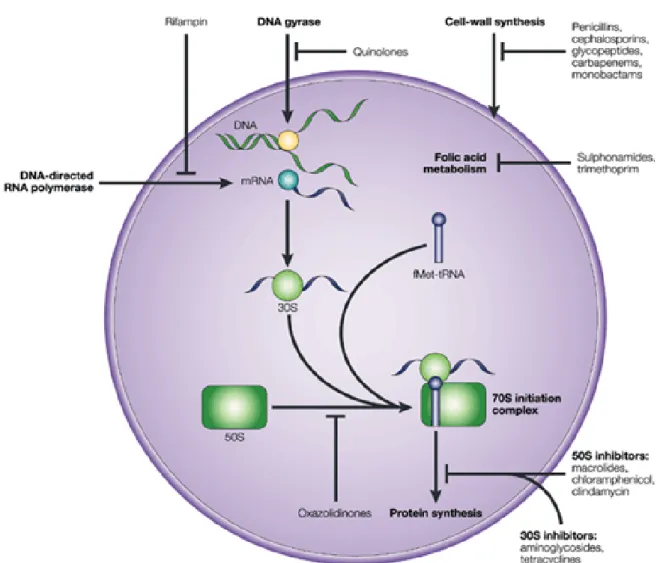
I.2.3. Mechanisms of antimicrobial action ... 32
I.2.3.1. Inhibition of cell wall synthesis ... 32 I.2.3.2. Inhibition of protein synthesis ... 32 I.2.3.3. Interference with nucleic acid synthesis ... 33 I.2.3.4. Inhibition of a metabolic pathway ... 33
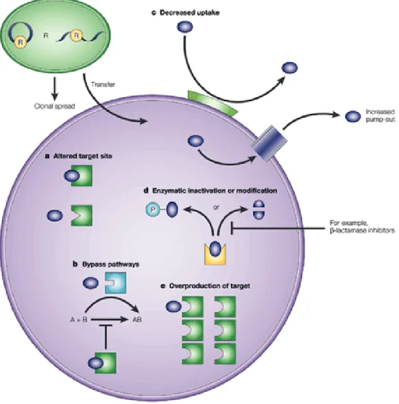
I.2.4. Mechanisms of antimicrobial resistance ... 33
I.2.4.1. Intrinsic resistance ... 38 I.2.4.2. Acquired resistance ... 39
I.2.5. Resistance to β-lactam antimicrobials ... 41 I.2.6. Resistance to glycopeptide antimicrobials ... 42 I.2.7. Virulence and pathogenicity ... 46
I.2.7.1. Expression of microbial virulence determinants ... 46 I.2.7.2. Bacterial Pathogenesis... 47
I.2.8. Genomic approaches as a tool against resistance ... 47
I.2.8.1. Genomics in surveillance and control of resistance... 49 I.2.8.2. Genomics in understanding of resistance mechanisms ... 50 I.2.8.3. Genomic knowledge and antibacterial drug discovery ... 50
I.2.9. Acknowledgements... 51 I.2.10. References ... 52 I.3. After genomics, what proteomics tools could help us understand the antimicrobial resistance of Escherichia coli? ... 55
I.3.1. Abstract ... 55 I.3.2. Introduction ... 56 I.3.3. The Omics era ... 58 I.3.4. Proteomic methods ... 62 I.3.5. Proteome mapping and the effects of stress environments on E. coli ... 66 I.3.6. Outlook of proteomic biomarkers ... 77
I.3.7. Acknowledgements ... 78 I.3.8. References... 79
Chapter II. Aims of the study ...83 PART II: GENOMICS APPROACH ... 87
Chapter III. Genomic characterization of antimicrobial-resistant Escherichia coli and enterococci isolates of wild animals with no antimicrobial selective plates ...89
III.1. Antimicrobial resistance and phylogenetic groups in isolates of Escherichia coli from seagulls at the Berlengas nature reserve ... 91
III.1.1. Abstract ... 91 III.1.2. Introduction ... 92 III.1.3. Material and Methods ... 93
III.1.3.1. Samples and bacterial isolates ... 93 III.1.3.2. Antimicrobial susceptibility testing ... 93 III.1.3.3. Extraction of DNA ... 93 III.1.3.4. Characterisation of antibiotic resistance genes ... 94 III.1.3.5. Detection of phylogenetic groups ... 94
III.1.4. Results ... 95
III.1.4.1. Percentages of antimicrobial resistance and resistance phenotypes ... 95 III.1.4.2. Mechanisms of antibiotic resistance ... 96 III.1.4.3. Phylogenetic group ... 97
III.1.5. Discussion ... 97 III.1.6. Acknowledgements ... 99 III.1.7. References ... 100 III.2. Wild birds as biological indicators of environmental pollution: antimicrobial resistance patterns of Escherichia coli and enterococci isolated from common buzzards (Buteo buteo) ... 103
III.2.1. Abstract ... 103 III.2.2. Introduction ... 104 III.2.3. Methods ... 105
III.2.3.1. Samples and bacteria... 105 III.2.3.2. Antimicrobial susceptibility test ... 105 III.2.3.3. Antibiotic resistance genes ... 106 III.2.3.4. Detection of phylogenetic groups ... 106
III.2.4. Results ... 107
III.2.4.1. Bacteria isolation ... 107 III.2.4.2. Antimicrobial resistance among E. coli isolates... 107 III.2.4.3. Phylogenetic groups and virulence factor genes among E. coli isolates ... 108 III.2.4.4. Antimicrobial resistance among enterococci isolates ... 109
III.2.5. Discussion ... 109 III.2.6. Acknowledgements ... 112 III.2.7. References ... 112 III.3. Antimicrobial resistance and virulence genes in Escherichia coli and enterococci from red foxes (Vulpes vulpes) ... 115
III.3.1. Abstract ... 115 III.3.2. Introduction ... 116 III.3.3. Material and methods ... 116
III.3.3.1. Samples and bacteria... 116 III.3.3.2. Antimicrobial susceptibility testing ... 117 III.3.3.3. Antimicrobial-resistance genes ... 117 III.3.3.4. Virulence factor genes ... 118 III.3.3.5. Detection of phylogenetic groups ... 118
III.3.4. Results and discussion ... 118 III.3.5. Conclusion ... 122 III.3.6. Acknowledgements ... 123 III.3.7. References ... 123
III.4. Molecular characterization of antibiotic resistance in enterococci recovered from seagulls (Larus cachinnans) representing an environmental health problem ... 125
III.4.1. Abstract ... 125 III.4.2. Introduction ... 126 III.4.3. Materials and methods ... 127
III.4.3.1. Samples and bacteria ... 127 III.4.3.2. Antimicrobial susceptibility testing ... 128 III.4.3.3. Antibiotic resistance genes ... 128 III.4.3.4. Statistical Analysis ... 128
III.4.4. Results ... 129
III.4.4.1. Percentages of antimicrobial resistance and phenotypes of resistance... 129 III.4.4.2. Mechanisms of antibiotic resistance ... 130 III.4.4.3. Data analysis ... 131
III.4.5. Discussion ... 132 III.4.6. Conclusion ... 134 III.4.7. Acknowledgements ... 135 III.4.8. References ... 135
Chapter IV. Genomic characterization of antimicrobial-resistant E. coli and enterococci isolates of wild animals with antimicrobial selective plates ... 137
IV.1. Seagulls of the Berlengas natural reserve of Portugal as carriers of faecal Escherichia coli harboring CTX-M and TEM extended-spectrum beta-lactamases ... 139
IV.1.1. Abstract ... 139 IV.1.2. Introduction ... 140 IV.1.3. Material and methods ... 140 IV.1.4. Results and discussion ... 141 IV.1.5. Conclusion ... 142 IV.1.6. Acknowledgements ... 142 IV.1.7. References ... 144 IV.2. Detection of Escherichia coli harbouring extended-spectrum β-lactamases of the CTX-M classes in faecal samples of common buzzards (Buteo buteo) ... 145
IV.2.1. Abstract ... 145 IV.2.2. Introduction ... 146 IV.2.3. Material and methods ... 146 IV.2.4. Results and discussion ... 147 IV.2.5. Conclusion ... 147 IV.2.6. Acknowledgements ... 148 IV.2.7. References ... 148 IV.3. Molecular characterization of extended-spectrum-beta-lactamase-producing Escherichia coli isolates from red foxes in Portugal ... 149
IV.3.1. Abstract ... 149 IV.3.2. Introduction ... 150 IV.3.3. Material and methods ... 150 IV.3.4. Results... 151 IV.3.5. Discussion ... 152 IV.3.6. Conclusion ... 153 IV.3.7. Supplementary material ... 153 IV.3.8. Acknowledgements ... 153 IV.3.9. References ... 154 IV.4. MLST and a genetic study of antibiotic resistance and virulence factors in vanA-containing Enterococcus from buzzards (Buteo buteo) ... 155
IV.4.1. Abstract ... 155 IV.4.2. Introduction ... 156 IV.4.3. Material and methods ... 156 IV.4.4. Results and discussion ... 158 IV.4.5. Conclusion ... 160
IV.4.6. References ... 160 IV.5. Clonal Lineages, antibiotic resistance and virulence factors in vancomycin-resistant enterococci isolated from faecal samples of red foxes (Vulpes vulpes) ... 161
IV.5.1. Abstract ... 161 IV.5.2. Introduction ... 162 IV.5.3. Material and methods ... 162 IV.5.4. Results and discussion ... 163 IV.5.5. Conclusion ... 165 IV.5.6. Acknowledgements ... 165 IV.5.7. References ... 165
PART III: PROTEOMICS APPROACH ... 167 Chapter V. Proteomic characterization of antimicrobial-resistant E. coli isolates in wild animals 169
V.1. Proteomic study in an Escherichia coli strain from seagulls of the Berlengas Natural Reserve of Portugal ... 171
V.1.1. Abstract ... 171 V.1.2. Introduction ... 172 V.1.3. Material and methods ... 173
V.1.3.1. Cell culture and purification of E. coli ... 173 V.1.3.2. Protein extraction ... 174 V.1.3.3. One-dimensional electrophoresis and staining ... 174 V.1.3.4. Two-dimensional electrophoresis and proteome analysis... 174 V.1.3.5. Protein identification by MALDI-TOF/TOF ... 175 V.1.3.6. Database search ... 175
V.1.4. Results and Discussion ... 176 V.1.5. Concluding remarks ... 179 V.1.6. Supplementary material ... 179 V.1.7. References ... 179 V.2. Proteomic changes in an extended-spectrum beta-lactamase-producing Escherichia coli strain under cefotaxime selection ... 181
V.2.1. Abstract ... 181 V.2.2. Introduction ... 182 V.2.3. Material and methods ... 183
V.2.3.1. Isolation of bacteria from fox faeces ... 183 V.2.3.2. Genetic characterization of E. coli C5478 ... 183 V.2.3.3. Culture conditions and total protein extraction ... 183 V.2.3.4. SDS-PAGE and staining ... 184 V.2.3.5. Two-dimensional electrophoresis and proteomics analysis ... 184 V.2.3.6. Protein digestion ... 185 V.2.3.7. Matrix formulation and sample deposition ... 185 V.2.3.8. MALDI-TOF MS analysis ... 185
V.2.4. Results ... 186 V.2.5. Discussion ... 189
V.2.5.1. Stress response ... 190 V.2.5.2. DNA damage, cell division, redox homeostasis and immune response ... 191 V.2.5.3. Antimicrobial resistance ... 192 V.2.5.4. Transport proteins ... 192 V.2.5.5. Amino acids and protein biosynthesis/metabolism ... 193
V.2.6. Conclusion... 194 V.2.7. Supplementary material ... 195 V.2.8. Acknowledgements ... 195 V.2.9. References ... 195
Chapter VI. Proteomic characterization of antimicrobial-resistant enterococci isolates in wild animals ... 199
VI.1. Proteomic characterization of vanA-containing Enterococcus recovered from Seagulls at the Berlengas Natural Reserve, W Portugal ... 201
VI.1.1. Abstract ... 201 VI.1.2. Introduction ... 202 VI.1.3. Material and methods ... 204
VI.1.3.1. Samples and bacteria ... 204 VI.1.3.2. Antimicrobial susceptibility testing ... 204 VI.1.3.3. Antimicrobial resistance genes ... 204 VI.1.3.4. MLST typing ... 205 VI.1.3.5. Protein extraction ... 205 VI.1.3.6. One-dimensional and coloration ... 205 VI.1.3.7. Two-dimensional electrophoresis and proteomics ... 206 VI.1.3.8. Protein identification by MALDI-TOF/TOF ... 206 VI.1.3.9. Database search ... 207 VI.1.3.10. Sequence alignments and construction of the phylogenetic tree... 207
VI.1.4. Results... 208
VI.1.4.1. Phenotypic and genetic characterization of enterococci isolates to antibiotic resistance 208
VI.1.4.2. One-dimensional electrophoresis ... 208
VI.1.5. Two-dimensional Electrophoresis... 209 VI.1.6. Discussion ... 212 VI.1.7. Supplementary material ... 215 VI.1.8. Conclusion ... 215 VI.1.9. References ... 216 VI.2. Comparative proteomic map among vanA-containing Enterococcus isolated from yellow-legged gulls ... 219
VI.2.1. Abstract ... 219 VI.2.2. Introduction ... 220 VI.2.3. Material and methods ... 221
VI.2.3.1. Samples and bacteria ... 221 VI.2.3.2. Virulence factor genes ... 222 VI.2.3.3. Assay of gelatinase activity ... 222 VI.2.3.4. Assay of haemolytic assay ... 222 VI.2.3.5. PCR amplification of pbp5 gene ... 222 VI.2.3.6. DNA sequence analysis... 222 VI.2.3.7. Protein extraction ... 223 VI.2.3.8. Two-dimensional electrophoresis and proteomics ... 223 VI.2.3.9. Protein identification by MALDI-TOF/TOF ... 224 VI.2.3.10. Database search ... 224 VI.2.3.11. Sequence alignments and construction of the phylogenetic tree... 225
VI.2.4. Results... 225
VI.2.4.1. Characteristics of the 2 vanA strains included in the study ... 225 VI.2.4.2. Two-dimensional Electrophoresis ... 226
VI.2.5. Discussion ... 226 VI.2.6. Concluding remarks ... 230 VI.2.7. Supplementary material ... 230 VI.2.8. Acknowledgements ... 232 VI.2.9. References ... 232
PART IV: CONCLUSION ... 235 Chapter VII. General discussion and conclusions ... 237 Chapter VIII. Future prospects ... 251 ATTACHMENTS ... 255
|
I
NDEX OF FIGURES
Figure I.1.1 | Phylogenetic group distributions of E. coli in wild animal from Portugal ... 17 Figure I.1.2 | Distribution of Enterococcus species in wild animals from Portugal ... 18 Figure I.2.1 | Dissemination of antibiotics and antibiotic resistance within agriculture, community,
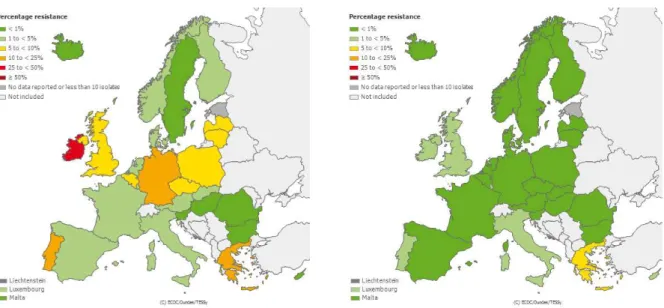
hospital, wastewater treatment, and associated environments ... 29 Figure I.2.2 | Mechanisms of antimicrobial action ... 34 Figure I.2.3 | Bacterial mechanisms of antimicrobial resistance ... 36 Figure I.2.4 | Horizontal gene transfer between bacteria ... 40 Figure I.2.5 | Chemical structure of β-lactam antimicrobials ... 41 Figure I.2.6 | Distribution of ESBL-producing E. coli resistant to 3rd generation cephalosporins in
Europe in 2011 ... 42 Figure I.2.7 | Chemical structure of glycopeptide antimicrobials ... 43 Figure I.2.8 | Distribution of vancomycin-resistant enterococci in Europe in 2011 ... 44 Figure I.3.1 | Circular genome map of E. coli O157:H7 EDL933 compared with MG1655 ... 59 Figure I.3.2 | Left side: Silver-stained 2-D PAGE pattern of E. coli K-12 type extracts, strain W3110
and Right side: 3.5-10 silver-stained 2-D master gel of E. coli K-12 type extracts, strain W3110 ... 60 Figure I.3.3 | Typical proteomic workflow representing the classical gel-based approach to protein
identification ... 63 Figure III.4.1 | Dendrogram based on the numerical analysis of the resistance patterns to 11
antimicrobials of 42 enterococci using simple matching coefficient and UPGMA for clustering ... 132 Figure IV.1.1 | Genetic environment of blaCTX-M genes in three E. coli isolates of seagulls ... 143
Figure V.1.1 | Seagulls (Larus cachinnans) in Berlengas archipelago natural reserve ... 173 Figure V.1.2 | 2-DE gel image of E. coli sample GV5. ... 178 Figure V.1.3 | Distribution of the biological processes related to the protein spots found in the 2-DE
gels of the E. coli GV5 ... 178 Figure V.2.1 | Typical genomic and proteomic workflow representing the classical components of
genetical study and protein identification ... 186 Figure V.2.2 | Distribution of biological functions of the two strain proteins ... 187 Figure V.2.3 | Percentage distribution by biological function of proteins identified in the wild type
and CTX stressed E.coli C5478 strains ... 188 Figure V.2.4 | Percentage distribution of stress response proteins identified in wild type and CTX
stressed E. coli C5478 strains... 189 Figure VI.1.1 | SDS-PAGE of vancomycin-resistant enterococcal strains ... 209 Figure VI.1.2 | 2-DE gel image of SG 3 VRE with IPG strips pH4-7 ... 210 Figure VI.1.3 | 2-DE gel image of SG 41 VRE with IPG strips pH4-7 ... 211 Figure VI.1.4 | Distribution of the biological processes related to the protein spots found in the 2-DE
gel of the vanA E. durans SG 3 isolate ... 211 Figure VI.1.5 | Distribution of the biological processes related to the protein spots found in the 2-DE
gel of the vanA E. faecium SG 41 isolate ... 212 Figure VI.1.6 | Phylogenetic tree of FASTA protein sequences of all proteins identified ... 213 Figure VI.2.1 | 2-DE gel image of SG 50 VRE with IPG strips pH4-7 ... 227 Figure VI.2.2 | MALDI/MS spectra obtained for Enolase from vanA E. durans SG 2 strain ... 228 Figure VI.2.3 | Phylogenetic tree of FASTA protein sequences of all proteins identified ... 231
|
I
NDEX OF TABLES
Table I.2.1 | Antimicrobial agents and their mechanism of antimicrobial action ... 35 Table I.2.2 | Biochemical mechanisms of antimicrobial resistance ... 37 Table I.3.1 | Protein spots identification of 2-DE gels and MALDI-TOF sequencing results from
different bacterial species and hosts ... 71 Table I.3.2 | Modes of action and resistance mechanisms of commonly used antibiotic for E. coli
infections ... 75 Table III.1.1 | Primers and annealing temperatures of PCR reactions used to detect antimicrobial resistant mechanisms ... 94 Table III.1.2 | Number and percentages of the 53 isolates of E. coli from faecal samples from
yellow-legged gulls that were resistant to different antibiotic agents ... 95 Table III.1.3 | Phenotypes of antibiotic resistance detected among the 53 isolates of E. coli recovered from yellow-legged gulls ... 96 Table III.1.4 | Genes of resistance detected among the 29 antimicrobial-resistant isolates of E. coli recovered from yellow-legged seagulls... 97 Table III.1.5 | Phenotypes of resistance and phylogenetic groups detected among the 29
antibiotic-resistant isolates of E coli recovered from yellow-legged seagulls ... 98 Table III.2.1 | Distribution of antibiotic resistance in E. coli and Enterococcus spp. isolated from faecal samples of common buzzards ... 108 Table III.2.2 | Virulence factor genes and phylogenetic groups detected among 36 E. coli isolates recovered from common buzzards ... 109 Table III.3.1 | Resistance genes detected in antibiotic resistant E. coli and enterococcal isolates obtained from red fox faecal samples ... 120 Table III.3.2 | Virulence genes and phylogenetic groups detected among 22 E. coli isolates recovered
from red fox faecal samples ... 121 Table III.4.1 | Target genes and primers used in the PCR reactions carried out in this studya ... 129
Table III.4.2 | Antibiotic resistance in 42 enterococci of different species showing resistance to one or more antibiotic agents isolated from faecal samples of yellow-legged gulls... 130 Table III.4.3 | Phenotypes of resistance detected among the 42 enterococci showing resistance to one
or more antibiotic agents recovered from yellow-legged gulls ... 131 Table IV.1.1 | Characteristics of the ESBL-positive faecal E. coli isolates recovered from seal gulls
of Berlengas island in Portugal ... 143 Table IV.2.1 | Characteristics of the ESBL-positive faecal E. coli isolates recovered from buzzards ... 148 Table IV.4.1 | Characteristics of vancomycin-resistant enterococcal strains recovered from buzzards
in Portugal ... 159 Table IV.5.1 | Characteristics of vancomycin-resistant enterococcal isolates recovered from Vulpes
I
NTRODUCTION
C
HAPTER
I
C
HAPTER
II
C
HAPTER
I
C
HAPTER
I
I.1. Radhouani H, Correia S, Torres C, et al. (2013) The genetic and evolutionary frontier of antimicrobial-resistant bacterial colonization and its implications for wildlife conservation.
I.2. Radhouani H, Correia S, Torres C, et al. (2013) Antimicrobial resistance: genomic insights of an old problem.
I.3. Radhouani H, Pinto L, Poeta P, et al. (2012) After genomics, what proteomics tools could help us to understand the antimicrobial resistance of Escherichia coli?
I.1.
The genetic and evolutionary frontier of antimicrobial-resistant bacterial
colonization and its implications for wildlife conservation
Hajer Radhouani, Susana Correia, Carmen Torres, Patrícia Poeta & Gilberto Igrejas
| Manuscript under revision
I.1.1. Abstract
Given the significant spatial and temporal heterogeneity in antimicrobial resistance distribution and the factors that affect its evolution, dissemination and persistence, it is important to highlight that antimicrobial resistance must be viewed as an ecological problem. Monitoring the resistance prevalence of an indicator bacteria such as Escherichia coli and enterococci in wild animals makes it possible to show that wildlife has the potential to serve as an environmental reservoir and melting pot of bacterial resistance. These researchers address the issue of antimicrobial-resistant microorganism proliferation in the environment and the related potential human health and environmental impact.
I.1.2. Wildlife conservation in Portugal
In Portugal, the nature conservation policy acquired importance in the 70s, when the Law no. 9/70 of June 19th regarding the establishment of Protected Areas was delivered. After April 25th
1974, with the Decree-Law no. 550/75 of September 30th, the Environment State Secretariat was
created, together with the National Park, Reserve, and Landscape Heritage Service, a body with legal competency, administrative and financial autonomy, that further in the 80s, with the Decree-Law no. 49/83 of January 31st, would become the National Park, Reserve, and Nature Conservation Service.
In 1993, with the Decree-Law no. 19/93 of 23rd January, the new legal regime for the classification
of the Protected Areas was elaborated and the Institute for Nature Conservation (ICN) was created with the Decree-Law no. 193/93 of May 24th (ICNB, 2012). This is a division of the Environment,
Territory, and Regional Development Ministry that focuses on the study and identification of endangered habitats and species, in addition to the management of protected area (ICNB, 2012).
Portugal is one of the European countries with the highest diversity of organisms and farming systems, which at the same time, is at most risk of losing this diversity (MEA, 2004). Portugal has a number of very diverse fauna and flora in relation to its size, and is considered one of the 25 biodiversity hotspots of the world (MEA, 2004).
Due to political and economic circumstances, the Portuguese society was predominantly rural until around twenty years ago (ICNB, 2012). Land use changes have produced modifications in the Portuguese landscape, ecosystems and environment. Coastal zones are a combination of complex ecological systems and intense human occupation. These areas are subjected to constant pressures, as they are the focus of increasingly intensive urbanization, tourism and countless leisure activities leading to habitat loss. During the last 20 years, the widening of roads has increased considerably and this has had an impact on all areas with protection status, be it ecological reserves, reserves of agriculture, protected areas, Natura 2000 sites or areas of water in the public domain (EEA, 2012).
Wild animals are also adversely affected by habitat change caused by increasing pressure from agribusiness practices, but also by changes in depopulation and therefore land use. Intensive agriculture, monoculture tree plantations, continued urban expansion, enlargement of the road network and excessive hunting also affect the survival of certain species (EEA, 2012).
Usually, wildlife is not exposed to clinical antimicrobial agents but can acquire antimicrobial resistant bacteria through contact with humans, animals and the environment, where water polluted with faeces appears to be the most significant vector of contamination. The incidence of commensal and pathogenic bacteria in faecal contaminations can be expected to be a connection between the environment and settings with regular or even constant antimicrobial pressure (aquaculture, livestock farming, human, and veterinary clinical settings), resulting in a constant release of antimicrobial-resistant human and animal bacteria into the environment through wastewater or manure (Martinez, 2009). Additionally, the detection of antimicrobial-resistant bacteria in aquatic environments
affected by human and animal wastewater and soil provides evidence for this hypothesis (Kummerer & Henninger, 2003). In this context the common use of antimicrobials in aquaculture is also of utmost importance due to possible direct influences on wild animals (Smith, 2008). As intestinal bacteria like Escherichia coli and enterococci can be easily disseminated in different ecosystems through water, they are intensively used as indicator species for faecal pollution (Guenther et al., 2011).
I.1.3. E. coli and enterococci as ubiquitous bacteria
I.1.3.1. Biology and description of E. coli and enterococci
E. coli, first described in 1885, is a Gram-negative straight rod, catalase-positive and non-sporulating. It is a facultative anaerobic chemoorganotroph capable of both respiratory and fermentative metabolism (Blattner et al., 1997). E. coli can survive on a wide variety of substrates. It uses mixed-acid fermentation in anaerobic conditions, producing acetate, carbon dioxide, ethanol, lactate and succinate. Since fermentation pathways yield very little energy, this is generally a last resort metabolic process (Madigan et al., 2006).
E. coli is a common inhabitant of the intestinal tract of animals and humans (Sørum & Sunde, 2001; Tannock, 1995). E. coli and related bacteria constitute about 0.1% of gut microbiota (Eckburg et al., 2005) and this species can be easily spread in different ecosystems through water, soil, food, and others. Because it can transit in water and sediment, it is regularly used as an indicator of faecal pollution of water; using intuitive calculations, it has been estimated that half of the E. coli population inhabits in these secondary habitats (Savageau, 1983).
E. coli is the most widely studied prokaryotic model organism. While pathogenic strains have been significantly studied, few reports have focused on commensal strains, resulting in a bias towards pathogenic strains in the data sets (Tenaillon et al., 2010).
The reference strain Escherichia coli K-12 became a favourite for geneticists and molecular biologists who discovered and elaborated fundamental genetic and biochemical processes by studying this organism. In the wild, the size of E. coli total population has been estimated to be 1020,
and it has the characteristic of being both a widespread gut commensal of vertebrates and a versatile pathogen (Kosek et al., 2003; Russo & Johnson, 2003).
With its large range of pathologies, E. coli is a major cause of human morbidity and mortality around the world. Each year E. coli kills more than two million humans due to infant diarrhea (Kotloff et al., 1999) and extraintestinal infections (mainly septicaemia derived from urinary tract infection) (Russo & Johnson, 2003), and also causes about 150 million cases of uncomplicated cystitis (Russo & Johnson, 2003; Touchon et al., 2009).
Concerning enterococci, these bacteria were first described as a group by Thiercelin in 1899, and the genus Enterococcus was suggested later by Thiercelin and Jouhaud (1903) for Gram-positive diplococci of intestinal origin. Enterococci fit within the general definition of lactic acid bacteria and modern classification methods resulted in the transfer of some members of the genus Streptococcus to the new genus Enterococcus (Franz et al., 2003).
Therefore, enterococci are Gram-positive, catalase-negative, gamma-hemoltyic, non-spore-forming, facultative anaerobic bacteria (Fisher & Phillips, 2009). They usually inhabit the gastrointestinal tract of humans being also isolated from environmental and animal sources. They are able to survive a range of stresses and hostile environments, including those of extreme temperature (5–65ºC), pH (4.5-10.0) and high NaCl concentration (Palmer et al., 2012). The capability of enterococci to withstand broad pH ranges is likely due to their membrane durability and impermeability to acid and alkali, while their resistance to temperature is due to membrane lipids and fatty acids (Fisher & Phillips, 2009). The extreme conditions by which enterococci can survive permit them to colonize a wide range of niches, which could have implications for their clinical importance (Vu & Carvalho, 2011).
The enterococci colonizes with lifestyles ranging from intestinal symbiont to environmental persister to multidrug-resistant nosocomial pathogen (Aarestrup et al., 2002; Malani et al., 2002; Palmer et al., 2012; Tannock & Cook, 2002), and became one of the most common nosocomial pathogens with a mortality rate of up to 61 % (Lopes Mde et al., 2006). The genus Enterococcus, after different taxonomical allocations that have identified more than 40 different species (Santagati et al., 2012), only those from humans and animals have been investigated in detail. The most important species are the potential human pathogens Enterococcus faecalis and E. faecium, though E. gallinarum and E. casseliflavus have also been studied because they are inherently vancomycin-resistant and colonize the intestinal tract (Murray et al., 2009).
Moreover, enterococci have been used extensively over the last decade in the food industry as probiotics or as starter cultures due to their capability to produce bacteriocins (Foulquié Moreno et al., 2006). As these microorganisms are used for tracking faecal contamination, they constitute regulatory and industrial concern (Fisher & Phillips, 2009).
I.1.3.2. Role in disease
Among the intestinal pathogenic E. coli, there are six well-described categories: Enterotoxigenic E. coli (ETEC), Enteropathogenic E. coli (EPEC), Enteroinvasive E. coli (EIEC), Enterohaemorrhagic E. coli (EHEC), Enteroaggregative E. coli (EAEC) and diffusely adherent E. coli (DAEC). Urinary tract infections (UTIs) are the most common extraintestinal E. coli infections and are caused by uropathogenic E. coli (UPEC). An increasingly common cause of extraintestinal
infections is the pathotype responsible for meningitis and sepsis – meningitis-associated E. coli (MNEC). The E. coli pathotypes implicated in extraintestinal infections have recently been named ExPEC, EPEC, EHEC and ETEC; and can also cause disease in animals using many of the same virulence factors that are detected in human strains and unique colonization factors that are not present in human strains. A further animal pathotype, known as avian pathogenic E. coli (APEC), causes extraintestinal infections – principally respiratory infections, pericarditis, and septicaemia of poultry (Kaper et al., 2004). In order to treat these infections, β-lactam antimicrobials are significantly used in human and veterinary medicine. In the last decade a variety β-lactamases emerged in Gram-negative bacteria, resulting in reduced susceptibility to broad-spectrum β-lactams (Faure et al., 2009).
Regarding to enterococci, they are considered emerging pathogens of humans and have become of major importance in community- and hospital-acquired infections such as endocarditis, bacteraemia, urinary tract, neonatal, central nervous system, intra-abdominal and pelvic infections. They are commonly source of infections in severely injured and immunocompromised patients who undergo prolonged and intensive antimicrobial therapy (Maki & Agger, 1988; Murray, 1990).
Within the last two decades enterococci became prominent as important hospital-acquired pathogens. Isolates of E. faecalis and E. faecium are the third- to fourth-most prevalent nosocomial pathogens worldwide (Werner et al., 2008). The first vancomycin-resistant enterococci (VRE) strains, E. faecium and E. faecalis, were isolated in 1986 in France (Leclercq et al., 1988) and in the United Kingdom (Uttley et al., 1989), and since then they have been found in many other countries (Bell et al., 1998; McGregor & Young, 2000; Suppola et al., 1999). In some countries, VRE may drastically contribute to enterococcal populations circulating in hospitals (Kawalec et al., 2001).
During the last years, a decrease of E. faecium resistant to vancomycin was observed in Portugal as in other geographical regions in Europe; efforts to control glycopeptide resistance seem to be successful and resulting in stabilisation or continuous decrease (EARS-Net results, 2011). Moreover, E. faecium have the unique ability of acquiring high-level resistance to aminoglycosides, ampicillin and vancomycin, the most efficient antienterococcal drugs (Amyes, 2007). Being part of the gastrointestinal flora, the enterococci are in a unique condition to receive resistance genes from other commensals, but also transfer these to other and more pathogenic bacteria located in the gastrointestinal tract (Engel et al., 1980). This highlights the clinical importance of enterococci as a reservoir for antimicrobial resistance determinants.
In this sense, it is essential to interpret the evolutionary and ecological forces that influence in the population structure of the commensal strains to fully understand the antimicrobial resistance and virulence of pathogenic strains. Certainly, the selective pressures in the habitats of commensal strains may coincidentally promote the emergence of antimicrobial resistance and virulence factors, rendering commensal strains reservoirs of virulent and resistant strains (Tenaillon et al., 2010).