Universidade Católica Portuguesa
Faculdade de Engenharia
Membrane-active Peptides from Structural Viral Proteins:
Identifying Novel Delivery Vectors for Gene therapy
Luís Rafael Pereira do Carmo Flores
Dissertation to obtain the Master of Science Degree in
Biomedical Engineering: Specialization in Biomolecular,
Tissue and Organ Engineering
Jury
Professor Doctor Manuel José Martinho Barata Marques (President)
Doctor Fábio Monteiro Fernandes
Professor Doctor Cecília Ribeiro da Cruz Calado
Professor Doctor Miguel Augusto Rico Botas Castanho (Supervisor)
Resumo
Péptidos activos em membranas são relevantes em diversos campos da biomedicina. Os péptidos translocadores de membranas (CPPs), em particular, são promissores na administração de fármacos, incluindo em terapia génica.
O presente trabalho teve por objectivo identificar novas sequências CPPs em proteínas estruturais de vírus utilizando técnicas bioinformáticas e validação experimental. 270 proteínas virais foram examinadas para reconhecimento de potenciais CPPs, tendo sido identificadas 2400 sequências putativas. 14 CPPs de vírus foram seleccionados para ensaios in vitro como vectores para carga génica, utilizando oligonucleótidos de ssDNA como modelo. A eficiência de entrega foi monitorizada por espectroscopia de fluorescência, citometria de fluxo e microscopia confocal. Adicionalmente, efectuaram-se ensaios biofísicos para compreender propriedades físico-químicas necessárias para entrega celular eficiente dos CPPs. Consequentemente, medidas de potencial de membrana com di-8-ANEPPS foram utilizados ao estudar afinidade de CPPs para membranas. Foi usado dicroísmo circular para inferir estruturas secundárias induzidas em CPPs por membranas lipídicas. A conjugação entre CPPs de vírus e oligonucleótidos foi também avaliada por dispersão dinâmica de luz para aferir a formação de complexos entre vectores e carga transportada.
Seis dos péptidos demonstraram eficiência na entrega de ssDNA a células. Dados biofísicos demonstraram que a eficiência da entrega de CPPs está dependente das interacções entre CPPs e lípidos, assim como da capacidade de conjugação com a carga a transportar. Dois CPPs foram particularmente eficientes e deverão continuar sob desenvolvimento e caracterização.
Proteínas estruturais de vírus são uma fonte viável de CPPs, e podem ser exploradas para outras biotecnologias de péptidos activos em membranas, nomeadamente péptidos antimicrobianos.
Palavras-chave: Proteínas estruturais de vírus, Péptidos translocadores de membranas, Administração controlada de fármacos, Terapia génica, Péptidos activos em membranas, Bioquímica física
Abstract
Membrane-active peptides provide wide therapeutic potential in several biomedical areas. Among these, cell-penetrating peptides (CPPs) are highly promising molecules in drug delivery, particularly when applied to gene therapy applications.
This work aimed to identify novel CPP sequences in structural viral proteins using bioinformatics, followed by experimental validation. 270 structural viral proteins were screened for the existence of potential CPP sequences, which resulted in the identification of 2400 putative sequences. A subset of 14 viral CPPs was selected for in vitro testing as gene cargo vectors using a 15-mer ssDNA oligonucleotide as a model. Delivery efficiency was monitored by fluorescence spectroscopy, flow cytometry and confocal microscopy. Furthermore, biophysical assays were conducted to understand the physical-chemical properties required for effective CPP cellular delivery. As such, membrane dipole potential sensing, using di-8-ANEPPS, was employed to study the affinity of CPPs towards lipid membranes. Circular dichroism was used to infer about lipid membrane-induced CPP secondary structure. Moreover, conjugation between each viral CPP and oligonucleotides was evaluated by dynamic light scattering to infer about the proper formation of vector:cargo complexes.
Six peptides demonstrated clear efficiency in delivering ssDNA into cells. Biophysical data showed that the molecular determinants required for an efficient CPP are dependent on CPP-lipid interactions and proper conjugation with the cargo to deliver. Thus, two CPPs were particularly efficient and should be considered for future development and characterization.
Structural viral proteins are a viable source for new CPPs, which may also be explored for other membrane-active peptide biotechnologies, namely antimicrobial peptides.
Keywords: Structural viral proteins, Cell-penetrating peptides, Drug delivery, Gene therapy, Membrane-active peptides, Physical biochemistry
Acknowledgements
I would like to start by thanking Professor Miguel Castanho for welcoming me into his laboratory. The Professor is an excellent supervisor at a professional and personal level, and I have been most fortunate to be able to conduct research under his inspirational guidance.
I must equally acknowledge Dr. Ana Salomé Veiga for the long hours she has put into helping me perfect this work. I have learned important lessons from her, for which I am very grateful.
A special acknowledgment is due to João Freire, who has introduced me into the exciting field of cell-penetrating peptides. He has taught me how to be an independent worker and his support has never faltered when needed. João is a dedicated individual, relentless in his research which he conducts with proficiency and passion. I am certain he has a very bright future in the academic world and it has been a wonderful opportunity to work alongside such a model student.
To Tiago Figueira, Sandra Pinto and Diana Gaspar, as well as to the remaining members of MCastanho lab I leave my best regards. They have been great colleagues, very welcoming, and although excellent professionals, they have made the office a relaxed environment and a great place to work in.
To the members of Faculdade de Engenharia da Universidade Católica Portuguesa, former professors and peers alike, I express my gratitude. You have been part an integral part of my growth as a student.
Finally, to my parents and family who have always supported me and who I deeply love and respect, and to Raquel Vaz, to whom I owe so much, I dedicate this dissertation to you. All the setbacks and strife I could not have surpassed if not for your loving support and wise words, for which I am truly thankful.
To all those in my life, you have never been any less than excellent, and I sincerely hope this work does justice to all I’ve learned with you during the past five years, and that it is but a small sample of all there is still to come.
Contents
1. INTRODUCTION ... 1
1.1. DRUG DELIVERY SYSTEMS ... 1
1.1.1. VIRAL VECTORS ... 2
1.1.2. NON-VIRAL VECTORS ... 2
1.2. CELL-PENETRATING PEPTIDES ... 5
1.2.1.HISTORY ... 5
1.2.2.BIOCHEMICAL REQUIREMENTS FOR CPPS ... 7
1.2.3.CLASSIFICATION ... 9
1.2.4.MECHANISMS OF CELLULAR INTERNALIZATION ... 9
1.2.5.CPP-MEDIATED GENE THERAPY ... 12
1.2.6.CURRENT DRAWBACKS OF CPPS ... 14
1.3. STRUCTURAL VIRAL PROTEINS AND CPPS ... 14
1.4. MOTIVATION ... 18
2. TECHNICAL OVERVIEW ... 20
2.1. COMPUTATIONAL TOOLS ... 20
2.1.1. EXPASY VIRALZONE ... 20
2.1.2. CELLPPD ... 20
2.2. FLOW CYTOMETRY ... 21
2.3. BIOPHYSICAL METHODOLOGIES ... 22
2.3.1. STEADY-STATE FLUORESCENCE SPECTROSCOPY ... 22
2.2.2.CIRCULAR DICHROISM SPECTROSCOPY ... 25
2.2.3.DYNAMIC LIGHT SCATTERING ... 26
3. MATERIALS AND METHODS ... 29
3.1. COMPUTATIONAL METHOD FOR IDENTIFYING CPPS IN STRUCTURAL VIRAL PROTEINS ... 29
3.1.1.IN SILICO ANALYSES OF VIRALCPP SEQUENCES ... 29
3.2. VIRALCPP SYNTHESIS AND RECONSTITUTION ... 30
3.3. CHEMICALS ... 30
3.4. DELIVERY OF SSDNA INTO CELLS BY VIRALCPPS ... 30
3.4.1.CELL CULTURE PROCEDURES ... 30
3.4.2.INCUBATION OF VIRALCPP:SSDNA CONJUGATES WITH HEK CELLS ... 31
3.4.3.FLUORESCENCE SPECTROSCOPY ... 32
3.4.4.MICROPLATE FLUORESCENCE READING AND FLOW CYTOMETRY ... 32
3.4.5.EFFECT OF TRYPSIN ON VIRALCPP:SSDNA-A488 DELIVERY ... 33
3.4.6.CONFOCAL LASER SCANNING MICROSCOPY OF CPP:SSDNA-A488 CELL INTERNALIZATION ... 33
3.4.7.LIVE/DEAD ASSAY ... 34
3.5. BIOPHYSICAL ASSAYS ... 34
3.5.1.PREPARATION OF LIPID VESICLES ... 34
3.5.2.DI-8-ANEPPS MEMBRANE DIPOLE POTENTIAL SENSING ASSAY ... 35
3.5.3.CIRCULAR DICHROISM SPECTROSCOPY ... 35
3.5.4.DYNAMIC LIGHT SCATTERING ... 36
4. RESULTS AND DISCUSSION ... 37
4.1. VIRALCPPS: IDENTIFYING NOVEL CPP SEQUENCES IN STRUCTURAL VIRAL PROTEINS ... 38
4.2. VALIDATION OF VIRALCPPS AS DDSS FOR GENE THERAPY... 41
4.2.1. DELIVERY OF VIRALCPP:SSDNA-A488 INTO HEK CELLS EVALUATED THROUGH FS AND FC ... 41
4.2.2. EFFECT OF TRYPSIN ON VIRALCPP MEDIATED SSDNA-A488 DELIVERY ... 47
4.2.3. LIVE CELL IMAGING OF VIRALCPP:SSDNA-A488 INTERNALIZATION BY CM ... 49
4.2.4. VIRALCPP CYTOTOXICITY DETERMINATION THROUGH THE LIVE/DEAD ASSAY ... 51
4.3. BIOPHYSICAL ASSESSMENT OF VIRALCPPS ... 53
4.3.1. MEMBRANE AFFINITY ASSAYED BY DI-8-ANEPPS DIPOLE POTENTIAL SENSING ... 53
4.3.3. VIRALCPP/SSDNA AGGREGATION STUDIED BY DLS ... 60
4.4. FROM VIRALCPPS TO VIRALMAPS ... 63
5. CONCLUSIONS AND FUTURE PROSPECTS ... 67
6. REFERENCES ... 70
ANNEXES ... 81
ANNEX A – VIRALCPP SIMULATIONS ... 81
ANNEX B – SUPPLEMENTARY DATA ... 89
B1–FLUORESCENCE SPECTROSCOPY... 89
B2–MICROPLATE READER AND FLOW CYTOMETRY ... 90
B3–CONFOCAL MICROSCOPY ... 92
B4–MEMBRANE DIPOLE POTENTIAL SENSING ... 93
Figure List
Figure 1.1 – CPPs in Drug Delivery ... 4
Figure 1.2 – CPP Timeline ... 6
Figure 1.3 - CPP cellular entry routes and models of uptake. ... 11
Figure 1.4 - Sources of CPP Sequences. ... 15
Figure 2.2- Alexa-Flour 488 Absorption and emission Spectra ... 23
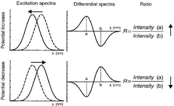
Figure 2.3 – Electrical potential across the cytoplasmic membrane (Left panel) Schematics of a di-8-ANEPPS molecule in membrane environment (Right panel). ... 24
Figure 2.4 – Voltage-sensitive probe dual wavelength ratiometric measurements. ... 25
Figure 4.1 – Example of viralCPP in silico analysis . ... 40
Figure 4.2 – Determination of viralCPP efficiency by FS and FC ... 42
Figure 4.3 - Correlation between viralCPP SVM scores and efficiency. ... 44
Figure 4.4 – Effect of protease treatment on viralCPP efficiency. ... 48
Figure 4.5 – CM of viralCPP-mediated ssDNA Delivery ... 50
Figure 4.6 – viralCPP toxicity assessment by FC Life/dead assay ... 52
Figure 4.7 – Di-8-ANEPPS dipole potential sensing of viralCPP-membrane interactions. ... 54
Figure 4.8 – viralCPP dual wavelength ratiometric measurements. ... 55
Figure 4.9 – CD spectra of viralCPP 2319 in POPC/POPS (4:1) LUVs ... 58
Figure 4.10 - CD spectra of viralCPPs in POPC/POPS (4:1) SUVs ... 59
Figure 4.11 - Sources of AMP Sequences. ... 65
Figure B.1 – A-488 emission spectra collected for a replicate of the viralCPP/ssDNA deliver ... 89
Figure B.2.1 – Mean of fluorescence measurements obtained with a PR ... 90
Figure B.2.2 – Mean of FC fluorescence intensity measurements ... 90
Figure B.2.3 – (Left panel) FC results from viralCPP mediated uptake of ssDNA-A488 (Right panel) Live/dead assay of a sample of live cells ... 91
Figure B.3 – Examples of erratic Cellmask Deep Red membrane staining in CM images ... 92
Figure B.4.1 – Excitation spectra of di-8-ANEPPS in LUVs before and after peptide additions. .... 93
Figure B.4.2 - Excitation spectra of di-8-ANEPPS in LUVs before and after viralCPP 0769 additions at increasing concentration... 94
Figure B.4.3 - (Upper panels) Differential spectra of interactions reported by di-8-ANEPPS in POPC LUVs (Lower panels) Differential spectra of interactions reported by di-8-ANEPPS in POPC:POPS (4:1) LUVs ... 95
Figure B.4.4 - Illustration of shifts in amplitude of viralCPP differential spectra due to increases in peptide concentration.. ... 96 Figure B.5.1 – Hydrodynamic diameter of poor viralCPP leads before (upper panel) and after ssDNA conjugation ... 97 Figure B.5.2 – Hydrodynamic diameter of good viralCPP leads before (upper panel) and after ssDNA conjugation ... 98
Table List
Table 1.1 – Nomenclature, origin, sequence and cargo type of representative CPPs. Adapted from
(35) ... 7
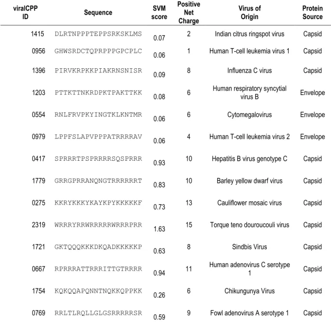
Table 4.1 – viralCPPs: sequences, SVM score, charge, viral and protein origin ... 39
Table 4.2 - Pearson correlation coefficient of viralCPP SVM scores and efficiency ... 44
Table 4.3 – K2D3 prediction of the helical content in CD spectra of viralCPP 2319 in POPC/POPS LUVs ... 58
Table 4.4 – K2D3 prediction of the helical content in CD spectra of viralCPPs in POPC/POPS SUVs ... 59
Table 4.5 – Hydrodynamic diameter of low efficiency viralCPPs ... 61
Table 4.6 - Hydrodynamic diameter of high efficiency viralCPPs ... 62
Acronyms and symbols
(∆) - Transmembrane potential (s) - Surface potential
(d) - Dipole potential
∆A –Difference in absorption of circularly polarized light components θ - Ellipticity
A-488 – Alexa Fluor 488 AMP - Antimicrobial peptide BHK 21 – Baby hamster kidney 21 C – Peptide concentration
CD – Circular dichroism
CM – Confocal laser scanning microscopy CPP – Cell-penetrating peptide
d - Optical path of light
di-8-ANEPPS – 4-[2-[6-(dioctylamino)-2-naphthalenyl]ethenyl]-1-(3- sulfopropyl)- pyridinium D – Diffusion constant
DDS - Drug Delivery System DENV – Dengue virus
DLS – Dynamic light scattering FBS – Fetal bovine serum FC - Flow cytometry
FS – Fluorescence spectroscopy GFP - Green fluorescent protein
HEK 293 – Human embryonic kidney 293
HEPES - N-2-hydroxyethylpiperazine-N′-2-ethanesulfonic acid Hr – Hydrodinamic radius
LUV - Large unillamelar vesicle MAP – Membrane-active peptide Mr – Mean residue molecular weight NLS – Nuclear location signal NP – Nanoparticle
P:L – Peptide-to-lipid ratio PBS – Phosphate buffer saline PC - Phosphatidylcholine PE - phosphatidyletanolamine
PMO - Phosphorodiamidate morpholino oligomer PNA -Peptide nucleic acid
POPC - 1-palmitoyl-2-oleoyl-sn-glycero-3-phosphocholine POPS - 1-palmitoyl-2-oleoyl-sn-glycero-3-phospho-L-serine PR – Microplate reader
PS - Phosphatidylserine
R - Ratio of fluorescence intensities
R0 –Ratio of fluorescence intensities in undisturbed di-8-ANEPPS LUV spectrum ssDNA - Single Strand DNA
ssDNA-A488 – ssDNA oligonucleotides labeled with A-488 ssRNA - Single Strand RNA
SCP - Supercharged protein SEM – Standard error of the mean SUV – Small unillamelar vesicle SVM – Support vector machine
TAT – HIV Trans-Activator of Transcription protein Tat – CPP derived from TAT
viralCPP – Putative CPP identified in a structural viral protein
1. Introduction
1.1. Drug delivery systems
The administration of classical drugs, subscribed to Lipinski’s rule of five, as well as of state-of-the art therapeutics, such as protein/peptide or gene therapies, are a major aspect of medical care. However, these treatments are often associated with undesirable outcomes. For instance, adverse off-target effects can result from an inherent lack of specificity of pharmaceutical molecules, and are associated with high dosages and toxicity (1). Additional downsides of these drugs include systemic degradation and clearance by the renal and reticulo-endothelial systems, poor solubility, stability, circulation time, and bioavailability/biodistribution in vivo (2-4). Furthermore, macromolecules cannot transpose the cytoplasmic membrane of cells, which is a limiting step in the development of biomolecular therapies aimed at intracellular targets (5, 6). This is particularly relevant as it has been estimated that only 10% of the known drugable genome can be targeted by conventional drugs (7). Altogether, these issues greatly diminish pharmacological potential and are major hindrances to the pharmaceutical and biotechnological industries (2).
In this context, the emergence of improved drug delivery systems (DDSs) is expected to be of seminal importance by providing tools to overcome such limitations. Drug delivery can be defined as a multidisciplinary field that deals with the construction of DDSs, also known as vectors, capable of carrying therapeutics to their sites of action. The action of DDSs should therefore improve the pharmacodynamic and pharmacokinetic profiles of therapeutics (2). Modern DDSs are descendants from Ehrlich’s concept of a “magic bullet” (1, 8). Ideally these “magic bullets” should promote drug efficacy and reduce the emergence of toxic effects, by protecting and retaining drug integrity in vivo, and by constraining its release into the site of therapeutic action. Such a system must be biocompatible/ biodegradable to be stably carried throughout the body, and be able to reach the site of drug release (3). However, any drug or delivery system is subjected to harsh conditions and must override imposing frontiers during their journey through the bloodstream and other tissues. For instance, epithelia and endothelia must be crossed by transcytosis, and recognition by many of the proteins and immune factors circulating in blood must be avoided (4, 6, 9). In addition, selectivity and appropriate drug release are very difficult to translate from the research bench into in
vivo environments and the transposition of the cell membrane is still an imposing obstacle to
delivery, even for vectors of latest generation (5, 9). DDSs can be classified into two main groups (5), namely viral vectors (10), which are employed in the context of gene therapy, and non-viral
vectors (11), which can be tailored to transport a wider range of cargoes such as classical drugs and macromolecules.
1.1.1. Viral vectors
While theoretically feasible, effective gene therapy practices have been quite difficult to achieve as nucleotides are very prone to enzyme degradation (DNAses and RNAses). Furthermore these molecules cannot penetrate cells due to high abundance of anionic charges (5). In this regard, viruses were the first carriers employed for gene therapy because viruses have developed the intrinsic ability to protect, carry and deliver nucleic acids through evolution. These vectors may belong to several virus families, for example Adenoviridae, Herpesviridae, or Retroviridae, and each one possesses certain advantages and drawbacks (10, 12). In general, viral vectors have the advantage to transfect cells with efficiency as they naturally bypass cytoplasmic and nuclear membranes using cellular internalization pathways. Viral vectors can also be genetically engineered to increase selectivity. However, there are two major drawbacks associated with the use of viral vectors: I) limited loading capacity which constrains the amount of genetic material that can be carried, but more importantly II) the propensity for eliciting immune responses and the risk for induction of mutagenesis, which may not only prevent re-administration, but can seriously compromise the patient’s health. Indeed, a past clinical trial with an adenoviral vector has resulted in the death of a patient, and two cases of the onset of leukemia in patients treated with gammaretrovirus were also reported in another trial (5, 10).
1.1.2. Non-viral vectors
Contrary to viral vectors, non-viral carriers may be devised to transport every type of pharmaceutical load, from nucleic acids, to proteins/peptides, and classical drugs (5). These vectors are constructed from an immense variety of structures and materials (2). Non-viral vectors can be assembled from organic molecules (e.g. liposomes, polymers, carbon nanostructures or peptides), or from inorganic colloids synthesized from such materials as iron, gold or cadmium selenide. Virus-like particles have also been constructed from chemical engineering of the natural scaffold provided by viral capsids (13).
Due to their fabrication at the nanoscale, these DDSs are collectively known as nanoparticles (NPs). NP development is usually divided into three generations. The first generation was constituted by simple colloids without surface modifications to achieve passive delivery. The
second generation includes the first targeted nanocarriers, functionalized with ligands for specific biological receptors (e.g. antibodies) in attempts to increase selectivity. The current and third generation is comprised by complex NP systems tuned to bypass physiological obstacles, to increase active targeting capacity, and to ensure some degree of temporal control of drug release through physical and chemical stimuli (2, 11). However, most of this technology must be further optimized before reaching the clinical setting, as it still exhibits considerable drawbacks. These include long term biocompatibility concerns, poor retention of colloidal stability in vivo, and the need for fine tuning of targeting efficiency and drug release capability (1, 9, 11).
Intracellular delivery is another aspect of NP systems that must be addressed. In fact, entering through the cell membrane, escaping endosomal compartments before enzymatic degradation, and distributing the cargo to a particular organelle are still some of the most challenging aspects of drug delivery (14). In this regard, cell uptake leading to internalization and intracellular traffic of biomolecules can occur by a diverse set of endocytosis mechanisms, divided into pathways which either proceed by non-specific entrapping of molecules on the cell surface, or which are mediated by cell-receptors. This receptor-mediated uptake is especially important for last-generation DDSs because it may be exploited for selective internalization of NPs into particular cell types. Active targeting thus involves coupling targeting moieties to NPs which are specifically recognized by particular cell-surface receptors (2). However, NP cell-targeting strategies are still quite inefficient (6, 14). Furthermore the uptake of non-viral DDSs is poorly characterized, as their internalization depends on diverse properties such as carrier size, charge, concentration or cell type (2, 6). Following cell entry by endocytosis, DDSs must escape from the endocytic vesicles. This is due to the fact that as endosomes mature, and eventually fuse with lysosomes, a subsequent drop in vesicle pH activates catabolic enzymes responsible for the degradation of molecules entrapped in the endocytic vesicles. Development of DDS escape strategies has been reported in the literature, such as pH-sensitive or fusogenic peptides (15), the construction of fusogenic lipoplexes (6), and the use of dynamic polyconjugates or proton-sponge polymers (9). Nonetheless great amounts of research must be conducted so these strategies can be properly adjusted to every NP system in order to ensure sufficient activity without eliciting cytotoxicity (6, 9, 15).
After three decades of development, few NP delivery systems are reaching the market, but more are currently under preclinical or clinical trials. Nonetheless many limitations still plague the majority of DDSs reported in the literature, which must be solved in order to increase the flux of novel developments to clinical settings (11). In this context, cell-penetrating peptides (CPPs) are an important and diverse category of biomolecules that can transport drugs through biological
membranes with high efficiency (16). CPPs have been extensively employed in the construction of third-generation NP delivery systems, as they can be chemically coupled to improve the efficiencies of uptake or to promote endosomal escape (17-20). In fact, these peptides have considerable advantages relative to other DDSs, such as being less prone to elicit immunological phenomena and not depending on receptor-mediated endocytosis as a principal pathway for uptake. Furthermore, some CPPs have demonstrated the potential to directly translocate cytoplasmic membranes independently of endocytosis, which is a rare trait in biological macromolecules (17). CPPs also present a vast repertoire of vector possibilities due to an immense variety of conceivable amino acid sequences which might be derived from natural or synthetic sources. These peptides can be extensively optimized by tailoring their sequences through mutation of selected amino acid residues (e.g. amino acid substitutions, shuffling or truncation of sequences). Several mutant variants of each CPP can thus be created, further expanding the variety of conceivable designs.
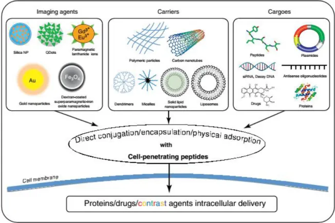
Figure 1.1 graphically depicts how CPP-based technology is deeply connected to the field of drug delivery (19, 20).
Figure 1.1 – CPPs in Drug Delivery – Representation of several biomolecular therapies and available DDSs.
In fact, it can be said that CPP history follows a parallel with that of biomedical nanotechnology, in a sense that only now, after decades of intense research and characterization by academia, have they been able to start realizing their therapeutic potential (20, 21).
For their relevance as core subjects in this work, the following chapter will address these molecular entities in detail, presenting their mechanisms of action, cargo loading strategies, and therapeutic successes and pitfalls.
1.2. Cell-penetrating peptides
Due to their remarkable properties, peptide-based drugs and drug candidates have been steadily increasing in the past decade, with more than 100 having reached the market and a few having already surpassed the global sales threshold of a 1000 million US$ (22, 23).
In this context of investment in peptide therapeutics, the acronym “CPP” refers to the most accepted nomenclature for the subset of peptides capable of interacting with, inserting into, and ultimately transposing lipid membranes independently of a need for chiral recognition by cell-surface receptors. In general, these molecules display net positive charge and are comprised by amino acid sequences between 5 and 30 residues in length, obtained from naturally occurring regions in proteins or designed by in silico methods. As a particular subset of leads for the development of vectors that can transport cargo molecules to the interior of cells, CPPs are already key players in the field of innovative DDSs (19).
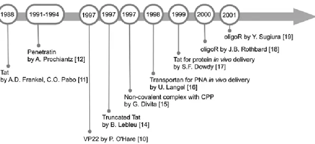
1.2.1. History
CPP history, illustrated in Figure 1.2, had its genesis 25 years ago with the observation by Frankel and Pabo (24) that the HIV-1 transcription-transactivating protein (TAT) could enter cells and translocate into the nucleus. Three years later, it was demonstrated that the Drosophila Antennapedia homeodomain could be internalized by neuronal cells, leading to the discovery, in 1994, of the first CPP, penetratin (25). Afterwards, in 1997, Lebleu et al. (26) identified a peptide (Tat), containing the minimum functional sequence from HIV-1 TAT required for cellular uptake. The first proofs-of-concept for in vivo application of CPPs were reported using peptides and proteins in 1999 (27).
Figure 1.2 – CPP Timeline – Main events in early CPP research. Adapted from (28).
Langel et al. (29) introduced the “cell-penetrating” nomenclature with their work on transportan, the first chimeric peptide carrier. Several other terms have existed to refer to CPPs, such as “protein transduction regions” and “membrane translocating peptides”, but those terminologies have been abandoned (30). Another cornerstone of CPP development, the strategy of using non-covalent binding between the molecular cargo and CPPs, dates back from 1997. It was first based on nucleic acid delivery mediated by MPG (a short peptide consisting of hydrophilic and hydrophobic regions), and later on the primary amphipathic peptide pep-1 to deliver peptides and proteins (16, 31, 32). A final relevant event in CPP history was the demonstration by Wender and Futaki et al. (33, 34) that octa-arginine sequences were sufficient for eliciting cellular and in
vivo peptide uptake, originating R8 and the family of poli-arginines, which are currently among the most extensively studied CPPs (30).
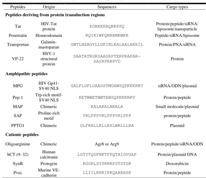
Ever since the discovery of penetratin and Tat, researchers have been steadily increasing the pool of known CPPs, originated from natural, synthetic or chimeric sources (28). Some representative examples are listed in Table 1.1. Furthermore, the CPPsite webpage (http://crdd.osdd.net/raghava/cppsite/) (35) constitutes a major curated database for CPPs comprising more than 800 peptide entries collected from literature.
Table 1.1 – Nomenclature, origin, sequence and cargo type of representative CPPs. Adapted from (36)
Peptides Origin Sequences Cargo types
Peptides deriving from protein transduction regions
Tat HIV-Tat
protein PGRKKRRQRRPPQ
Protein/peptide/siRNA/ liposome/nanoparticle
Penetratin Homeodomain RQIKIWFQNRRMKWKK Peptide/siRNA/liposome
Transportan
Galanin-mastoparan GWTLNSAGYLLGKINLKALAALAKKIL Protein/PNA/siRNA
VP-22 HSV-1 structural protein DAATATRGRSAASRPTERPRAPAR-SASRPRRPVD Protein Amphipathic peptides MPG HIV Gp41-SV40 NLS GALFLGFLGAAGSTMGAWSQPKKKRKV siRNA/ODN/plasmid
Pep-1 Trp-rich
motif-SV40 NLS KETWWETWWTEWSQPKKKRKV Protein/peptide
MAP Chimeric KALAKALAKALA Small molecule/plasmid
SAP Proline-rich
motif VRLPPPVRLPPPVRLPPP protein/peptide
PPTG1 Chimeric GLFRALLRLLRSLWRLLLRA Plasmid
Cationic peptides
Oligoarginine Chimeric Agr8 or Arg9 Protein/peptide/siRNA/ODN
hCT (9–32) Human
calcitonin LGTYTQDFNKTFPQTAIGVGAP Protein/plasmid DNA
SynB Protegrin RGGRLSYSRRRFSTSTGR Doxorubicin
Pvec Murine
VE-cadherin LLIILRRRIRKQAHAHSK Protein/peptide
1.2.2. Biochemical requirements for CPPs
The exact biochemical properties that confer cell-penetrating activity to a peptide have not yet been identified. This is due to a great variety in the amino acid sequences of CPPs and the fact that they are divided into families which do not share sequence similarity (30, 37). However, general biochemical profiles have been found to correlate with CPP activity, such as particular configurations in charge, hydrophobic amino acid residue content, and a propensity for folding into secondary structures in membrane environments.
In terms of the first biochemical property, net positive charge at physiological pH is the most recognizable feature of these peptides. Although there are rare exceptions (e.g. the anionic peptide SAPE (38)), the vast majority of CPP sequences are enriched in basic amino acid residues,
namely arginines and lysines, which are determinant for their activity. For example, Tat is highly cationic with 6 arginine and 2 lysine residues, and it was found that substituting any of its basic residues by a neutral alanine would markedly reduce uptake, while substitution of uncharged residues would have no effect on Tat activity (39). Furthermore, it has been found that arginines potentiate internalization relative to lysines, although often at a cost of an increase in toxicity (39, 40). Arginine is the most basic of all amino acids, because its side chain ends with a guanidinium group which allows multiple hydrogen bonding with anionic and polar molecules. Thus, arginine can form bi-dentate hydrogen bonds with phosphates moieties of more than one lipid headgroup. On the other hand, lysine can only form monodentate hydrogen bonds with molecules and therefore can only interact with a single lipid. As such, arginine rich peptides can bond with more zwitterionic and anionic lipids than their lysine rich counterparts (39, 40). The magnitude of charge is equally linked to internalization efficiency, and it has been demonstrated that in some peptides a minimum of 8 positive charges was required for efficient translocation (33).
Hydrophobic residues are also regularly present in CPP sequences, and in these cases may be critical for internalization. For instance, a study in the CPP pVEC demonstrated that single amino acid substitutions in a terminal hydrophobic patch decreased cellular uptake of the peptide (41). Moreover, other studies showed that the uptake of penetratin was abolished by substitution of tryptophan residues by a phenylalanine (7) or that the uptake of peptide R7 was enhanced by the addition of a C-terminal tryptophan (40).
The relative importance of secondary structure in membrane insertion is still under debate. Structure polymorphism has been observed for peptides such as penetratin, reported to assume α-helical and β-sheet conformations under different conditions (e.g. different model vesicle compositions, salt concentrations, or peptide to lipid ratios (P:L)) (40, 42-44). Conversely, studies in Circular Dichroism (45), Nuclear Magnetic Resonance spectroscopy (46) and Molecular Dynamics simulations(47) have been concordant in finding that the peptide BP100 remains unstructured in aqueous environment, but promptly adopts a α-helical conformation in different lipid membranes (48). Other peptides, such as Tat and R9, are proposed to be inherently disordered (42). Therefore structural flexibility and polymorphism may be important for amphipathic peptides (49), without being obligatory in cationic CPPs (50).
Altogether, these general considerations suggest that CPP uptake does not depend on a single parameter, but on the conjugation of biochemical conditions that render internalization energetically favorable (51), especially an equilibrium between charge and hydrophobicity.
1.2.3. Classification
Because of the high degree of heterogeneity in CPPs, conceiving straightforward classification methods has proven to be hard. Several approaches can be found in the literature, namely classification through origin, cargo-loading strategy or physical-chemical characteristics of the sequences.
In terms of origin, CPPs are classified as protein-derived, when originated from regions of protein amino acid sequences; chimeric, when resulting from the conjugation of two previously existing amino acid motifs; or synthetic, when constructed de novo (52).
Relative to linkage to the therapeutic agent, CPP-cargo systems can be divided into the classes of covalent bonding and electrostatic affinity (53). In the first case, CPPs are attached to their cargo via covalent conjugation through cross-linking chemistry or through fusion of CPP tags in cloned proteins. In contrast, electrostatic affinity involves non-covalent formation of complexes due to attraction between drugs and CPPs of symmetric charges.
Concerning the physical-chemical classification criteria, Francesca Milletti (7) has recently proposed a comprehensive and systematic summary of so-far described CPPs. A representative set of 100 well-characterized CPPs, the majority (83%) of which had positive net charge, was divided into the cationic, amphipathic and hydrophobic categories (7, 17, 52). Amphipathic peptides comprised the largest of these classes, accounting for 44% of samples. Conversely, hydrophobic sequences were the rarest (15%). According to Milletti, a CPP is considered of the cationic group if it contains a stretch of positive charges that is essential for uptake, and if its secondary conformation does not lead to formation of an amphipathic structure. This class included Tat, poli-arginines and nuclear location signal (NLS) peptides. The category of amphipathic CPPs includes peptides with opposing hydrophobic and hydrophilic regions, and is subdivided into primary (amphipatic through sequence) or secondary (amphipatic through spatial conformation) α-helical, β-sheet and proline-rich peptides, according to the secondary structure that is thought to mediate their uptake. Finally, hydrophobic CPPs include peptides mainly composed by apolar amino acid residues, either having very low net charge (less than 20% of sequence) or a hydrophobic motif determined to be crucial for uptake.
1.2.4. Mechanisms of cellular internalization
The mechanisms by which CPPs enter cells are still far from resolved. Biophysical assays (43) and computational simulations (54) have shed some light on this issue at the molecular scale,
by revealing that these peptides can exert profound effects on lipid surfaces. Therefore, it has been proposed that the initial steps of CPP uptake involve electrostatic binding between basic amino acid residues and negatively charged proteoglycans or phospholipid moieties on the cell surface (39). This is followed by peptide insertion into the aliphatic regions of membrane leaflets. In many CPPs this insertion step is thought to be related to the influence of hydrophobic residues, or to conformational shifts which lead the peptides to acquire an amphipathic structure (30, 39). Proceeding stages of the molecular mechanism of cellular entry of CPPs are equally unclear.
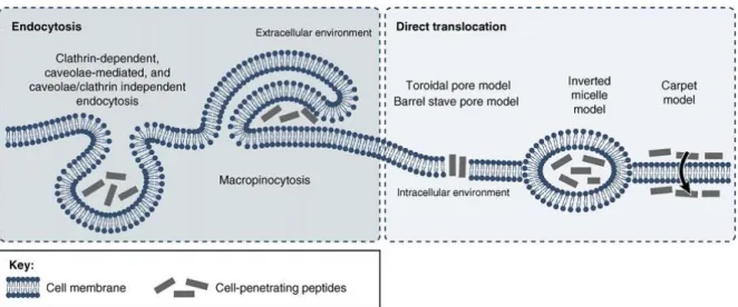
The first studies conducted to ascertain the biological entryway for Tat, penetratin and R9 used microscopy observations, and fluorescently activated cell sorting, on cells incubated with fluorophore-labeled peptides (40). These studies suggested that CPP in vitro uptake could take place despite treatment of cells with endocytosis-blocking drugs. Such findings lead to the conclusion that these peptides could directly translocate membranes through an energy-independent, non-endocytic process (36, 39). However, in 2003, Richard et al. (55) provided evidence that formaldehyde fixation prior to microscopic analysis drastically changed the intracellular distribution of CPPs. This finding, along with the fact that flow cytometry cannot distinguish between membrane-bound and internalized fluorophores, demanded a reevaluation of the mechanisms that had been previously described (7, 54, 56). Furthermore, it is now recognized that peptide-membrane interactions, cellular uptake and cytosolic distribution, are influenced by such factors as the concentration of the peptides, lipids and salts in the solvent, medium pH, lipid membrane composition or cell type (57, 58) and that cargo molecules, including fluorescent probes, also substantially impact CPP internalization routes (59, 60). Thus each CPP/cargo DDS has its own particular behavior and must be studied independently. Despite this drawback in unearthing the biological pathways responsible for CPP internalization, the vast number of publications in the literature concerning the biological and biophysical characterization of CPPs has resulted in the formulation of general models for the mechanisms of uptake. Figure 1.3 illustrates some of these models. Thus, CPP cellular uptake is thought to proceed through endocytosis or through direct translocation of cell membranes.
The left panel of Figure 1.3 comprises several endocytic mechanisms which have been implied in CPP internalization. Endocytic mechanisms are natural processes occurring in living cells that can be triggered by electrostatic interactions with proteoglycans in the extracellular matrix, by direct interaction with the plasma membrane or through specific binding to cell-surface receptors (40). The influence of CPP interactions with proteoglycans and the plasma membrane as triggers for endocytosis remains unresolved (61-63).
Figure 1.3 - CPP cellular entry routes and models of uptake - CPP internalization can be divided into
energy-dependent and energy-inenergy-dependent pathways. The first type comprises clathrin-energy-dependent, caveolae-mediated and caveolae/clathrin independent endocytosis, and macropinocytosis. Direct translocation may proceed by the torroidal pore model, the barrel-stave model, the carpet model, or via inverted micelles. Adapted from (64).
On the other hand, receptor recognition of target molecules is dependent on their chirality, and both L- and D- stereoisomers of a CPP can be equally internalized under the same testing conditions (52), i.e it is clear that receptor-mediated endocytosis does not contribute for CPP internalization. As illustrated in Figure 1.3, all of the currently recognized pinocytic routes have been implicated in CPP uptake (40). For instance, macropinocytosis involves an actin-driven invagination of the plasma membrane which forms large irregular vesicles, and several studies have supported its involvement in the uptake of poly-arginines, penetratin and Tat (40). Clathrin-mediated endocytosis, formed by energy-dependent assembly and pinch-off of clathrin-coated pits from the plasma membrane, has also been connected to Tat and penetratin (39). Caveolin-dependent endocytosis, on the other hand, is lipid raft-mediated and depends on hydrophobic membrane cholesterol-rich microregions, the caveolae, which bud off the membrane to form caveosomes (40). In this case, co-localization studies of fluorescently-labeled Tat with cholera toxins, which are known to internalize via caveolae-mediated endocytosis, supported the uptake of Tat through this entryway. However, contrary results were reported by authors who found that treatment with inhibitors for caveolae-mediated uptake was unable to abolish Tat internalization (39). Examples like these reflect the often contradictory results that have been obtained from different CPP uptake studies. In fact, no single endocytosis pathway could yet be resolved as a predominant contributor
for CPP internalization, and it is now though that several different endocytic mechanisms may act in concert to promote the uptake of each CPP or CPP/cargo system.
The rightward panel of Figure 1.3 concerns non-endocytic internalization. Although direct translocation had been related to artifacts of cell fixation in early studies of CPP uptake, more recent assays on live cells have re-established its involvement in the internalization of some CPPs (39). In this regard, direct translocation involves peptide action in transiently destabilizing and bypassing cell membranes independently of ATP consumption or endocytosis. Figure 1.3 displays four pathways which have been proposed for direct translocation of CPPs, namely the torroidal pore, barrel stave and carpet models, as well as the inverted micelle model. According to the barrel-stave model, translocation happens when a number of peptides form a membrane channel by self-assembly into a barrel-like ring around an aqueous pore. The toroidal pore model is similar as it involves the formation of another type of pore, triggered by peptide induction of high curvature folds in the bilayer. On the other hand, the carpet model results from an accumulation of peptide molecules electrostatically bound to the membrane surface so that when a P:L threshold is reached, the barrier is locally destabilized without the formation of pores (65). Finally, the inverted micelle model states that disturbances in lipid bilayers lead to the formation of inverted micelles that trap the CPPs in their hydrophilic core until further destabilization, effectively releasing the molecules into the cytosol (64, 66). Additional models for peptide transposition mechanisms, e.g. driven by membrane potential differences, can also be found in the literature (67). Nonetheless, it should be noted that, although informative, such models are limited approximations, and thus may not fully describe the complex molecular interactions that take place during CPP activity (68).
As a consequence of the former paragraphs, it should be realized that several kinds of endocytosis and translocation mechanisms may act in concert to promote the internalization of each CPP/cargo system. Indeed, this notion is gaining increasing support and acceptance in CPP-related literature (20, 39).
1.2.5. CPP-mediated gene therapy
CPP delivery strategies have gained considerable popularity in the past decade, and more than 1000 applications have already been attempted both in vitro and in vivo (54). In fact, since the first in vivo proofs-of-concept, CPPs have been used in therapies to target multiple disorders including asthma, ischemia, diabetes, or inflammation (54, 56). Therapies for cancer have also been intensively studied, and CPPs are equally investigated for treatment of brain related illnesses
because of their ability to cross the blood-brain barrier (69, 70). In this regard, the application of CPPs as gene and oligonucleotide therapy vectors is a particularly promising and widely researched area (28).
Gene therapy is a powerful form of treatment by which cells are supplied with exogenous genetic material that complement, substitute, or suppress functions linked to disorders (5). Cancers (e.g. melanomas, carcinomas, leukaemia), genetic (e.g. cystic dibrosis, X-linked severe combined immunodefficiency disorder), auto-immune (e.g. diabetes type 1), neurodegenerative (Parkinson’s disease, multiple sclerosis) and cardiovascular disorders and viral infections are just some of the highly problematic illnesses that may be overcome by a continued investment in this type of therapy (5, 10, 71). Diverse approaches to gene therapy have been developed in the past twenty years (72), namely the use of plasmids containing genes of therapeutic interest, or splicing correction and silencing of deleterious genes via antisense oligonucleotide technology. These strategies may be achieved using single strand DNA (ssDNA), small interfering RNAs (siRNA) or microRNAs, or by steric blocking DNA mimics, such as phosphorodiamidate morpholino oligomers (PMO) and peptide nucleic acids (PNA) (72-75). Extensive examples of CPP conjugation to all of these molecules for gene delivery, antisense knockdown or mRNA splice correction can be found in the literature (36).
For instance, CPP -mediated delivery of plasmid DNA has been achieved in cultured cells, chiefly through non -covalent approaches that have included CPPs such as MPG, Tat and poly-arginines (36). In vivo results have also been reported. For instance, the intravenous injection of a luciferase expression plasmid conjugated with the peptide PPTG led to significant gene expression in the mouse lung (76). In another study, a recombinant form of Tat was mixed with a therapeutic plasmid encoding α-galactosidase A and injected into muscles of knockout mice models for a lysosomal storage disease. In this latter case the skeletal muscle expression of galactosidase was significantly enhanced in comparison to injection of DNA alone (58).
Likewise, CPP strategies have been developed for the administration of siRNA in vitro and
in vivo. For example, MPG has been reported to enhance siRNA delivery in several cell lines, but
has also been applied for in vivo delivery of OCT-4 targeting siRNA into mouse blastocysts (77). Furthermore, Tat conjugated with an RNA-binding motif was reported to block epidermal growth factor in vivo (78), and the fusion of a small peptide from rabies virus glycoprotein to R9 was used to deliver siRNA to the CNS of mice (79).
Finally, the conjugation of oligonucleotide mimics to CPPs has also been extensively assayed in peptides such as transportan Tat, penetratin and oligo-arginines (36). In vivo results have
also been reported. Prominent examples include several synthetic and chimeric peptides which have been tested in the delivery of therapeutic PMOs to animals models of Duchenne muscular dystrophy (80).
These are only a few instances of a large amount of proofs-of-concept related to the viability of CPP-mediated gene delivery. Although encouraging results have been attained, CPP research still has an important role in the translation of these applications from the work bench to the clinical setting (21).
1.2.6. Current drawbacks of CPPs
Despite demonstrating remarkable properties and constituting promising vectors, CPPs encounter obstacles to clinical translation. Lack of selectivity, and concerns about toxicity, are perhaps obvious, but problems like reduced in vivo efficacy, poor bioavailability, instability, and short time-span, must also be considered (7). A number of studies have assessed the toxicity of CPP
in vitro, while in vivo information from animal assays remains more scarce (58, 81-84). In general
CPP have demonstrated low levels of cytotoxicity (85, 86). The kinetics and biodistribution of some CPPs have also been studied in vivo establishing that these therapeutics can potentially be safe and target most tissues in the body (64, 87-89). However, downsides such as rapid blood clearance and preferable distribution to the liver, kidneys, spleen, bowels and lungs, have also been reported (90, 91). In this regard, phagocytosis by the reticuloendothelial system, proteolysis by intestinal enzymes, as well as interactions with blood components can severely decrease the circulation of CPPs.
However several strategies are being investigated to counter CPP drawbacks, such as substitution of biological L- amino acids for protease resistant R- stereoisomers, chemical optimization of structure, or covalent linkage to shielding molecules, such as poly-ethylene glycol (21, 64). Clearly CPPs have demonstrated great therapeutic potential but there is still the need and ample opportunities to expand the known pool of peptide leads and to continue studying the biochemical properties which are determinant for their activity.
1.3. Structural viral proteins and CPPs
It was previously mentioned that the discovery of TAT protein’s natural translocation ability is often held at the beginning of CPP research. Additionally the Tat peptide was one of the first CPPs to be discovered and one of the most extensively studied peptide DDSs (40). However,
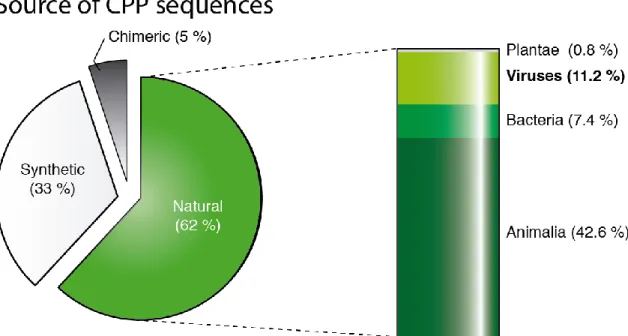
more examples exist of CPPs derived from viruses, concretely from structural components of viral shells. Vp22 is one such example, found in a Herpes Simplex virus tegument protein capable of translocating membranes (92). Additional CPPs have been originated from the glycoprotein H of the same virus (93), or from the k8 protein of Herpesvirus 8 (94). Other sources of CPPs from structural viral proteins have included the classical swine fever virus’ envelope protein (95), the human respiratory syncytial virus surface glycoprotein G (94), the hepatitis-B surface glycoprotein (96) and a N-terminal domain of its X protein (97), or the capsid units of the flock house virus (98), the brome mosaic virus (99), and the alphavirus (94). Furthermore the extensively studied chimeric CPPs pep-1 and MPG comprise a NLS from the Large T antigen from simian virus 40 (16), and the hydrophobic motif of MPG is derived from the fusion sequence of the HIV protein gp41. Nonetheless, despite these well-established precedents, the percentage of CPPs derived from viruses is still very limited when compared to the total number of currently known CPP sequences, as indicated in Figure 1.4. This suggests that structural viral proteins are an underexplored source of therapeutic peptides.
Figure 1.4 - Sources of CPP Sequences – Each percentage was calculated based on the amount of hits on
CPPsite (35).
In fact, viruses are fascinating, complex and very diverse molecular entities that have evolved, through aeons of selective pressure, a wide array of strategies to protect and deliver
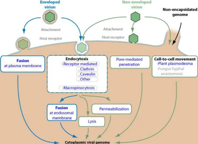
genomes to their replication sites in the interior of obligatory cellular hosts (100). Moreover, viruses persist throughout all of life’s regions and exhibit a large number of taxa with differing sizes and morphologies. Simple viruses can possess a single genome molecule contained within a protective structure, the capsid or nucleocapsid. These structures may be organized in a variety of arrangements, such as helical or icosahedral patterns (101). More complex virions have additional elements, including non-structural proteins with diverse roles in the viral cycle (102) (e.g. genome replication, modulation of host gene expression), and the envelope (103), a lipid bilayer surrounding the viral core in association with a protein shell.A distinction between enveloped and naked viruses is important because the strategies to transpose cell barriers employed by species within each group may be quite distinct (104), as illustrated in Figure 1.5.
Figure 1.5 - Diversity in viral cellular entry mechanisms – Structural proteins from capsid, envelope or
membrane elements mediate the uptake of virions into host cells through a variety of strategies. Adapted from Expasy ViralZone (105).
As the figure indicates, some viruses gain access to the cytosol through the cytoplasmic membrane (106), while others are first internalized and transported through endocytic vesicles (107). In addition, viruses have also been shown to exploit the proximity between neighboring cells, spreading from the interior of a host directly through the membrane to the cytosol of contacting targets (100, 107). Therefore, a fundamental consequence of viral uptake is that virions must interact with lipid bilayers to reach their replication sites. In this regard, several mechanisms have already been characterized, including the induction of membrane lysis or of fusion events, and the formation of pore structures through which genomes can escape (108).
Due to the propensity that structural proteins from viruses display towards lipid membranes, some authors have recognized the similarities between selected regions of these proteins and a broad group of peptides, collectively known as membrane-active peptides (MAPs) (109-111). These peptides share the intrinsic property of exerting biological effects (antimicrobial (45), antiviral (112), anticancer (113)) based on molecular interactions with lipid membranes. Indeed, CPPs can equally be classified as MAPs because their activity is related to an affinity for lipid structures (109, 114). Given this fact, and the data presented in Figure 1.4, it is surprising that viruses have not been more intensively studied in the field of CPPs, and in a wider perspective, of therapeutic MAPs.
Professor Castanho’s lab has combined the subjects of structural proteins from viruses and MAPs in the determination of Flavivirus entrance mechanisms through the study of the dengue virus (DENV) capsid protein. DENV, an enveloped virus, has three structural proteins termed as envelope, membrane, and capsid, relating to the structures from which they are originated. There is ample evidence that this virus enters cells through endocytosis and that endossomal acidification triggers a fusion type II mechanism (115). This entry strategy consists in the formation of envelope protein trimers and their insertion into the host membrane, but consecutive stages of the process are still left up to speculation (116). Recently, Freire et al., from Professor Castanho’s group, have found that the C protein is implicated in Flavivirus fusion by demonstration of its inherent capacity to permeate cells. For this end, peptides were first derived from the two putative functional domains of the C protein monomer, the RNA binding domain – pepR, and the membrane binding domain- pepM. Both peptides were found to form stable electrostatic aggregates with ssDNA and to strongly favor partition into model membranes, even when conjugated with nucleic acids (111, 117). Furthermore pepR and pepM were confirmed as full-fledged CPPs capable of penetrating cells and delivering ssDNA with high efficiency (118). Uptake studies suggested that pepR internalization is dependent on an endocytic pathway, and that pepM can directly translocate cell membranes in
accordance with the inverted micelle model (118). PepR had also previously been reported as an antimicrobial peptide (AMP) capable of severely disrupting the outer surface of Escherichia coli (119). Furthermore, the full-length DENV C protein was also studied and shown to directly translocate cells while delivering nucleic acids in vitro with high efficiency (120). This work also proposed a relation between the capsid of Flavivirus and a recent concept of super charged proteins (SCP). Liu et al. have found that engineering natural proteins like green fluorescent protein (GFP) to express a very high density of positive charges per molecular weight (e.g. GFP wt: -8 to SCP GFP: +36 charges) can impart them with the ability to deliver functional cargo to cells, outperforming even cationic CPPs (121-124). The authors also found SCPs expressed in the human genome, and tested them as DDSs in vitro, effectively establishing that SCPs can occur spontaneously in nature (125). Freire et al. have further expanded this idea by identifying the DENV C monomer as a SCP. In fact, DENV C protein has a charge to weight ratio of +1.97 kDa/MW (120) and this value greatly surpasses the cutoff for a protein to be considered super charged, set by Liu et al. (of > +0.75 kDa/MW)(125). Freire et al. also studied the prevalence of natural SPCs in structural proteins from additional viral families (120) (categorized as elements from envelope, membrane or capsid). They found that the great majority of SCPs in the structural viral proteome corresponded to capsid elements, including capsids from all other members of the Flavivirus family. Additionally it was found that, excluding Tat, all virus-derived CPPs did not belong to a natural SCP (120).
1.4. Motivation
The work described in this document further explores the findings of Freire et al, that viral proteins and/or derived peptides are efficient intracellular carriers of genetic cargo. Thus, the main objective was to identify specific peptide sequences in structural proteins from viruses with the inherent property of interacting with lipid bilayers, and exploit them as delivery vectors for gene therapy. To do so, the project assumed a tripartite configuration, as described in the following paragraphs.
The first stage of the work concerned the identification of putative CPP regions (viralCPPs) in capsid, envelope or membrane proteins of viral proteomes. This was achieved by collecting a large sample of structural viral protein sequences from online databases. These were then uploaded to the CellPPD website (126), containing an algorithm for prediction of CPP regions in protein sequences. This process resulted in thousands of putative CPP sequences, or viralCPPs. These
sequences were grouped together based on pairwise similarity using the software package CLANS (127). A total of 150 viralCPPs were selected from the several clusters produced by CLANS. A final set of 14 viralCPPs was then chosen from the previous group, to be assayed in vitro and validated as CPPs.
As such, the second stage of the project involved the execution of a protocol for in vitro assessment of the 14 viralCPPs to evaluate their capacity in the delivery of ssDNA oligonucleotides into cells. Consequently, the peptides were tested as possible DDSs for gene therapy applications. This protocol consisted on mixing each viralCPP with Alexa-488 labeled ssDNA oligonucleotides to create electrostatically bound complexes, adding them to cells, and incubating the system for one hour. After washing, to remove non-internalized molecules, the fluorescence intensity of probe retained by cells was evaluated by fluorescence spectroscopy and flow cytometry. Therefore, this protocol allowed that the efficiency of viralCPP-mediated ssDNA delivery be evaluated. Confocal microscopy was employed to visualize viralCPP delivery of oligonucleotide probes. The effect of trypsin on viralCPP activity was also studied, and cytotoxicity assays were conducted to establish the influence of viralCPPs on cell viability.
Finally, in the third stage of the work, biophysical methodologies were conducted to assay biochemical properties of these peptides that could be related to their biological activity. Di-8-ANEPPS membrane dipole potential sensing experiments were made to assay the degree of interaction between each viralCPP and lipid model vesicles. This study was important because membrane affinity is linked to CPP activity. Furthermore, circular dichroism spectroscopy was employed to detect conformational shifts in viralCPPs in the presence or absence of lipids, a common biochemical trait in CPPs. A last set of experiments used dynamic light scattering spectroscopy to calculate the hydrodynamic diameter of viralCPP/ssDNA electrostatic complexes. Hydrodynamic diameters relay the apparent size of peptide/cargo conjugates which can be linked to the efficiency of delivery.
This work is of relevance in several areas of biomedical research. Indeed, new CPP leads could be identified and isolated from structural viral proteins, a previously underexplored source. In this regard, the project marked a “return to origins” in the context of CPP research due to the fact that its cornerstone - HIV’s TAT– was a viral protein. Furthermore, proof-of-concept was provided for viralCPP development as DDSs for gene therapy, a state-of-the-art application in medicine.
2. Technical overview
Experimental procedures conducted in this work concerned: I) the use computational tools to identify novel CPP sequences, II) an in cellulo approach to study CPPs combining flow cytometry (FC) and fluorescence spectroscopy (FS), III) biophysical studies to assay interactions between CPPs and lipid membranes or oligonucleotides. This section provides a brief theoretical background for the techniques that were employed during the project.
2.1. Computational tools
2.1.1. Expasy ViralZone
ViralZone (http://viralzone.expasy.org/) (105) is a web resource which provides fact sheets on all known virus families, combining textbook knowledge with genomic and proteomic sequences. For each genus within these families, reference strains have well documented and annotated functional and structural data.
In this work, ViralZone was employed to easily locate a wide range of structural viral proteins with clear functional and structural annotated information. Using the Baltimore classification system as a starting point, viruses from all known families were accessed. For each virus the amino acid sequences corresponding to its structural proteins were collected from the UniProtKB (128). These structural proteins included elements from capsid, envelope or membrane, according to individual virion morphologies.
2.1.2. CellPPD
Understanding if a peptide can act as a CPP based on its amino-acid sequence is a non-trivial problem. This is due to a lack of clear homology in the hundreds of very diverse sequences recognized as CPPs, despite the fact that several biophysical studies have revealed biochemical features which are common to the majority of CPPs (129). These include properties such as positive net charge, relative degree of hydrophobicity and short length, which can be used as general rules of thumb for predicting CPP potential (130). Recently, important developments have been made in the field of CPP prediction. The onset of CPPsite (35), a database for CPP sequences, as well as a growing number of CPPs reported in the literature, has allowed the refinement of prediction algorithms. As a consequence, there are now two online tools for predicting CPP potential in amino acid sequences, CPPpred (131) and CellPPD (126).
CellPPD (http://crdd.osdd.net/raghava/cellppd/) (126) has been used in this work to identify putative CPP regions in structural proteins from viruses. The algorithm implemented in CellPPD divides proteins into sequences of a length defined by the user (e.g. sequences of 20 amino acids) and each sequence is analysed for its CPP propensity. Therefore, the algorithm predicts and relays the likelihood that each of those sequences is a CPP. This prediction is conducted by a machine learning algorithm instructed by a main dataset of 700 peptides. The algorithm accounts for factors such as total amino acid constitution, and relative position of particular amino acid residues in the sequence. The evaluation conducted by the CellPPD algorithm also attributes an estimator of CPP likelihood, the Support Vector Machine (SVM) score, to each sequence. Per standard definitions, protein sequences scoring positive SVM score values are considered CPP regions, and can be isolated as potential novel CPP sequences. Additionally, higher SVM scores are related to increased CPP likelihood while protein sequences with negative SVM score are considered non-CPPs. This algorithm has a probabilistic nature, and the authors report that it can predict CPPs with over 80% accuracy. Therefore, each positive prediction by CellPPD must be subjected to experimental validation to ascertain that it is a true positive hit, i.e. validate CPP activity.
2.2. Flow cytometry
FC is a technology that measures several physical characteristics of populations of living cells by means of spectroscopic data (132). To fulfill this function, a standard FC apparatus is equipped with a fluidics system able to transport single cells through the optical path of excitation lasers. The signals generated by the optical system are then individually recorded by a set of detectors and digitally processed. Data from FC allows the quantification of particle size and relative granularity/internal complexity through light scattering (as represented in Figure 2.1). Forward scattered light, scattered off the axis of an incident beam, is proportional to cell size. Side-scattering, which is refracted or reflected light, can be related to granularity and cellular complexity. The correlation between forward and side scattering allows the identification of different populations in a heterogeneous pool of cells (133). Fluorescence intensity can also be recorded by FC when cells or biomolecules are labeled with a fluorescent probe. In this case, different populations can be recognized based on fluorescent staining.
Data from a FC assays can assume different graphical representations, but histograms and 2D-scatter plots are the most frequent. In scatter-plots the concept of gating is especially important because thresholds can be defined to establish cell populations. Thus, a gate is a graphical/numeric
boundary that defines the range of values in which each population is included, so that positive and negative events may be distinguished and analyzed.
In the context of this work, FC was used to quantify the effect of CPPs on the delivery of ssDNA labeled with a fluorescent probe Alexa-Fluor 488 (A488). This technique was also employed to conduct toxicity evaluations through a Live/Dead assay (134).
Figure 2.1 - Schematics of basic components of a flow cytometer – Samples are loaded and carried by a sheath fluid. Hydrodynamic focusing produces a single file in which particles are individually probed by a system of lasers. Fluorescence intensity and forward/side scattering of light can thus be recorded and transduced to the electronic component of the device. Adapted from (132).
2.3. Biophysical methodologies
Biophysics is an interdisciplinary field concerning the study of living systems by employing theories and techniques from physics. Thus it provides a quantitative analytical approach to many biological questions. In this context, an assortment of spectroscopic methods can be used to inquire relationships between the physical properties of molecules, such as structure, dynamics and function, in order to determine their role in cells, tissues and organs.
2.3.1. Steady-state fluorescence spectroscopy
Fluorescence results from the absorption of light by a molecule, its excitation, and the subsequent emission of light of higher wavelength after loss of energy by nonradiative processes