DEPARTAMENTO DE GENÉTICA E BIOTECNOLOGIA
Development of human osteogenic cell sheets
co-cultured with endothelial and pericyte-like
cells.
Luís Filipe Freitas Mendes
Dissertação orientada por:
Doutora Alexandra Pinto Marques
Doutor Rui Luís Gonçalves dos Reis
Mestrado em Genética Molecular Comparativa e Tecnológica
Vila Real, 2011
I
First of all I would like to thank Drª Alexandra Marques, my supervisor, for all the
support, perseverance and fruitful discussions. I am especially grateful for her always
ready availability and good relationship.
To Prof. Rui Reis, my co-supervisor, for giving me the chance to complete this
importance step of my academic years working in 3B’s research group and for all the
unconditional support during that time.
To Dr. Rogério Pirraco for his scientific aid in everyday lab routine, constant
encouragement and, above all, for his friendship.
To Dr. Wojciech Szymczyk, Drª Tírcia Santos, Drª Ana Frias, Drª Mariana Cerqueira
and Drª Ana R. Martins for sharing their scientific knowledge and contribute for the
development of this work.
To all the people of the 3B’s research group including technicians, colleagues and friends, for their good work environment and friendship.
II
with some studies showing that it is a reliable alternative for the traditional scaffold-based approaches by avoiding their associated shortcomings. This technology permits not only to eliminate the exogenous scaffolding biomaterials but also to create ex vivo tissue-like substitutes with organized cellular entities and cohesive cell-to-cell and cell-extracellular matrix (ECM) interactions. However, the progression of this technology is being limited by some of the barriers which have been hampering the evolution of other TE strategies, such as the lack of appropriate vasculature to supply thicker constructs in vivo. It is herein proposed the creation of a cell sheet construct, by co-culturing osteogenic, endothelial and pericyte-like cells, with the purpose of enhancing the vascularization of newly formed bone tissue and also the degree of maturation and stability of the vascular network.
The optimization of the culture conditions to fabricate osteogenic cell sheets derived from human bone marrow stromal cells (hBMSCs) was the first step of this work. This optimization allowed furthering evolving to the design of a co-culture system with human umbilical cord vein endothelial cells (HUVECs) and pericyte-like cells as a model for the vascularization of in vitro cultured constructs with potential for bone tissue engineering applications. Bone marrow was previously proposed as a source of perivascular-like cells, in particular those expressing CD146, therefore hBMSCs were isolated, cultured under different conditions and screened for the expression of CD146 as well as for the Mesenchymal Stem Cells (MSCs) markers CD90, CD73 and CD105. Flow cytometry results demonstrated that supplementation of standard hBMSCs culture medium with TGF-β1 promote an increase of CD146 expression in hBMSCs, from approximately 50% to more than 97%. Moreover, changes in CD146 expression are associated with different cellular morphologies. Immunocytochemistry assays performed on the co-cultures showed that induced CD146+ hBMSCs and HUVECs migrated and organized themselves over a thin, collagen-rich, osteogenic cell sheet, suggesting the existence of an efficient cross-talk involving all the co-cultured cell types. Further studies concerning the ability of these constructs to form functional, vascularized and osteo-committed tissue in vivo were performed using a well described protocol for subcutaneous cell sheets transplantation on mice. Immunohistochemistry analysis of transplanted cell sheets revealed the integration of HUVECs with host network vasculature as well as the osteogenic potential of the created construct, as shown by the expression of osteocalcin. Additionally, analysis of the diameter of CD146 positive blood vessels showed a higher mean vessel diameter for the experimental condition, reinforcing the advantage of the proposed model regarding blood vessels maturation and stability.
III
existindo alguns estudos que provam a viabilidade desta tecnologia como alternativa aos tradicionais métodos baseados no uso de scaffolds. O uso de cell sheets permite não só dispensar o uso de biomateriais de suporte mas também criar substitutos de tecidos, ex-vivo, com uma organização celular própria mantendo altamente coesivas as adesões célula-célula e as interacções com a matriz extracelular. Contudo, a evolução desta tecnologia tem sido limitado por algumas das barreiras que têm vindo a impedir o progresso das estratégias tradicionais de engenharia de tecidos, nomeadamente a ausência de uma vasculatura apropriada que permita a integração dos constructs in vivo. Deste modo, este trabalho propõe a criação de um construct baseado na tecnologia de cell sheets, obtido co-cultivando células osteogénicas, endoteliais e com marcadores pericíticos, com o objetivo de promover a vascularização de tecido ósseo novo e o grau de maturação e estabilidade da rede vascular formada.
Numa primeira fase deste trabalho começou-se por otimizar as condições de cultura celular para o fabrico de cell sheets osteogénicas a partir de células mesenquimais humanas derivadas da medula óssea (hBMSCs). Esta otimização permitiu evoluir em seguida para o design de um sistema de co-cultura de células endoteliais da veia do cordão umbilical (HUVECs) e células com potencial perivascular (CD146+), criando um modelo para a vascularização de constructs produzidos in vitro com potencial para aplicação em engenharia de tecido ósseo. A medula óssea foi anteriormente proposta como uma possível fonte de células com potencial perivascular, nomeadamente as que expressam o marcador celular CD146. Deste modo, as hBMSCs isoladas da medula óssea foram cultivadas em diferentes meios de cultura e caracterizadas tendo em conta a expressão de CD146 e dos marcadores associados a células mesenquimais, CD90, CD73 e CD105. Os resultados de citometria de fluxo demonstraram que a adição de TGF-β1 ao meio de cultura provoca um aumento de expressão do marcador CD146 nas hBMSCs, de aproximadamente 50% para um valor superior a 97%. Simultaneamente, verificou-se que alterações na expressão de CD146 estão associadas a alterações da morfologia celular. A análise das co-culturas por imunocitoquímica mostrou que as células induzidas a expressar CD146 e as HUVECs foram capazes de migrar naturalmente e de se organizar sobre uma superfície rica em colagénio composta por células osteogénicas confluentes, sugerindo a existência de um cross-talk eficiente entre todas as células que compõem o sistema de co-cultura. A capacidade do construct formar, in vivo, tecido osteo-comprometido, vascularizado e funcional foi avaliada após a transplantação subcutânea do construct em ratinhos. A análise de imunohistoquímica das cell sheets transplantadas revelou a
IV
Adicionalmente, a análise do diâmetro dos vasos positivos para o marcador CD146 revelou um aumento da média do diâmetro dos vasos na condição experimental, o que confirma a hipótese colocada e reforça a relevância do modelo proposto na maturação e estabilização dos vasos sanguíneos formados.
V
1 - Regenerative Medicine ... 1
2 - Bone Tissue Engineering ... 2
2.1– Bone Tissue Biology and Regeneration Process ... 3
3 - Mesenchymal Stromal/Stem Cells: From one to multiple sources ... 3
4 - The Vascularization Problem in Tissue Engineering ... 5
5 – Pericytes Functions, Hallmarks and Origin ... 7
6 – Cell sheet engineering technology ... 9
6.1 - The intelligent cell detachment ... 10
6.2 – Applications in Regenerative Medicine ... 11
7 - References ... 13
II - Material and Methods ... 20
1. Cell culture... 20
1.1 – Isolation and culture of hBMSCs ... 20
1.2 – Osteogenic differentiation of hBMSCs ... 21
1.3 – Induction of CD146+ phenotype ... 21
1.4 –Isolation and culture of endothelial cells (ECs) ... 21
1.5 – Co-culture of hMBSCs, ECs and pericyte-like (CD146+) cells ... 22
2. Cell sheets fabrication ... 23
2.1 Monocultured and co-cultured cell sheets ... 23
2.2 Stacked cell sheets ... 23
3. In vivo transplantation of cell sheets ... 24
4. Characterization Techniques and Methodologies ... 26
4.1 - Flow cytometry Analysis ... 26
4.2 – Fluorescence immunocytochemistry ... 28
4.3 – Alizarin Red Staining ... 28
4.4 – Cell sheets characterization ... 29
5 - References ... 33
Development of human osteogenic cell sheets co-cultured with endothelial and pericyte-like cells ... 34
Abstract ... 34
1 - Introduction ... 36
2 - Material and Methods ... 37
2.1 - Cell Isolation and culture ... 37
VI
2.5 - Immunofluorescence ... 39
2.6 - Immunohistochemistry ... 39
2.7 - Cell sheet stacking and transplantation ... 40
2.8 - Statistical analysis ... 41
3 – Results ... 41
3.1 - Osteogenic cell sheets derived from hBMSCs ... 41
3.2 - Effect of TGF-β1 on CD146 expression and cellular morphology ... 42
3.3 - Characterization of co-cultured cell sheets ... 43
3.3 - Characterization of implanted cell sheets ... 45
3.4 - Analysis of the number and diameter of blood vessels ... 47
4 - Discussion ... 47 5 - Conclusions ... 51 6 - References ... 52 IV – General Conclusions ... 56 References ... 57 V – Annex ... 58
1 – Morphologic analysis of hBMSCs cultured into different media ... 58
2 – Morphological similarities between pericyte-like (CD146+) cells and reported pericytes 59 3 – Calcium deposits at different days of hBMSCs culture in osteogenic medium. ... 59
4 – Fluorescence Immunocytochemistry ... 60
4.1 – Immunocytochemistry for CD146 on HDMECs in monoculture ... 60
4.2 – Fluorescence immunohistochemistry for CD146 on co-cultures of osteoblastic cells, HDMECs and pericyte-like (CD146+) cells. ... 60
VII
I – Introduction
Figure 1 – Features and functioning of TR surfaces. Adapted from Thermo Scientific Nunc UpCell Surface manual. ... 11 Figure 2 - Examples of CS engineering to create tissue-like substitutes. (A) Application of Single CS transplantation for cornea replacement and regeneration of skin, periodontal ligament and bladder. (B) Stacking of several CS for the improved performance of cardiac tissue. (C and D) Liver and kidney, tissues with laminar and higher-order structures, can also be recreated with CS engineering. Adapted from (5). ... 12
II – Material and Methods
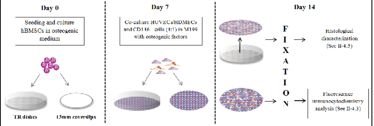
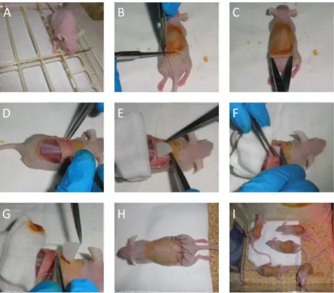
Figure 1 – Schematic representation of the steps to set-up the co-cultures. ... 23 Figure 2 – Overview of the cell sheets implantation process ... 25 Figure 3 – Overview to the process of implanted cell sheets recovery ... 25
III - Development of human osteogenic cell sheets co-cultured with endothelial and
pericyte-like cells
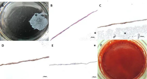
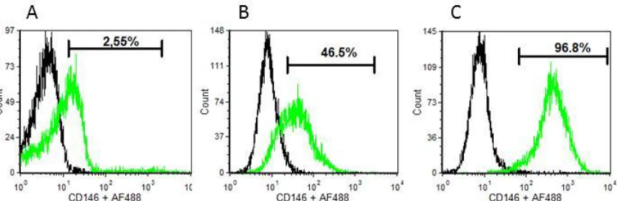
Figure 1 - Macroscopic view (A,F) and histological characterization (B-E) of hBMSCs cell sheets cultured for 14 days in osteogenic medium; (A) Osteogenic cell sheets derived from hBMSCs after detachment and contraction. (B) H&E staining; (C) immunostaining for osteocalcin and (D) type-I collagen; (E) Control for immunostaining; (F) Osteogenic character of CSs after 21 days in culture revealed by AR-S staining. * PVDF membrane used to protect CS during processing step. ... 42 Figure 2 - Representative flow cytometry analysis of hBMSCs at different passages and cultured in different culture medium. (A) CD146 expression of bone marrow mononuclear fraction at isolation day; (B) CD146 expression on hBMSCs (P5) cultured in complete α-MEM; (C) CD146 expression on hBMSCs (P5) cultured 7 days in complete α-MEM supplemented with 1 ng/mL TGF-β1. ... 43 Figure 3 – Expression of CD146 (green) (A) and evolution of cell morphology of hBMSCs before (B) and after culture for 7 days in α-MEM + 1ng/mL TGF-β1 (C). For immunocytochemistry
VIII
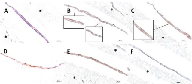
Figure 4 - Immunocytochemistry for CD146 (green) expression on hBMSCs, pericyte-like (CD146+) cells and HUVECs co-cultured for 7 days. Dil-AcLDL (red) was used to assess LDL uptake and identify HUVECs. DAPI (blue) was used as nuclear staining. A, B and C represent successive close ups. ... 44 Figure 5 - Histological analysis of co-cultured cell sheets after H&E staining (A) and immunostaining for CD31 (B), CD146 (C); osteocalcin (D) and type-I collagen (E). Squares represent close up views of specific regions of the cell sheets showing expression of CD31 (B) and CD146 (C). Identification of positive signal was determined in comparison to immunocytochemistry negative control (F). * PVDF membrane used to protect cell sheet during processing. ... 44 Figure 6 - Representative flow cytometry analysis of CD146 and CD31 expression on (A;B) HUVECs (P5) and (C) CD146 expression on pericyte-like (CD146+) cells (P5) cultured for 7 days in M199 supplemented with osteogenic factors. High expression (>98%) of CD31 and CD146 on HUVECs confirmed the absence of an effect of the osteogenic factors on their native phenotype. A reduction on CD146 expression of pericyte-like (CD146+) cells (84%) was verified after 7 days in culture without TGF-β1. ... 45 Figure 7 - H&E (A;B;C;D) and Alizarin Red-S (E;F;G;H) staining on cell sheets after 7 (A;C;E;G) and 21 days (B;D;F;H) of subcutaneous implantation. (A;B;E;F) control group (C;D;G;H) experimental group. ... 45 Figure 8 - Immunohistochemistry for Osteocalcin on experimental (A,C) and control (B,D) conditions at 7 (A,B) and 21 (C,D) days of implantation. (E,F,G,H) immunostaining negative control. ... 46 Figure 9 – Immunohistochemistry for CD31 (A,F) and CD146 ( B,G,D, I) on experimental (A,B,C,F,G,H) and control (D,E,I,J) conditions at days 7 (A,B,C,D,E) and 21 (F,G,H,I,J) of implantation. (C,H,I,J) Immunostaining negative control. → negative blood vessels for CD146; ► positive blood vessel for CD146 ... 46 Figure 10 - Co-localization for CD146 and human-specific anti-mitochondria performed on experimental condition after 7 days of implantation. DAPI (blue) was used as nuclear staining. (A) Human cells (green) detected by human-specific anti-mitochondria antibodies. (B) Double labelled cells for CD146 (green) and anti-mitochondria (red) assembled in a blood vessel-like structure (arrow). ... 47 Figure 11 – Representation of the number of blood vessels and the mean diameter for control and experimental conditions at days 7 and 21 of implantation. *p ≤ 0.05; **p ≤ 0.01.. ... 47
IX
Figure 1 - hBMSCs cultured in different conditions. (A) hBMSCs (P1) in α-MEM+2 ng/mL FGF-β; (B) confluent hBMSCs (P3) cultured in osteogenic medium for 7days; (C) pericyte-like (CD146+) cells (P3) cultured in M199+osteogenic factors for 7 days; (D) pericyte-like (CD146+) cells (P3)
cultured for 7 days with EGM-2+osteogenic factors. ... 58
Figure 2 – Morphological similarities between pericyte-like (CD146+) cells cultured in our lab (A) and perivascular cells sorted from skeletal muscle ([B] ×100) (1). ... 59
Figure 3 – Representative Alizarin red staining on hBMSCs cultured in osteogenic medium during (A) 7days, (B) 14 days and (C) 21 days. ... 59
Figure 4 – Immunocytochemistry for CD146 performed on monocultured HDMECs capable of uptaking Dil-AcLDL (red). (A) Low magnification revealing heterogeneity of CD146 expression (green) on HDMECs. (B) High magnification image showing HDMECs morphologic profile and interconnections. ... 60
Figure 5 - Immunocytochemistry for CD146 (green) on hBMSCs, pericyte-like (CD146+) cells and HDMECs co-cultures using CD146, mouse:anti-human primary antibody and AF488-linked secondary antibody (green). Dil-AcLDL (red) was used to assess LDL incorporation. DAPI (blue) was used as nuclear counterstain. EGM-2MV supplemented with osteogenic factors was used. A, B, C, and D are successive magnifications. ... 61
List of Tables
II – Material and Methods
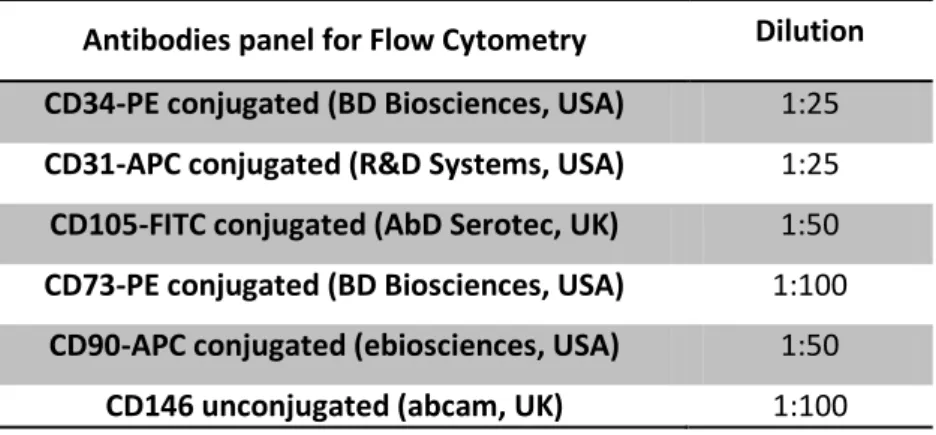
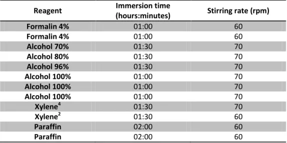
Table 1 - List of antibodies and dilutions used to perform flow cytometry analysis ... 27Table 2 – Automatic program used for samples deparaffinisation ... 30
Table 3 – Automatic program used on spin tissue processor ... 30
X 3D – Three dimensional
α-MEM – Minimum Essential Medium Alpha
α-SMA – Alpha smooth muscle actin °C – Celsius degrees
AF – Alexa Fluor AR-S – Alizarin Red-S
BMP – Bone morphogenetic protein BSA – Bovine serum albumin CAM – Cell adhesion molecule CD – Cluster of differentiation CECs – Circulating endothelial cells CO2 - Carbon dioxide
CS – Cell sheet
MSCs – Mesenchymal Stem Cells DAB – 3, 3'- diaminobenzidine DAPI – 4',6-diamidino-2-phenylindole Dil-AcLDL – 1,1'-dioctadecyl-3,3,3',3'-tetramethylindocarbocyanine perchlorate Acetylated-low density lipoprotein
DNA – Deoxyribonucleic acid ECs – Endothelial cells ECM – Extracellular matrix
EGM-2 MV – Microvascular Endothelial Cell Growth Medium-2
EOCs – Endothelial outgrowth cells EPC – Endothelial progenitor cells FBS – Fetal bovine serum
FGF-β – Basic fibroblast growth factor H&E – Hematoxylin and Eosin
hBMSCs – Bone Marrow-derived Mesenchymal Stromal/Stem Cells
HRP – Horseradish peroxidase
HUVECs – Human Umbilical Vein Endothelial Cells
IGF – Insulin-like growth factor
ISCT – International Society for Cellular Therapy
LCST – Lower critical solution temperature MAPC – Multipotent adult progenitor cells NG2 – Surfatide or nerve/glial antigen-2 (NG2) proteoglycan
NHS – Normal horse serum PBS – Phosphate Buffered Saline PCR – Polymerase Chain Reaction
PDGF-β – Platelet-derived growth factor beta
PDGFR-β – Platelet-derived growth factor receptor beta PIPAAm – Poly(N-isopropylacrylamide) PVDF – Poly(vinylidene difluoride) qRT-PCR – Quantitative reverse transcription PCR RM – Regenarative Medicine RT – Room temperature Shh – Sonic hedgehog TE – Tissue Engineering
TCPS – Tissue culture polystyrene
TGF-β1 – Transforming growth factor beta-1
TR – Thermoresponsive
vSMC – Vascular smooth muscle cells VEGF – Vascular endothelial growth factor
1
I
–
I
NTRODUCTION1 - Regenerative Medicine
The Regenerative Medicine (RM) field, in particular the area of tissue engineering (TE), holds a great promise for creating substitutes to repair congenital/trauma defects or diseased tissue. The utopian idea is to create whole organs or parts of organs in laboratory to replace and/or repair damaged parts of our body (1). Cell therapy, a RM approach, takes advantage of the systemic infusion of cells, namely mesenchymal stem cells (MSCs), to treat diseases or disorders using minimal invasive procedures. This concept has been proposed for a variety of applications namely to regenerate damaged tissue and to treat inflammation resulting from cardiovascular disease, myocardial infarction, brain, spinal cord, cartilage and bone injuries and Crohn’s disease (2). A major issue regarding cell therapy is that only less than 1% of the total injected cells reach and home at the desired tissue due to the lack of MSCs homing receptors (3). On the contrary, the most common TE approach comprehends the seeding of cells, as an autologous approach, on a biodegradable scaffold with desired mechanical properties, and the in vitro culturing of the construct prior to in vivo implantation (4). Although it looks simple and valuable, this strategy also has some associated issues, namely cases of pathological fibrosis after scaffolds biodegradation, strong inflammatory reaction due to non-specific responses to the polymers and consequent significant pH drop in host tissues, and the existence of a necrotic core at the centre of large constructs (5, 6). A common issue both for cell therapy and TE in general concerns the in vitro cell expansion conditions before use. In the case of therapies using MSCs, the overall impact of in vitro culture conditions over those cells is largely unknown however, some studies have shown that confluence and high passage number have undesirable impact on MSCs function (7, 8). It is also possible that stem cells cultured in vitro acquire new DNA mutations that promote cell growth under in vitro conditions, which might increase the risk of tumour developing (benign or malignant) after transplantation (1).
Despite all the considered limitations and cautions, several clinical trials of stem cell therapies, namely for the treatment of muscular dystrophy (9) and to regenerate myocardium de novo after myocardial infarction (10) have shown good results. Furthermore, successful TE approaches were also reported after the transplantation of bioartifial bladders and trachea (1). A different TE perspective has arisen with the advent of the cell sheets (CS) engineering technology. CS engineering intends to be an alternative to the traditional TE approaches by avoiding their associated shortcomings, not only by eliminating the scaffolding structures but also by creating artificial tissue-like substitutes with organized cellular entities and cohesive
2
cell-to-cell and cell-extracellular matrix (ECM) interactions (5). This relatively new concept is taking advantage of temperature-responsive cultured dishes to create those intact CSs which have already demonstrated to possess great potential as cardiac (11), and skin (12) and corneal epithelium (13) grafts, as well as to regenerate urothelium (14) and periodontal ligament (15) without the use of biodegradable scaffolds.
2 - Bone Tissue Engineering
Bone tissue deficiencies, caused either by malformations, trauma, or medical treatments, have a great impact over the patient’s life quality. Since the skeleton offers structure, posture and protection, damaged bones will hamper their physical function and appearance. Bone tissue engineering aims to be an alternative for skeletal reconstitution using metal plates or allogeneic and autologous bone grafts, decreasing the time and the associated costs, nonetheless it still remains a clinical challenge (16). Meijer et al. have considered that bone tissue engineering research comprises two promising approaches: the first one is based on three dimensional scaffolds that act as growth factors carriers, while the second one comprehends the combination of living osteogenic cells with three dimensional (3D) scaffolds (17). These structures are expected to provide a 3D environment where the cells are able to migrate, organize, differentiate and eventually, after implantation, successfully regenerate the desired tissue (18). However, despite the significant number of works (19-21) proving the feasibility of use the combination of scaffolds and MSCs in mouse models the fact is that few (22, 23) report the orthotropic application of bone-tissue engineering constructs in large animal models. The reality is that the four major causes for the stagnation of the bone tissue engineering field are: insufficient number of cells with osteogenic capacity; inappropriate scaffolds to seed the cells; uncontrolled delivery and unknown mechanisms of action of factors to stimulate osteogenic differentiation in vivo; and insufficient vascular supply (24). In fact, this last one is considered the principal cause of failure of bone tissue engineering constructs (25) and more considerations about this subject are discussed later (see I-3). MSCs-based bone tissue engineering therapies rely on the capacity to pre-differentiate those cells in vitro, usually by the addition of dexamethasone, ascorbic acid and β- glycerophosphate. In addition to the time of expansion, cell’s full differentiation has been achieved between 21 to 28 days, depending on the MSCs source, therefore, other strategies are being developed to decrease the time and increase the efficiency of osteogenic differentiation. Among them, the supplementation of culture media with growth factors (Platelet-derived growth factor (PDGF); Bone morphogenetic proteins (BMPs); transforming growth factor (TGF); Insulin-like growth factor (IGFs)) (26), gene delivery using gene therapy techniques to induce production of
3
growth factors (27), or the development of new bioreactor designs with capacity to enhance osteogenic performance of osteogenic cells (26), have been significantly explored.
2.1– Bone Tissue Biology and Regeneration Process
Bone is a remarkable organ with an interesting hierarchical structure. It combines an intimate interplay between an organic matrix and a mineralized phase with an extraordinary capacity to dealwith mechanical stress (28). In bone it is also possible to find several distinct tissues and organs, including mineralized osseous tissue, marrow, endosteum and periosteum, nervous tissue, and cartilage (26). Moreover, it is constituted by three distinct cell types which contribute for a constant self-remodelling through a controlled balance between bone resorption and bone formation. In this process, osteoblasts are responsible for bone extracellular matrix production and mineralization, which become calcified and entrap the osteocytes (osteoblasts in a fully differentiated state), the most abundant cell type in bone, which play an important role in bone homeostasis. In opposition, osteoclasts, the third cell type, are highly specialized cells with special importance for bone resorbing (29). Adequate and balanced bone remodelling assures maintenance of skeletal integrity, healing, blood calcium regulation and accommodation of chances in bone stress profiles (30).
After wounding, the way by which multicellular organisms can restore the architecture and functions of injured tissues is called regeneration. This leads to the reactivation of complex development pathways restoring homeostasis of the damaged area (31). Bone fracture healing is a dynamic process where a diversity of molecular and cellular processes occur along the subsequent healing phases: haematoma formation, inflammation, angiogenesis, cartilage formation and bone remodelling (32). Being key players in the regulation of the inflammatory cascade it is clear the importance of several growth factors, such as interleukin (IL) 1 and 6, PDGF; TGFs; IGF and BMPs during bone healing. By releasing earlier BMPs, MSCs are also important players on that process (32). In addition to the inflammatory phase, the angiogenic response during healing is also a critical process. In fact, several studies have established a connection between inadequate or inappropriate bone vascularization and a decreased bone formation, resulting in the formation of fibrous tissue (33-35).
3 - Mesenchymal Stromal/Stem Cells: From one to multiple sources
Stem cells, in general sense, bare potential not only to repair and regenerate damaged or loss tissues, but also to treat several diseases including metabolic, degenerative and inflammatory ones (36). In mammals, several stem cells have been identified, such as muscle-derived stem
4
cells (37, 38), germline stem cells (39), epithelial stem cells (40, 41), neural stem cells (42) and hematopoietic stem cells (43). All of them are believed to be precisely located in a diversity of organs where they are maintained and regulated within a well-controlled microenvironment, the stem cell niches (44). At present, stem cells under investigation are: embryonic stem cells, embryonic stem cells created by somatic cell nuclear transfer and adult stem cells (45). However, lack of efficient strategies to control lineage-specific differentiation of embryonic stem cells has been directing the research focus to adult, proliferative but lineage restricted stem cells (45).
Multipotent mesenchymal stromal cells, generally called Mesenchymal Stem Cells (MSCs), have the ability to differentiate into more than one cell type of the body but committed to the mesenchymal lineage (36). Besides their potential to differentiate in vitro along adipocytic, osteoblastic and chondrocytic lineages (24, 46), this type of cells was also previously recognized as critical to the support of hematopoiesis (47), providing, within the bone marrow stroma, an environment for homing, maintenance, proliferation, and maturation of hematopoietic progenitors (48). An immunoprivileged status was also attributed to MSCs (46), meaning that these cells are able to evade immune recognition and modulate immune responses after in vivo transplantation, which might have significant impact over the translation into the clinic of allogeneic cell based therapies (49-51).
Unfortunately, there was a lack of common definition for these cells, which lead the Mesenchymal and Tissue Stem Cell Committee of the International Society for Cellular Therapy (ISCT) to define the criteria to standardise the characterisation and nomenclature of MSCs populations. According to those, MSCs must be plastic-adherent in standard culture conditions using tissue culture flasks and must express CD105, CD73 and CD90 (≥95%) without traces (≤2%) of CD34, CD45, CD14 or CD11b, CD79α or CD19 and HLA class II antigens (52).
However, it is clear that over a MSC population selected by adherence to plastic, heterogeneity and variable expression of surface markers are inevitable (46). Moreover, accordingly to Horwitz et al. not all MSCs are true stem cells (considering the current definition of stem cells, i.e. a long term self-renewing cell that is capable of differentiation into specific, multiplecell types in vivo) (53), and the results of several studies suggest that cellular senescence is induced in MSCs due to long-term culture in vitro (54, 55), resulting in the loss of their proliferation and differentiation potential (56). Apart from the limitations described above, Jiang et al. reported the existence of a very small subset of mesenchymal cells from the bone marrow, termed multipotent adult progenitor cells (MAPC), which are capable of extensive self-renewal and possess pluripotency (57). Thus, it seems wise to consider MSCs as a mix of progenitor cells
5
with varying degrees of replicative/differentiation potential, rather than a homogeneous population of stem cells (45).
The existence of MSCs was first proposed in 1976 by Friedenstein who suggested the bone marrow as a source of adult stem cells (58). Twelve years after, Owen and Friedenstein proposed a model (59), later termed mesengenesis (60), of MSC differentiation into fibroblastic, reticular, adipogenic, osteogenic, and eventually other cell lineages. Since then, and for a long time, bone marrow stroma was the most commonly used MSCs’ source (61). However, new sources of MSCs, with similar characteristics to bone marrow MSCs, have been discovered and proposed to fill the regenerative medicine requirements. Thus, MSCs have been isolated from multiple mouse and human organs and tissues, such as skeletal muscle, skin, pancreas, fat, dental pulp, placenta and umbilical cord, all of them with identical characteristics (62). Among these, MSCs from adipose tissue, the so called adipose-derived stem cells (ASCs), seem to be a good alternative to bone marrow-derived MSCs due to their easier accessibility, abundance and similar capacity to differentiate into adipogenic, osteogenic and chondrogenic cells (63, 64).
Despite all the generated knowledge along the years, one of the most exciting discoveries in the field of MSCs homing and origin was made by Crisan and colleagues which proposed a perivascular origin for MSCs in multiple human organs (65). It was demonstrated that human perivascular cells sorted from diverse human tissues and cultured over plastic adherent conditions are multilineage progenitor cells that exhibit MSCs features (for more considerations about this subject see I-5). In addition, da Silva Meirelles et al. directly correlated the amount of MSCs with lipoaspirates vascular density which is in agreement with previous considerations (66).
4 - The Vascularization Problem in Tissue Engineering
Most of the tissues in human body require a functional vascular network for the efficient delivery of oxygen and nutrients and removal of waste materials (4). In adults, the formation of quiescent and stable new blood vessels from pre-existent ones, i.e. angiogenesis, is a rare event, observed only in cases of pregnancy, female reproductive cycle and after prolonged and heavy physical exercise (67). For that reason and because passive diffusion of nutrients and the removal of metabolic waste are limited, the use of TE constructs thicker than approximately 150 μm (68) lead to loss of cell viability, due to nutrient deficiencies and/or hypoxia, and consequently, unsatisfactory tissue regeneration (5). During in vitro culture, it is possible to supply and diffuse nutrients through larger tissue engineered constructs using for example
6
perfusion bioreactors (69), however constructs vascularization is a demand to maintain viability and attain tissue regeneration after implantation. Interestingly, the signals released and respective response by the implanted cells, as a reaction to hypoxia, is sufficient for the blood vessels from the host start to invade new tissue and form a capillary-like network. A major issue is the time that such networks take to develop and invade dipper sections of the implanted construct, which significantly contributes to the limited efficiency of the process (70). In an attempt to overcome this limitation alternative paths, using different scaffolds designs or patterned techniques to re-create the microvasculature of normal tissues in vitro or angiogenic factors to potentiate angiogenesis in vivo, have been explored. In the field of bone tissue engineering one of the most common approaches is to co-culture endothelial or endothelial progenitor cells, and osteoblastic or osteoprogenitor cells on three-dimensional biomaterials in order to achieve a pre-vascularized construct prior to implantation. All of those strategies have used Human umbilical Vein Endothelial Cells (HUVECs), Human Dermal Microvascular Endothelial Cells (HDMECs) or Endothelial Outgrowth Cells (EOCs) in combination with osteoprogenitor cells from bone marrow, primary osteoblast cells or osteoblast-like cell lines, seeded on 3D scaffolds or arranged in co-culture spheroids (71-73). Independently of the combined source and type of cells, those works demonstrated that the proposed strategy has potential to improve vascularization in vivo however there are no solid evidences to consider it as the solution for what the TE field is claiming.
Moreover, the establishment of co-culture systems implies to consider several issues, such as the source(s) of cells and the culturing conditions, that from a clinical perspective are determinant to validate the proposed strategy (74). During in vitro culture stage, questions related with the choice of culture medium, the phenotypic characterization of all the cell types involved in the system (75), the ratio of different cells between the co-culture systems and the use of static or dynamic culture conditions are important considerations for co-cultures setting up (74). In the case of co-cultures aimed at attain a pre-vascularized bone TE constructs the obvious cells sources are osteoprogenitor cells derived from bone marrow and endothelial cells. Despite the source of osteoblasts seems evident, the choice for the correct source of endothelial cells has intrigued scientific community. Endothelial cells are ubiquitous in the entire vascular system and constitute the innermost layer of blood vessels (76), although they display remarkable heterogeneity in different organs and also within the same organ (77). HUVECs and human aortic endothelial cells are two commonly studied cell populations concerning the analysis of endothelial cell functions in vitro (78), however they might not be the ideal model since they are considered mature and fully differentiated endothelial cells close to senescence and derived from hypoxic and probably activated vessels (77). In this
7
context, a promise source of cells to enhance the neovascularization of tissue engineered constructs, called endothelial progenitor cells (EPC), has attracted general interest. EPCs have been described as capable to form vascular structures in pro-angiogenic matrices in vitro, as well as to contribute for vascularization in vivo (79). Although the concept of have an EPC that can differentiate into a true endothelial cells with high proliferation potential is attractive, its application revealed more difficult than originally anticipated (80). The main problem has been to define strategies to efficiently select and characterize those cells and subsequently understand their particular roles in vascularization. As reviewed by Ingram et al. in 2005, EPCs displays distinct phenotypes and can be classified by their specific antigens as hematopoietic derived EPCs, circulating endothelial cells (CECs) and EOCs (78). Also, after the first studies have described the cord and peripheral blood as possible sources of EPCs (81-83), Ingram and colleagues suggested that endotelial cells (ECs) surrounding umbilical veins and human aorta combine resident EPCs at different stages of maturation and levels of proliferative potential (84). The specific function of each progenitor cell type described above during new vessel formation is still to understand but has a remarkable importance for TE as it might be the key to create co-cultured TE constructs. Despite those questions, at present, there are arguments supporting the potential of EOCs for bone tissue engineering. These cells are able to grown in different types of scaffolds and to organize in pre-vascular structures when co-cultured with primary human osteoblasts at in vitro (73, 85) or in vivo levels (86).
Also, co-culture systems of osteoblasts and ECs appear to naturally fulfil all the requirements that a successful co-culture model for pre-vascularization should have, since several studies has proved that, in a co-culture environment, ECs stimulate the osteoblasts to upregulate vascular endotelial growth factor (VEGF) secretion (87), while at the same time ECs release several BMPs which contribute for bone formation and repair (74). These findings suggest that co-cultures provide a pro-angiogenic matrix based on components such as collagen and a network of signals based on intercellular cross-talks that lead to the activation of ECs triggering angiogenesis (79).
In sum, clinical application of co-cultured TE constructs outcomes is still dependent on several technical issues such as improvements of ECs or progenitor’s isolation and culture protocols and on understanding the specific needs of each application.
5 – Pericytes Functions, Hallmarks and Origin
The cardiovascular system is the first functional organ system required for the development of mammalian embryo (88). Despite the huge contribution of ECs on that process, mural cells are
8
present since the first vascular network is formed (89). According to some reports, mural cells are also named vascular smooth muscle cells (vSMCs) while pericytes are described as a phenotypic variant of vSMC (89, 90). However, recent studies have questioned this simplified definition and, beyond their undeniable contribution for angiogenesis, vascular stabilization and blood flow regulation, pericytes have been looked as key players in response to injury and as precursors of MSCs (91, 92).
Regarding their localization, some studies have been correlating pericytes with the microvasculature (89, 90, 93) while others admit the presence of these cells around both capillaries (diameter < 10 µm) and arterioles (diameter from 10 to 100 µm) in all organs of human body, based on their ubiquitous expression of NG2 and CD146 (65). About this subject, Caplan and Correa have suggested the existence of a continuum of phenotypic similarities across various vessels types; pure pericytic cells are present in the microvessels while vSMCs, retaining the expression of some pericytic markers, can be found around larger vessels (91). Nevertheless, and independently of the pericytes origin, their recruitment appears to be regulated both by inductive and selective modes (89). Pericytes are induced to differentiate from immature mesenchymal cells surrounding blood vessels and/or are recruited, according to the selective model and as pre-existing mural cell or mural cells progenitors, from other locations (89). Taking into account new insights regarding pericytes multipotency and MSCs origin (65), it seems that the inductive model fails to explain the mechanism by which blood vessels become wrapped by pericytes. In what concerns the pericytes recruitment model, several in vivo experiments with knockout animals for PDGF-β or PDGFR-β have confirmed the involvement of PDGF-β (94-96). However, the absence of PDGF receptors on pericytes derived from sinusoidal vessels of liver represents evidence of the existence of other mechanisms in pericytes recruitment (97).
Once recruited into the correct place and at the appropriate time, pericytes play an important role in blood vessels maturation and stability through a fine balance between sphingosine-1-phosphate, TGF-β and angiopoietins (Ang1 and Ang2) secretion, and Ang1 and Ang2 Tie receptors expression (67). Nonetheless, vessels stability is not conferred by the mere presence of mural cells, but it was suggested to be highly dependent on the pericyte/endothelial interaction using the intermediate filament desmin, the so called Desmin Ensheathment Ratio (98).
The co-expression of several surface markers by pericytes and MSCs lead Caplan to suggest in 2008 (26), in part supported by other findings (65), that all the MSCs are pericytes, changing the MSCs application perspective. In fact, since then, pericytes are seen as the precursors of MSCs, and the perivascular space as a stem cell niche with cells carrying mesenchymal
9
differentiation capabilities and with implications in tissue response to injury (91). A controversy about the correct marker or the combination of markers for pure pericytes selection is installed. Pericytes were isolated from skeletal muscle based on alkaline phosphatase expression (99), but markers such as WAT7, CD146, NG2, α-SMA or PDGFR-β in the absence of hematopoietic, endothelial and myogenic cell markers, have been also considered to select pericytes from biological samples (91). Nevertheless, it seems that CD146, which is expressed by a subpopulation of bone marrow MSCs (100-102), is a useful marker to select MSCs with increased pericytic characteristics. The CD146 is a transmembrane glycoprotein, which belongs to a class of adhesion molecules (CAMs), and has important functions in early and late development. Moreover, CD146 has been suggested to play an important role in cancer, angiogenesis, cardiovascular diseases and placentation (103). The existence of potential recognition sites for protein kinases on its cytoplasmic domain suggests an involvement in signal transduction (104) however, the CD146 unknown ligand and the impact of CD146 signalling on cellular transcription is poorly understood (103). Studies performed by Yoshioka and colleagues suggest that CD146 mediate cell-endothelium adhesion and might play a role in neovascularization (105). Moreover, a significant number of studies in the field of cancer research have assigned to CD146 a critical role in tumour growth and metastasis, as well as in tumour angiogenesis, suggesting an anti-angiogenic effect in several types of tumour vessels by CD146 inhibition (106, 107). In summary, it seems that CD146 displays different expression patterns, structures and even biological functions depending on the circumstances, but these are certainly in the centre of the mechanisms of endothelial phenotype modulation and angiogenesis (108) and therefore constitute valuable elements to consider and empower bone TE vascularisation strategies.
6 – Cell sheet engineering technology
The use of scaffolds for the reconstitution of 3D tissues has been considered a potential solution and, at the same time, a limiting factor for further clinical applications. Because scaffolds are usually made of biomaterials with no or limited biological activity, those supports might function as a barrier for tissue regeneration (109). CS engineering technology has been proposed as a way to avoid this problem. The concept of this new technology can be placed in between the traditional regenerative medicine approaches, the systemic infusion of stem cells and the use of biodegradable scaffolds to create TE constructs.
10
6.1 - The intelligent cell detachment
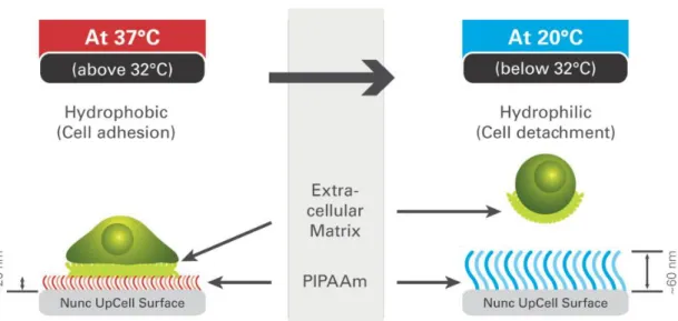
CS engineering is taking advantage of thermoresponsive (TR) surfaces prepared by covalent grafting of a TR polymer, poly(N-isopropylacrylamide) (PIPAAm), into ordinary polystyrene surfaces such as Petri dishes. PIPAAm has a reversible temperature-dependent phase transition, in aqueous solutions, also called lower critical solution temperature (LCST), at approximately 32°C (110, 111) and can be used to produce PIPPAm-modified intelligent surfaces, for in vitro cell culture (112, 113). Under normal 37°C culture conditions, the relatively hydrophobic surface allows cells to attach, proliferate and differentiate if desired. However, changing temperature for values below LCST turns the surface hydrophilic and by hydration of the PIPPAm, the polymeric chains extend allowing the spontaneous detachment of cells (Figure I-1) (5, 114, 115). Besides the “deadhesion” being inherent to the TR surface, cellular detachment is an active process, dependent of ATP and led by intracellular signalling and cytoskeleton reconstitution (116).
TR surfaces as well as the CS concept are expanding. The original idea was proposed during the 90’s in Japan by Okano and co-workers (117), but other groups were attracted by the potential of this approach and have been trying to create other responsive surfaces for non-invasive two or three-dimensional CS harvesting. Alternative TR culture surfaces, grafted with poly(N-isopropylacrylamide-co-acrylic acid)-b-poly( L-lactic acid) (118), were proposed as promoters of faster cell detachment, however osteoblastic cells growth and respective alkaline phosphatase activity are low (109). Other stimuli responsive polymers have also been used to fabricate “smart” surfaces with the capacity to regulate cell adhesion and detachment. Gold surfaces controlled on a voltage dependent manner (119), pH-dependent (120) and ionic strength (121) controlled surfaces are some examples. Nevertheless, these surfaces seem to be more useful for the understanding of cellular dynamics in anchorage-dependent cells than to produce CS with potential in TE.
A “next generation” of TR surfaces led by micropatterned (122-125) and biomolecule– immobilized (126, 127) cell culture dishes have also been developed. Micropatterning methods permitted to combine, within the same surface, two TR polymers with different LCSTs, allowing selective cell adhesion under temperature controlled environments. Furthermore the introduction of bioactive molecules onto the surface of a TR culture dishes constitute a first step to avoid the presence in culture of components from mammalian sources and allow the fabrication of CSs under serum-free conditions.
CS engineering avoids the traditional proteolytic treatment for cellular detachment, thus allowing preserving cell-cell interactions and maintaining their deposited ECM (114), which has
11
been considered and demonstrated as an achievement of great value for regenerative medicine applications (128).
Figure 1 – Features and functioning of TR surfaces. Adapted from Thermo Scientific Nunc UpCell Surface manual.
6.2 – Applications in Regenerative Medicine
A major feature of CS engineering is the maintenance of the deposited ECM after cell detachment as a sheet. After CS harvesting by temperature decrease, the ECM is totally recovered with the intact sheet of cells, which is being presented as the main reason for its adherence onto other surfaces, including other CSs or host tissues (5, 6, 114, 116, 129). Because the parenchyma end epithelia of many tissues consists of several cell layers associated with ECM, the use of CSs as the starting unit to follow a bottom-up approach for tissue reconstitution (6), mainly for thick, cell-dense tissues such as heart, liver, muscle and kidney (116), where biodegradable scaffolds has been failing, seems to be promising. In addition to the preservation of the ECM, the non-invasive cell-sheet harvesting by temperature decrease preserves ion channels, growth factor receptors and cell-to-cell junctions, as well as the integrity of cell surface markers, therefore cells retain higher differentiation functions in comparison to similar cells recovered by trypsinization (5).
The first clinical application of CS engineering was in cornea replacement. Corneal epithelial stem cells were isolated, expanded in TR dishes and recovered by temperature decrease to treat patients with ocular trauma. As a result, significant improvement in visual acuity was observed in all cases, which represents an alternative strategy to the use of scaffolds or substrate carriers that, due to their opacity, hampers proper reconstitution of that specific
12
tissue (5, 13). Tissue regeneration by direct transplantation of single CSs has been also proposed for skin, periodontal ligaments and bladder, using the base strategy of autologous cells isolation and proliferation on TR dishes, harvesting and subsequent application in the host (Figure 2 A).
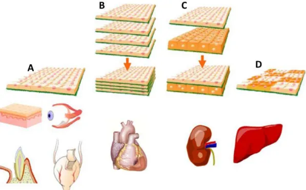
Figure 2 - Examples of CS engineering to create tissue-like substitutes. (A) Application of Single CS transplantation for cornea replacement and regeneration of skin, periodontal ligament and bladder. (B) Stacking of several CS for the improved performance of cardiac tissue. (C and D) Liver and kidney, tissues with laminar and higher-order structures, can also be recreated with CS engineering. Adapted from (5).
When replacement/regeneration of cell dense tissues is required, CSs can be manipulated to create 3D constructs with the desirable characteristics. A good example of CS engineering for complex tissue regeneration is the recreation of cardiac tissue by layering several cardiomyocyte CSs (11) (Figure 2 B). Once again, avoiding the inflexible and bulky properties of scaffolds, which significantly hamper the dynamic pulsation of cardiac myocytes, it was possible to recreate cell dense constructs in vitro with functional and synchronized pulsations. This phenomena is explained by the presence of gap junctions, specially connexin 43, formed during CSs adherence and by mediation through deposited ECM, which allows electrical communication between them similarly to what occurs in the heart (115). The four-layered construct fabricated with neonatal rat cardiac myocyte sheets was subcutaneously transplanted into rats and shown to be successfully integrated with the host tissue, as the formation of microvascular networks and maintenance of spontaneous beating proved (5).
13
Since this first attempt, improved, more complex, vascularized and thicker constructs for improving heart performance have been created. The successful implantation of 30 cardiac myocyte sheets stacked (approx. 1 mm) avoiding hypoxia, nutrient insufficiency or waste accumulation was achieved by the use of polysurgery (130). More recently, another approach was designed to treat congenital heart defects (129). Patterned TR surfaces permitted a controlled tissue organization and the creation of complex structures for vascular reconstruction.
It is expected that with the “next generation” of TR surfaces with dual phase transition temperatures it will be possible to create heterotypic CSs useful to reproduce higher order structures such as the liver (116). Using these surfaces the aim is to control the periphery contact between different cells on a CS co-culture system, mimicking the heterotypic cell–cell interactions occurring in in vivo structures (131).
Nevertheless, another important field where CS technology has been shown promising results is bone tissue regeneration. Both CS obtained by temperature decrease (132) and by mechanical means using a cell scraper (109, 133) have already demonstrated capacity to promote new bone tissue formation. However, a significant considering limitation of this approach for hard tissue regeneration concerns the mechanical properties of the construct. It is clear that a TE construct must possess mechanical properties capable of withstand the mechanical environment of the defect to be regenerated. Osteogenic cell sheets certainly comply with those demands when aimed at regenerating flat bone defects, however a “next generation” of osteogenic cell sheets-based constructs are expected to allow the creation of thicker and stiffer bone tissue substitutes. These can potentially generalize the application of the cell sheet technology to other bone defects with other mechanical demanding.
7 - References
1. Mummery C, Wilmut SI, van de Stolpe A, & Roelen BAJ (2010) Regenerative Medicine: Clinical Applications of Stem Cells. STEM CELLS, (Academic Press, San Diego), pp 133-195.
2. Phinney DG & Prockop DJ (2007) Concise review: mesenchymal stem/multipotent stromal cells: the state of transdifferentiation and modes of tissue repair--current views. STEM CELLS 25(11):2896-2902.
3. Sarkar D, et al. (2010) Engineered mesenchymal stem cells with self-assembled vesicles for systemic cell targeting. Biomaterials 31(19):5266-5274.
4. Melero-martin JM & Bischoff JE (2010) Corporation CsMC.
5. Yang J, et al. (2005) Cell sheet engineering: recreating tissues without biodegradable scaffolds. Biomaterials 26(33):6415-6422.
6. Yamato M, et al. (2007) Temperature-responsive cell culture surfaces for regenerative medicine with cell sheet engineering. Progress in Polymer Science 32(8-9):1123-1133.
7. Karp JM & Leng Teo GS (2009) Mesenchymal Stem Cell Homing: The Devil Is in the Details. Cell Stem Cell 4(3):206-216.
14
8. De Becker A, et al. (2007) Migration of culture-expanded human mesenchymal stem cells through bone marrow endothelium is regulated by matrix metalloproteinase-2 and tissue inhibitor of metalloproteinase-3. Haematologica 92(4):440-449.
9. Price FD, Kuroda K, & Rudnicki MA (2007) Stem cell based therapies to treat muscular dystrophy. Biochimica et biophysica acta 1772(2):272-283.
10. Tolar J, Le Blanc K, Keating A, & Blazar BR (2010) Concise review: hitting the right spot with mesenchymal stromal cells. STEM CELLS 28(8):1446-1455.
11. Shimizu T, et al. (2002) Fabrication of Pulsatile Cardiac Tissue Grafts Using a Novel 3-Dimensional Cell Sheet Manipulation Technique and Temperature-Responsive Cell Culture Surfaces. Circulation Research 90(3):e40-e48.
12. Yamato M, et al. (2001) Thermo-responsive culture dishes allow the intact harvest of multilayered keratinocyte sheets without dispase by reducing temperature. Tissue Eng 7(4):473-480.
13. Nishida K, et al. (2004) Functional bioengineered corneal epithelial sheet grafts from corneal stem cells expanded ex vivo on a temperature-responsive cell culture surface. Transplantation 77(3):379-385.
14. Shiroyanagi Y, Yamato M, Yamazaki Y, Toma H, & Okano T (2004) Urothelium regeneration using viable cultured urothelial cell sheets grafted on demucosalized gastric flaps. BJU international 93(7):1069-1075.
15. Flores MG, et al. (2008) Periodontal ligament cell sheet promotes periodontal regeneration in athymic rats. Journal of clinical periodontology 35(12):1066-1072.
16. Schönmeyr B (2010) Advances in bone tissue engineering. PhD (Skane University Hospital, Lund).
17. Meijer GJ, de Bruijn JD, Koole R, & van Blitterswijk CA (2007) Cell-based bone tissue engineering. PLoS Med 4(2):e9.
18. Khan Y, Yaszemski MJ, Mikos AG, & Laurencin CT (2008) Tissue engineering of bone: material and matrix considerations. The Journal of bone and joint surgery. American volume 90 Suppl 1:36-42.
19. Goshima J, Goldberg VM, & Caplan AI (1991) The osteogenic potential of culture-expanded rat marrow mesenchymal cells assayed in vivo in calcium phosphate ceramic blocks. Clinical orthopaedics and related research (262):298-311.
20. Ohgushi H, Goldberg VM, & Caplan AI (1989) Repair of bone defects with marrow cells and porous ceramic. Experiments in rats. Acta orthopaedica Scandinavica 60(3):334-339.
21. Yoshikawa T, et al. (2000) In vivo osteogenic durability of cultured bone in porous ceramics: a novel method for autogenous bone graft substitution. Transplantation 69(1):128-134.
22. Kon E, et al. (2000) Autologous bone marrow stromal cells loaded onto porous hydroxyapatite ceramic accelerate bone repair in critical-size defects of sheep long bones. J Biomed Mater Res 49(3):328-337.
23. Petite H, et al. (2000) Tissue-engineered bone regeneration. Nature biotechnology 18(9):959-963.
24. Caplan AI (1991) Mesenchymal stem cells. Journal of Orthopaedic Research 9(5):641-650. 25. James J & Steijn-Myagkaya GL (1986) Death of osteocytes. Electron microscopy after in vitro
ischaemia. The Journal of bone and joint surgery. British volume 68(4):620-624.
26. Porter JR, Ruckh TT, & Popat KC (2009) Bone tissue engineering: A review in bone biomimetics and drug delivery strategies. Biotechnology Progress 25(6):1539-1560.
27. Kimelman N, et al. (2007) Review: gene- and stem cell-based therapeutics for bone regeneration and repair. Tissue Eng 13(6):1135-1150.
28. Gupta HS, et al. (2006) Cooperative deformation of mineral and collagen in bone at the nanoscale. Proceedings of the National Academy of Sciences 103(47):17741-17746.
29. Pirraco RP, Marques AP, & Reis RL (2009) Cell interactions in bone tissue engineering. J Cell Mol Med 14(1-2):93-102.
30. Hadjidakis DJ & Androulakis, II (2006) Bone remodeling. Ann N Y Acad Sci 1092:385-396. 31. Gurtner GC, Werner S, Barrandon Y, & Longaker MT (2008) Wound repair and regeneration.
Nature 453(7193):314-321.
15
33. Burkhardt R, et al. (1987) Changes in trabecular bone, hematopoiesis and bone marrow vessels in aplastic anemia, primary osteoporosis, and old age: a comparative histomorphometric study. Bone 8(3):157-164.
34. Glowacki J (1998) Angiogenesis in fracture repair. Clinical orthopaedics and related research (355 Suppl):S82-89.
35. Hausman MR, Schaffler MB, & Majeska RJ (2001) Prevention of fracture healing in rats by an inhibitor of angiogenesis. Bone 29(6):560-564.
36. EMA (2011) Reflection paper on stem cell-based medical products. Commitee for Advanced Therapies:14.
37. Qu-Petersen Z, et al. (2002) Identification of a novel population of muscle stem cells in mice. The Journal of Cell Biology 157(5):851-864.
38. Peault B, et al. (2007) Stem and progenitor cells in skeletal muscle development, maintenance, and therapy. Molecular therapy : the journal of the American Society of Gene Therapy 15(5):867-877.
39. de Rooij D (2001) Proliferation and differentiation of spermatogonial stem cells. Reproduction 121(3):347-354.
40. Cotsarelis G, Sun TT, & Lavker RM (1990) Label-retaining cells reside in the bulge area of pilosebaceous unit: implications for follicular stem cells, hair cycle, and skin carcinogenesis. Cell 61(7):1329-1337.
41. Tumbar T, et al. (2004) Defining the epithelial stem cell niche in skin. Science 303(5656):359-363.
42. Doetsch F (2003) A niche for adult neural stem cells. Current opinion in genetics & development 13(5):543-550.
43. Adams GB & Scadden DT (2006) The hematopoietic stem cell in its place. Nature immunology 7(4):333-337.
44. Morrison SJ & Spradling AC (2008) Stem Cells and Niches: Mechanisms That Promote Stem Cell Maintenance throughout Life. Cell 132(4):598-611.
45. Mark E & Anthony A (2007) Future Perspectives. Principles of Tissue Engineering eds Lanza R, Langer R, & Vacanti J (Elsevier, Inc.), 3nd Ed.
46. Keating A (2006) Mesenchymal stromal cells. Current Opinion in Hematology 13(6):419-425 410.1097/1001.moh.0000245697.0000254887.0000245696f.
47. Dexter TM, Spooncer E, Schofield R, Lord BI, & Simmons P (1984) Haemopoietic stem cells and the problem of self-renewal. Blood cells 10(2-3):315-339.
48. Meregalli M, Farini A, & Torrente Y (2011) Mesenchymal Stem Cells as Mucle Reservoir. Stem Cell Research & Therapy 1(2):9.
49. Bartholomew A, et al. (2002) Mesenchymal stem cells suppress lymphocyte proliferation in vitro and prolong skin graft survival in vivo. Exp Hematol 30(1):42-48.
50. Le Blanc K, Tammik C, Rosendahl K, Zetterberg E, & Ringden O (2003) HLA expression and immunologic properties of differentiated and undifferentiated mesenchymal stem cells. Exp Hematol 31(10):890-896.
51. Maitra B, et al. (2004) Human mesenchymal stem cells support unrelated donor hematopoietic stem cells and suppress T-cell activation. Bone marrow transplantation 33(6):597-604.
52. Dominici M, et al. (2006) Minimal criteria for defining multipotent mesenchymal stromal cells. The International Society for Cellular Therapy position statement. Cytotherapy 8(4):315-317. 53. Horwitz EM, et al. (2005) Clarification of the nomenclature for MSC: The International Society
for Cellular Therapy position statement. Cytotherapy 7(5):393-395.
54. Ito T, Sawada R, Fujiwara Y, Seyama Y, & Tsuchiya T (2007) FGF-2 suppresses cellular senescence of human mesenchymal stem cells by down-regulation of TGF-[beta]2. Biochemical and Biophysical Research Communications 359(1):108-114.
55. Conget PA & Minguell JJ (1999) Phenotypical and functional properties of human bone marrow mesenchymal progenitor cells. J Cell Physiol 181(1):67-73.
56. Banfi A, et al. (2002) Replicative aging and gene expression in long-term cultures of human bone marrow stromal cells. Tissue Eng 8(6):901-910.
57. Jiang Y, et al. (2002) Pluripotency of mesenchymal stem cells derived from adult marrow. Nature 418(6893):41-49.
58. Friedenstein AJ, Gorskaja JF, & Kulagina NN (1976) Fibroblast precursors in normal and irradiated mouse hematopoietic organs. Exp Hematol 4(5):267-274.
16
59. Friedenstein AJ, Chailakhyan RK, & Gerasimov UV (1987) Bone marrow osteogenic stem cells: in vitro cultivation and transplantation in diffusion chambers. Cell and tissue kinetics 20(3):263-272.
60. Caplan AI (1994) The mesengenic process. Clinics in plastic surgery 21(3):429-435.
61. Minguell JJ, Erices A, & Conget P (2001) Mesenchymal stem cells. Exp Biol Med (Maywood) 226(6):507-520.
62. Corselli M, Chen C-W, Crisan M, Lazzari L, & Péault B (2010) Perivascular Ancestors of Adult Multipotent Stem Cells. Arteriosclerosis, Thrombosis, and Vascular Biology 30(6):1104-1109. 63. Zuk PA, et al. (2002) Human adipose tissue is a source of multipotent stem cells. Molecular
biology of the cell 13(12):4279-4295.
64. Zuk PA, et al. (2001) Multilineage cells from human adipose tissue: implications for cell-based therapies. Tissue Eng 7(2):211-228.
65. Crisan M, et al. (2008) A perivascular origin for mesenchymal stem cells in multiple human organs. Cell Stem Cell 3(3):301-313.
66. da Silva Meirelles L, Caplan AI, & Nardi NB (2008) In search of the in vivo identity of mesenchymal stem cells. STEM CELLS 26(9):2287-2299.
67. von Tell D, Armulik A, & Betsholtz C (2006) Pericytes and vascular stability. Exp Cell Res 312(5):623-629.
68. Folkman J & Hochberg M (1973) Self-regulation of growth in three dimensions. The Journal of experimental medicine 138(4):745-753.
69. Janssen FW, Oostra J, Oorschot A, & van Blitterswijk CA (2006) A perfusion bioreactor system capable of producing clinically relevant volumes of tissue-engineered bone: in vivo bone formation showing proof of concept. Biomaterials 27(3):315-323.
70. Rouwkema J, Rivron NC, & van Blitterswijk CA (2008) Vascularization in tissue engineering. Trends Biotechnol 26(8):434-441.
71. Unger RE, et al. (2007) Tissue-like self-assembly in cocultures of endothelial cells and osteoblasts and the formation of microcapillary-like structures on three-dimensional porous biomaterials. Biomaterials 28(27):3965-3976.
72. Rouwkema J, Boer JD, & Blitterswijk CAV (2006) Endothelial Cells Assemble into a 3-Dimensional Prevascular Network in a Bone Tissue Engineering Construct. Tissue Engineering 12(9):2685-2693.
73. Fuchs S, Hofmann A, & Kirkpatrick CJ (2007) Microvessel-Like Structures from Outgrowth Endothelial Cells from Human Peripheral Blood in 2-Dimensional and 3-Dimensional Co-Cultures with Osteoblastic Lineage Cells. Tissue Engineering 13(10):2577-2588.
74. Kirkpatrick CJ, Fuchs S, & Unger RE (2011) Co-culture systems for vascularization -- Learning from nature. Advanced Drug Delivery Reviews In Press, Corrected Proof.
75. Kirkpatrick CJ, et al. (1997) Physiology and cell biology of the endothelium: a dynamic interface for cell communication. International journal of microcirculation, clinical and experimental / sponsored by the European Society for Microcirculation 17(5):231-240.
76. Hirschi KK, Rohovsky SA, & D'Amore PA (1998) PDGF, TGF-beta, and heterotypic cell-cell interactions mediate endothelial cell-induced recruitment of 10T1/2 cells and their differentiation to a smooth muscle fate. J Cell Biol 141(3):805-814.
77. Garlanda C & Dejana E (1997) Heterogeneity of Endothelial Cells : Specific Markers. Arteriosclerosis, Thrombosis, and Vascular Biology 17(7):1193-1202.
78. Ingram DA, Caplice NM, & Yoder MC (2005) Unresolved questions, changing definitions, and novel paradigms for defining endothelial progenitor cells. Blood 106(5):1525-1531.
79. Fuchs S, Dohle E, Kolbe M, & Kirkpatrick CJ (2010) Outgrowth endothelial cells: sources, characteristics and potential applications in tissue engineering and regenerative medicine. Advances in biochemical engineering/biotechnology 123:201-217.
80. Verloop RE, Koolwijk P, van Zonneveld AJ, & van Hinsbergh VW (2009) Proteases and receptors in the recruitment of endothelial progenitor cells in neovascularization. European cytokine network 20(4):207-219.
81. Asahara T, et al. (1997) Isolation of putative progenitor endothelial cells for angiogenesis. Science 275(5302):964-967.
82. Gulati R, et al. (2003) Diverse Origin and Function of Cells With Endothelial Phenotype Obtained From Adult Human Blood. Circulation Research 93(11):1023-1025.