Combined effect
of α-lipoic acid
and
N-acetylcysteine:
influence on the
prooxidant state of
Fanconi anemia
cells
Ana Sofia Afonso Gomes
Mestrado em Bioquímica
Química e Bioquímica 2015
Orientador
Beatriz Porto, Professora Auxiliar, Instituto de Ciências
Biomédicas de Abel Salazar
Coorientador
Pedro Fontes Oliveira, Professor Afiliado, Instituto de
Ciências Biomédicas de Abel Salazar
We cannot solve our problems with the same thinking
we used when we created them – Albert Einstein
I.
ACKNOWLEDGEMENTS
É nesta altura que faço uma reflexão sobre este intenso ano de dissertação do mestrado em bioquímica. Como tal, gostaria de agradecer a todos os que caminharam comigo ao meu lado.
Tudo começou em fevereiro do ano passado quando me inscrevi na unidade curricular de Genética Médica. E se meu interesse pela genética já existia, assim que tive estas aulas despertou ainda mais este fascínio por esta área. Principalmente, quando tive o prazer de conhecer a professora Beatriz Porto, que com a sua paixão e o seu entusiasmo ao falar do seu trabalho, me cativou desde o inicio. Deste modo, gostaria de agradecer à minha orientadora Professora Beatriz Porto, por me ter acolhido no seu laboratório de citogenética e criado este projeto de investigação para mim. Obrigada por ter partilhado comigo os seus conhecimentos científicos e por estar sempre presente em todas as minhas dificuldades, ajudando a superá-las.
Queria agradecer ao meu coorientador professor Pedro Fontes Oliveira por se disponibilizar e fazer parte deste projeto. Obrigada pela partilha de conhecimentos e pelo fornecimento de muitos dos testes de stress oxidativo utilizados neste projeto. Agradeço também a todos os seus colaboradores: Ana Martins, Maria Meneses, Raquel Bernadino, Tânia Dias e Tito Jesus.
À técnica Rosa Sousa que esteve presente nesta minha caminhada e que me transferiu conhecimentos científicos e não só, agradeço imenso todo o seu tempo disponibilizado comigo. Obrigada por confiar as suas amostras, recolhidas ao longo de vários anos, nesta investigação. Agradeço também por me ter ajudado a ultrapassar alguns obstáculos que foram aparecendo, foi um prazer enorme estar ao seu lado e crescer consigo.
À Filipa Ponte que me apoiou e forneceu para este projeto um dos testes de stress oxidativo. Também quero agradecer técnica à Lara Andrade e à técnica Ana Paula Fernandes pelo suporte prestado no laboratório de citogenética.
A todos os pacientes de anemia de Fanconi e dadores de sangue do Hospital Geral de Santo António, obrigada pelo vosso fornecimento e consentimento para a realização deste projeto, sem vocês tal não seria possível.
A todos os meus colegas e amigos, especialmente à Cíntia Barreira, Diogo Neves, André Nadais e Margarida Moura, por estarem sempre presentes e juntos ultrapassarmos todos os obstáculos. O vosso incentivo e amizade foram essenciais.
Por último, mas não por isso menos importante, quero agradecer a toda a minha família pelo apoio, incentivo, carinho e compreensão prestados. Aos meus pais por estarem sempre presentes, por me ajudarem a percorrer este caminho académico e me tornarem na pessoa que sou hoje. Sem todos vocês nada disto seria possível.
II.
ABSTRACT
Fanconi Anemia (FA) is a complex genetic disorder, with mutations in one of the 16 genes so far characterized that function in a common pathway for the maintenance of genomic stability. FA is commonly characterized by bone marrow failure, increased risk to malignancies and excessive sensitivity to DNA crosslink agents, such as diepoxybutane (DEB). A number of independent studies support the relationship between hypersensitivity to DEB and oxidative stress (OS), and emphasized that glutathione (GSH)-based pathways have an important role in this system.
It has been extensively described that FA cells are in a continuous cellular prooxidant state that results in chromosome instability (CI). It is also known that mutations in the FA pathway result in down regulation of the endogenous antioxidant defense, including the red blood cells (RBC), which are considered one of the most important endogenous antioxidant defense systems. Therefore, FA cells may benefit with an antioxidant therapy independent of the FA pathway control.
In a previous work, based on the hypersensitivity of FA cells to DEB, the authors searched for antioxidant molecules with ability to counteract the DEB related OS; this study lead to the selection of two orthodox antioxidant small molecules: N-acetylcysteine (NAC) and α-lipoic acid (ALA). Afterwards, in vitro studies were performed confirming that the ALA and NAC improve genetic stability, decreasing CI in cultured lymphocytes from FA patients.
The aim of the present study was to evaluate the influence of ALA and NAC in the redox state of RBC in blood cultures from FA patients, comparatively to the influence of the same antioxidants in DEB-induced cultures from healthy donors. For that purpose, the following OS parameters we evaluated: levels of GSH and glutathione peroxidase (GPx) activity; expression of protein oxidative damage, by the determination of carbonyl group content; the antioxidant potential, by the ferric reduction antioxidant potential (FRAP) assay.
The results showed that exposure to the antioxidants increased GSH levels and decreased GPx activity in RBC from FA patients. In RBCs from healthy donors no differences were observed both in GSH levels and GPx activity. ALA and NAC had no effect on the expression of protein oxidative damage and on the antioxidant potential, either in RBC from FA patients and healthy donors. Knowing that FA cells are in a
continuous prooxidant state, our results, based on GSH levels, suggest that the addition of ALA and NAC may improve the endogenous antioxidant defense and, consequently, improve the redox state of FA RBCs. The simultaneous increase in GSH levels and decrease in GPx activity may point to a combined redox dependent mechanism of ALA and NAC to improve antioxidant defense in FA RBC, by supplying an extra source of GSH and decreasing the requirement of GPx activity.
Based on these and previous results, we can hypothesize that NAC and ALA may be good prophylactic candidates to decrease the adverse effects of OS-related damage, having potential to be used as an extra source of exogenous antioxidant defense to FA patients.
KEYWORDS
Fanconi anemia; Oxidative stress; Red blood cells; Antioxidants; α-lipoic acid; N-acetylcysteine; Chromosome instability.
III.
RESUMO
A anemia de Fanconi (FA) é uma doença genética complexa, com mutações em um dos 17 genes até agora caracterizados que funcionam numa via comum para a manutenção da estabilidade genómica. A FA é caracterizada por falência medular progressiva, um aumento do risco para neoplasias e uma sensibilidade excessiva a agentes indutores de DNA crosslink1, tais como o diepoxibutano (DEB). Vários estudos independentes apoiam a relação entre a hipersensibilidade ao DEB e o stress oxidativo (OS), e salientam que as vias que envolvem glutationa (GSH) têm um papel importante neste sistema.
Tem sido extensamente descrito que as células FA estão num contínuo estado prooxidante que resulta em instabilidade cromossómica (CI). É também sabido que mutações na via FA resultam numa regulação negativa na defesa antioxidante endógena, incluindo em eritrócitos, que são considerados um dos mais importantes sistemas de defesa antioxidante endógeno. Assim sendo, as células FA poderão beneficiar com uma terapia antioxidante independente do controlo da via FA.
Num trabalho prévio, com base na hipersensibilidade das células FA ao DEB, foram pesquisados antioxidantes com capacidade para proteger as células do OS relacionado com o DEB. Esse estudo levou à seleção de duas pequenas moléculas antioxidantes: o ácido α-lipoico (ALA) e a N-acetilcisteína (NAC). Mais tarde, estudos in vitro confirmaram que o ALA e o NAC melhoram a estabilidade genómica, diminuindo a CI em culturas de linfócitos de pacientes FA.
O objetivo do presente estudo foi avaliar a influência do ALA e NAC no estado redox dos eritrócitos de pacientes FA obtidos a partir de culturas de sangue com e sem tratamento antioxidante, comparativamente com a influência dos mesmos antioxidantes em eritrócitos de dadores saudáveis obtidos a partir de culturas de sangue induzidas por induzidas por DEB. Para essa finalidade foram avaliados os seguintes parâmetros de OS: níveis de GSH e atividade de glutationa peroxidase (GPx); expressão de dano oxidativo em proteínas, quantificada pelo conteúdo de grupos carbonilo; o potencial antioxidante, medido pelo método do potencial antioxidante de ferro redutor (FRAP).
Os resultados mostraram que a exposição aos antioxidantes aumentou os níveis de GSH e diminuiu a atividade de GPx nos eritrócitos de pacientes FA. Nos eritrócitos dos dadores saudáveis não foram observadas diferenças quer nos níveis de GSH quer na atividade da GPx. O ALA e o NAC não tiveram efeito na expressão de dano oxidativo em proteínas nem no potencial antioxidante, tanto em eritrócitos de pacientes FA como de dadores saudáveis.
Os resultados, baseados nos níveis de GSH, sugerem que a adição dos antioxidantes ALA e NAC podem melhorar a defesa antioxidante endógena e, consequentemente, melhorar o estado redox dos eritrócitos de pacientes FA. O efeito simultâneo de um aumento nos níveis de GSH e de uma diminuição da atividade GPx sugere a ocorrência de um mecanismo protetor do ALA e NAC que melhora a defesa antioxidante dos eritrócitos dos doentes FA, através de uma fonte extra de GSH e de uma diminuição no requerimento da atividade GPx. Com base nestes resultados e em estudos anteriores propõe-se que o ALA e o NAC podem ser bons candidatos profiláticos para diminuir os efeitos adversos relacionados com OS, tendo potencial para serem usados como uma fonte extra de defesa antioxidante para os pacientes FA.
PALAVRAS-CHAVE
Anemia de Fanconi; Stress oxidativo; Eritrócitos; Antioxidantes; Ácido α-lipóico; N-acetilcisteína; Instabilidade Cromossómica
INDEX
I. ACKNOWLEDGEMENTS ... 1
II. ABSTRACT ... 3
III. RESUMO ... 5
IV. LIST OF TABLES AND FIGURES ... 10
V. ABBREVIATIONS ... 10
1. INTRODUCTION ... 14
1.1 The Fanconi Anemia (FA) Disease ... 14
1.2 Relationship between Oxidative Stress (OS) and Fanconi Anemia ... 15
1.3 Relationship between Oxidative Stress and diepoxybutane (DEB) toxicity ... 15
1.4 Importance of red blood cells (RBCs) in measurement of oxidative stress (OS) 16 1.5 Link between FA pathway and antioxidant defense system ... 17
1.6 Antioxidant defense ... 17
1.6.1 Alpha Lipoic Acid (ALA) ... 18
1.6.2 N-acetylcysteine (NAC) ... 19
1.7 Aim of the study ... 19
2 MATERIAL AND REAGENTS ... 20
3. METHODS ... 20
3.2 Experimental procedures ... 20
3.2.1 Isolation of RBC ... 20
3.2.2 Whole blood cultures ... 20
3.2.3 Exposure to DEB ... 21
3.2.4. Antioxidant incorporation ... 21
3.2.5. Antioxidant treatments ... 21
3.3 Cytogenetic evaluation ... 21
3.4 Characterization of oxidative stress parameters ... 22
3.4.1 Red blood cell processing for the study of oxidative stress parameters ... 22
3.4.2 Sample processing for glutathione ... 22
3.4.3. Glutathione quantification ... 22
3.4.4 Protein quantification ... 23
3.4.5 Glutathione peroxidase activity determination ... 23
3.4.6 Ferric reducing antioxidant power assay ... 23
3.4.7 Carbonyl Group levels... 24
3.5 Statistical analysis ... 24
4. RESULTS ... 25
4.1 Effect of α-lipoic acid and N-acetylcysteine on the frequency of chromosome instability in lymphocytes from healthy donors ... 25
4.2 Effect of α-lipoic acid and N-acetylcysteine on glutathione levels in red blood cells from healthy donors and Fanconi anemia patients ... 26
4.3 Effect of α-lipoic acid and N-acetylcysteine on glutathione peroxidase activity in
red blood cells from healthy donors and Fanconi anemia patients ... 28
4.4 Effect of α-lipoic acid and N-acetylcysteine on the antioxidant potential in red blood cells from healthy donors and Fanconi anemia patients ... 30
4.5 Effect α-lipoic acid and N-acetylcysteine on protein oxidative damage in red blood cells from healthy donors and Fanconi anemia patients ... 32
5. DISCUSSION ... 34
6. BIBLIOGRAPHY ... 36
IV.
LIST OF TABLES AND FIGURES
Figure 1: Diepoxybutane (DEB)-induced chromosome instability (CI) in Fanconi anemia(FA) patient. ... 14 Graphic 1: Effect of α-lipoic acid (ALA) and N-acetylcysteine (NAC) on the frequency of 1,2:3,4-diepoxibutane (DEB)-induced (0,4 µg/mL) chromosome instability in cultured lymphocytes from healthy donors... 25 Graphic 2: Effect of α-lipoic acid (ALA) and N-acetylcysteine (NAC) on glutathione (GSH) levels in red blood cells from healthy donors blood cultures. ... 26 Graphic 3 Effect α-lipoic acid (ALA) and N-acetylcysteine (NAC) on glutathione (GSH) levels in red blood cells (RBCs) obtained from Fanconi anemia blood cultures. ... 27 Graphic 4: Effect of α-lipoic acid (ALA) and N-acetylcysteine (NAC) on glutathione peroxidase (GPx) activity of 1,2:3,4-diepoxybutane (DEB)-induced (0,4 µg/mL) red blood cells (RBCs) obtained from healthy ... 28 Graphic 5: Effect α-lipoic acid (ALA) and N-acetylcysteine (NAC) on glutathione peroxidase (GPx) activity in red blood cells (RBCs) obtained from Fanconi anemia patients blood cultures. ... 29 Graphic 6: Effect of α-lipoic acid (ALA) and N-acetylcysteine (NAC) on the antioxidant potential of 1,2:3,4-diepoxybutane (DEB)-induced (0,4 µg/mL) red blood cells (RBCs) obtained from healthy donors blood cultures. ... 30 Graphic 7: Effect α-lipoic acid (ALA) and N-acetylcysteine (NAC) on the antioxidant potential of red blood cells (RBCs) obtained from FA patients blood cultures... 31 Graphic 8: Effect α-lipoic acid (ALA) and N-acetylcysteine (NAC) on protein oxidative damage in 1,2:3,4-diepoxybutane (DEB)-induced (0,4 µg/mL) red blood cells (RBCs) obtained from healthy donors blood culture. ... 32 Graphic 9: Effect α-lipoic acid (ALA) and N-acetylcysteine (NAC) on protein oxidative damage of red blood cells (RBCs) obtained from FA patients blood cultures. ... 33
V.
ABBREVIATIONS
AML – acute myelogenous leucemia ALA – α-lipoic acid
CAT – Catalase
CHP – Hospital Geral de Santo António CI – Chromosome instability
DEB – 1,2:3,4-Diepoxybutane FA – Fanconi Anemia
FRAP – Ferric Reducing Antioxidant Power GSH – Glutathione
GPx – Glutathione peroxidase GR – Glutathione reductase HD – Healthy donors
Hb – Hemoglobin
ICBAS – Instituto de Ciências Biomédicas Abel Salazar ICL – Interstrand crosslink
metHb – Methemoglobin NAC – N-acetilcysteine OS – Oxidative Stress
ROS – Reactive oxygen species RBC – Red blood cell
RT – Room temperature
SCT – Stem cell transplantation SOD - Superoxide dismutase UV – Ultraviolet
1. INTRODUCTION
1.1 The Fanconi Anemia (FA) Disease
Fanconi anemia (FA) is a rare autosomic or X-linked recessive disorder reported for the first time by Guido Fanconi, a Swiss pediatrician, who recognized siblings with “peculiar phenotypic abnormalities and anaemia” (Fanconi, 1927). Nowadays it is known that FA is clinically characterized by diverse congenital malformations, bone marrow failure, and predisposition to cancer, in particular acute myelogenous leukemia (AML) (Kutler et al., 2003). At molecular level, seventeen genes have been characterized so far (FANCA, B, C, D1 (BRCA2), D2, E, F, G, I, J (BRIP1), L, -M, -N (PALB2), -O (RAD51C), -P (SLX4), -Q (ERCC4) and –S (BRCA1)) (Sawyer et al., 2015; Virts et al., 2015). It is known that these genes function together in an ordinary pathway, the FA-BRCA pathway, involved in the repair of interstrand DNA crosslinks (ICLs) Therefore, a mutation in one of those genes originates genomic instability, by lack of ICL repair (reviewed in Schneider, Chandler, Tischkowitz, & Meyer, 2014).
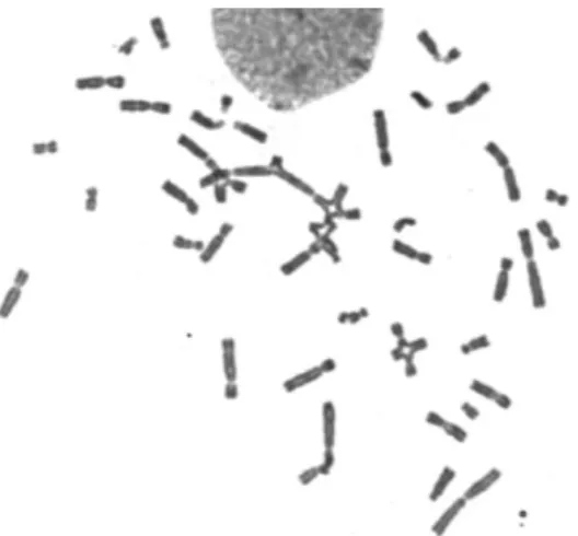
Although FA patients may be clinically and genetically variable, they are characterized by a common cellular feature: hypersensitivity to ICL agents. ICLs are very toxic lesions that block DNA replication and transcription. The lack of ICL repair results in increased genomic instability, represented, at cellular level, by increased chromosome instability (CI), including breaks, tri- and tetra-radial figures, that can be observed in metaphases obtained from cultured lymphocytes (Figure 1) (Auerbach, 1989).
Figure 1: Diepoxybutane (DEB)-induced chromosome instability (CI) in Fanconi anemia(FA) patient.
Selected metaphase from a FA lymphocyte culture exposed to 0.1 µg/ml of DEB for 48 h. Chromosomes were stained with 4% Giemsa solution. High levels of CI can be visualized; the presence of tri- and tetra-radial figures is particularly important for the diagnosis, since they can discriminate FA from the other CI syndromes. The image was captured with an optical microscope (500x amplification). Ilustration from chapter II of Giovanni, Pagano. Fanconi Anemia and Oxidative Stress: Mechanistic Background and Clinical Prospects. New York United States: Nova Science Publishers Inc, 2015
1.2 Relationship between Oxidative Stress (OS) and Fanconi
Anemia
Oxidative stress (OS) mediated by reactive oxygen species (ROS) is a major cause of DNA ICLs. Some of them produce or are free radicals. Free radicals are defined by any species containing unpaired electrons being alone in the orbital (Halliwell, 1991). Increased levels of OS are very prejudicial to the organism because they can damage membranes, proteins, and DNA. This effect is a factor involved in the development of many chronic and degenerative aging diseases (Packer, 1995).
Nordenson described for the first time the role of OS in FA cells damage (Nordenson, 1977), showing that superoxide dismutase (SOD) and catalase (CAT) decreased the frequency of spontaneous CI in cultured lymphocytes. Joenje and Oostra described for the first time a direct correlation between high tensions of oxygen and increased damage in FA lymphocytes (Joenje and Oostra, 1983). Another study demonstrated an overproduction of ROS by FA leukocytes, due to NADPH oxidase activation, decrease in cellular antioxidant defense systems, increase in cellular free iron levels and a deficiency in SOD measured in red blood cells (RBCs) (Korkina et al., 1992). Takeuchi and Morimoto suggested that a high sensibility of FA lymphoblastoid cells to oxidative DNA damage is possible, resulting from a decrease in CAT activity (Takeuchi and Morimoto, 1993). All these studies provide evidence that OS is an important element responsible for the FA cellular phenotype.
1.3 Relationship between Oxidative Stress and diepoxybutane
(DEB) toxicity
The hypersensitivity of FA cultured lymphocytes to ICL agents, that leads to a an increase of CI, supplies a diagnostic test for the disease (D’Andrea and Grompe, 2003). Since 1989, the alkylating agent 1,2:3,4-diepoxybutane (DEB) is used as a certified diagnostic tool for FA (D’Andrea and Grompe, 2003); Kutler et al. 2003). Its mode of action implies alkylation of nucleic acid bases producing monoadducts (Gherezghiher et al., 2013; Wen et al., 2011). This monoadducts can suffer again alkylation or hydrolysis with neighboring nucleobases to form DNA-DNA cross-links (Gherezghiher et al., 2013). Evidence points to the association of DEB toxicity with redox mechanisms. In fact, it was described that the epoxide structure of DEB implies redox-mediated catalysis in the rearrangement of oxygen bonds (Bartók and Láng,
1980). Additionally, it became known that glutathione (GSH)-based pathways are involved in the metabolism and detoxification of DEB. Korkina and co-workers (2000) showed that DEB induces high mortality and modulates CAT activity, affecting hydrogen peroxide (H2O2) balance in sea urchin embryos (Korkina et al., 2000). All
these studies support the association between DEB toxicity and OS, and emphasize that GSH-based pathways have an important role in this system. Assuming that, the increase of CI induced by DEB may be an indirect consequence of OS damage.
1.4 Importance of red blood cells (RBCs) in measurement of
oxidative stress (OS)
The most abundant cell in the human body is the red blood cell (RBC). RBCs are very important and highly specialized in the transport of O2 from lungs to tissues through
hemoglobin (Hb). Because of the elevated amount of O2 in contact with Hb, specifically
to ferrous iron, oxidate Hb to methemoglobin (metHb) which is incapable to bind O2.
MetHb levels can reach toxic levels under OS conditions. For that reason, RBCs have strong enzymatic and non-enzymatic antioxidant machineries, that are capable to move and scavenger free radicals from any tissue. So, they protect themselves, other tissues and organs (Arbos et al., 2008; Siems et al., 2000). Reduced GSH, ascorbic acid, α-tocopherol and other thiol groups are the major non-enzymatic antioxidants. Some of the main endogenous enzymatic protectors are SOD, glutathione peroxidase (GPx), glutathione reductase (GR) and CAT (Pandey and Rizvi, 2011).
Previous reports have shown that FA patients present RBCs damage by deficiency in SOD activity, although other enzymes such as GPx and CAT, may present normal activities (Joenje et al., 1979; Okahata et al., n.d.; Yoshimitsu and Kobayashi, 1984). In other study led by Gille and co-workers (1987) it was reported that FA fibroblasts exhibit normal CuZn-SOD activities and normal levels of GSH, but in the case of Mn-SOD, CAT, and GPx activities were consistently higher. They hypothesized that the increase of these activities might reflect a cellular prooxidant state in FA caused by an increased formation of endogenous oxidizing molecular species that induce an increased synthesis of enzymatic antioxidant defenses (Gille et al., 1987).
The biconcave disc form of mature RBC is also essential for its normal function. It is determined by membrane protein and minimal changes in the surface may manage to morphological and functional abnormalities. Lipid peroxidation caused by ROS can injury RBC shape. In a morphometric study presented by Malorni and co-workers
(2000) it was shown that RBCs from FA patients were significantly altered, and they hypothesized that those morphologic changes could be the result of the redox levels alterations (Malorni et al., 2000).
Another study by Porto and co-workers (2010) showed that normal RBC had a significant protective effect when compared with FA RBC, by testing the effect of normal RBC on the protection against DEB-induced CI in FA primary lymphocytes (Porto et al., 2010).
1.5 Link between FA pathway and antioxidant defense system
The influence of OS in the FA pathway has been extensively studied. Du and co-workers (2012) have reported that this pathway is involved in safeguarding the cellular antioxidant defense system. Specifically, FA patients showed a down-regulation of antioxidant defense genes, correlated with increased DNA damage in the promoters of those genes. They proposed that, in the presence of OS damage, the function of FA proteins is to regulate gene expression of antioxidant defense in order to protect the promoters from oxidative DNA damage. Accordingly, the loss of protective gene function causes an increment of OS damage (Du et al., 2012). These findings may suggest that the hypersensitivity of FA cells to OS is due to a down-regulation of genetic antioxidant control. Therefore, other enzymatic and non-enzymatic antioxidant defenses, working apart from the FA genetic control, may be important in protecting cells from OS damage.In summary, all these studies supply direct and indirect evidence that OS damage involved in FA phenotype is a direct cause for the increased CI, which may be counteracted by antioxidant agents other than the endogenous systems dependent of the FA pathway control.
1.6 Antioxidant defense
Some antioxidants are included in food as vitamin E, vitamin C, beta-carotine, leutin, α-lipoic acid (ALA), coenzyme Q10, lycopene, zeaxanthines, and selenium (I. Jialal and Devaraj, 2005; Lonn et al., 2002; Pruthi et al., 2001). Several of in vitro studies have been done to test the use of antioxidants to decrease chromosome damage in FA cells. Dallapiccola and co-workers (1985) described a partial correction of CI in FA lymphocytes treated with several antioxidants, like L-cysteine, 2-mercaptoethanol,
α-mercaptopropionyl-glycine and GSH, all of them known to be donors of the SH group (Dallapiccola et al., 1985). Later, the same group (Porfirio et al., 1989) obtained a 50% reduction of the spontaneous CI with desferoxamine, an iron chelator.
Researchers have found that combinations of antioxidants can be profitable in cases of pathology associated with increased OS levels (Age-Related Eye Disease Study Research Group, 2001; Milton et al., 2006; Rodriguez et al., 2007). In the last few years, several studies have been performed with a combined treatment of N-acetylcysteine (NAC) with α-lipoic acid (ALA), suggesting a powerful antioxidant effect (Zembron-Lacny et al., 2009). Some of them have shown an increased cellular viability and also in GSH and ATP percentages (Columbaro et al., 2014). Due to the unique hypersensitivity of FA cells to DEB, Ponte and co-workers (2012) have used NAC and ALA to reduce the clastogenic effect of DEB and prevent spontaneous CI. These antioxidants have shown protective roles against GSH depletion (Ponte et al., 2011) and, mostly, they demonstrated an increased capacity to reduce CI in DEB-induced primary lymphocytes of both FA patients and healthy individuals (Ponte et al. 2012). This group have also found that both molecules could act together, as a cocktail ALA+NAC, and it was shown a protective effect reducing significantly CI in spontaneous and DEB-induced FA lymphocyte cultures (between 60% and 80%). Presuming that this in vitro studies could occur also in vivo, it can be suggested that this cocktail ALA+NAC is a promising prophylactic way to decrease the adverse effects of oxidative damage in FA patients. This supposition is based on the particular properties of both antioxidants.
1.6.1 Alpha Lipoic Acid (ALA)
ALA is a disulphide natural and endogenous compound that occurs in mitochondria (Ponte et al., 2012) as a cofactor in metabolic reactions involving enzyme complexes such as pyruvate dehydrogenase, 2-oxo-glutarate dehydrogenase and branched chain oxo acid dehydrogenase (Tarnopolsky, 2008). ALA has beneficial effects against OS because of its synergistic action with other antioxidants (Prahalathan et al., 2006). This antioxidant exhibits a cyclic disulfide moiety which exists with its reduced form, dihydrolipoic acid (Ponte et al., 2012). This dihydrolipoic acid can reduce glutathione (GSH), vitamin C, vitamin E and coenzyme Q10 (Ponte et al., 2012; Tarnopolsky, 2008; Zembron-Lacny et al., 2009). It has been reported as well that ALA can increase these reduced GSH and restore these levels in GSH deficient cells (Mantovani et al., 2003).
1.6.2 N-acetylcysteine (NAC)
NAC is a sulphydryl group donor which acts as a precursor of reduced GSH and as direct scavenger of ROS. As a consequence of OS and inflammation, intracellular reduced GSH is exhausted. NAC can reverse this situation by regulating the redox equilibrium (Columbaro et al., 2014; Mantovani et al., 2003). A study demonstrated that NAC can inhibit ROS production and apoptosis induced by DEB in human lymphoblasts (Ponte et al., 2012).
Considering the above mentioned properties of ALA and NAC, we suggest that these two antioxidants may be good candidates to counteract the endogenous antioxidant defense systems dependent of the FA pathway control.
1.7 Aim of the study
FA cells are in a continuous prooxidant state, which results in increased CI, possibly due to a down regulation of the endogenous antioxidant defense, including the RBC system. Therefore, protection of FA cells against CI may be improved with an antioxidant therapy independent of the FA pathway control. Considering this, the aim of the present study is to evaluate the influence of the two selected antioxidants, ALA and NAC, in the redox state of FA RBCs. For that purpose, OS parameters will be evaluated in RBCs obtained from FA and healthy donors blood cultures with and without ALA and NAC treatments. Blood cultures from healthy donors will be exposed to DEB, in order to induce OS damage and consequently, a prooxidant state.
For the accomplishment of these objectives, the following studies will be performed:
- Effect of ALA and NAC on the frequency of CI in lymphocytes from healthy donors. This effect was already evaluated in in lymphocytes from FA patients. - Effect ALA and NAC on GSH levels, GPx activity, antioxidant potential and protein oxidative damage in RBCs from healthy donors and FA patients
2.
MATERIAL AND REAGENTS
See all the material and reagents used in the table 42 in appendix.
3. METHODS
3.1 Subjects
In this study 11, healthy donors (HD) recruited from the blood bank of Hospital Geral de Santo António (CHP) and 5 FA outpatients from CHP, with the diagnosis confirmed by DEB test in the Laboratory of Cytogenetics, ICBAS, were included in this study. All procedures were performed with the informed written consent of the participants.
For each experiment, 6-12 ml of blood was collected by venipuncture into vacuum tubes with lithium heparin, from each patient or control.
3.2 Experimental procedures
3.2.1 Isolation of RBC
HD RBC from day 0 and day 3 were isolated from peripheral blood and blood cultures, respectively, by a gradient density centrifugation method, using Histopaque solution 1077 in polypropylene centrifuge tubes. Briefly, 3 ml of collected blood was carefully layered on top of 3 ml of Histopaque 1077 in each 15 ml polypropylene tube. The tube was centrifuged at 890×g for 30 min at room temperature (RT). The RBC layer was removed to a separate tube. FA RBCs were previously isolated by the same method described and stored at -80 ºC.
3.2.2 Whole blood cultures
Whole blood (0.5 mL) was cultured in RPMI 1640 complete medium supplemented with 15% fetal calf serum, antibiotics (10,000 units/mL of penicillin and 10,000 μg/mL of streptomycin) and 29 mg/mL of L-glutamine. Cultures were stimulated with 5 μg/mL of
phytohemagglutinin and placed in an incubator at 37°C with 5% CO2 atmosphere, for 72 h.
3.2.3 Exposure to DEB
DEB ((±)-1,2:3,4-diepoxybutane, prepared in RPMI 1640, was added to appropriate cell cultures from HD, 24 h after their initiation, thus exposing cells to the chemical for 48 h. DEB was added at the final concentration of 0,4 μg/mL. Since DEB is a genotoxic agent specific precautions were taken. All culture procedures were handled using appropriate gloves and in a vertical laminar flow hood.
3.2.4. Antioxidant incorporation
Fresh solutions of the antioxidants ALA and NAC, at the final concentrations of 20 μM (Sigma) and 500 μM (Sigma) respectively, were added to cell cultures as described by Ponte et al 2012 (Ponte et al., 2012).
3.2.5. Antioxidant treatments
Three sets of peripheral blood cultures were performed for each condition (HD and FA patients). In the first set, cultures were pre-treated with α-LA 20 μM. In the second set, cultures were pre-treated with NAC 500 μM. In the third set, both antioxidants were added simultaneously, at the same concentrations. In all experiments from HD the antioxidants were added 1.5 h before DEB exposure. In FA experiments no DEB was added to the cultures, and the antioxidants were added 24h after culture initiation.
All cultures were performed in duplicate for cytogenetic evaluation and study of OS parameters.
3.3 Cytogenetic evaluation
After 3 days, cultures for cytogenetic evaluation were harvested after 1 h of incubation with colcemid (4 μg/mL), followed by hypotonic treatment with 75 mM KCl and fixed 3 times in a 1:3 iced solution of acetic acid : methanol. The resulting suspensions were dropped onto microscope slides and stained for 5 min in a 4% Giemsa solution diluted in phosphate buffer saline solution.
Analysis was performed on 50–100 metaphases from each experiment, by an independent scorer and in a blinded manner. Each cell was scored for chromosome number and the number and type of structural abnormalities. Gaps (acromatic areas less than a chromatid in width) were excluded in the calculation of chromosome breakage frequencies, and rearrangements (triradials and quadriradials, dicentrics and ring chromosomes) were scored as two breaks.
3.4 Characterization of oxidative stress parameters
3.4.1 Red blood cell processing for the study of oxidative
stress parameters
In an aliquot with 250 µL of isolated RBC it was added 1 mL of RBC lysis solution (1 M Tris-base HCL in a pH of 7.2, 5 M NaCl and 1 M MgCl2.6H2O) during 15 min. This
suspension was centrifuged at 180×g for 5 min at RT. The RBC pellet was dispensed and the supernatant (hemolysate) was transferred to another aliquot. The hemolysates were immediately stored at -20 ºC for posterior determination of OS parameters (GSH and GPx quantifications, ferric reducing antioxidant power (FRAP) and carbonyl group levels).
3.4.2 Sample processing for glutathione
Cell suspension aliquots were de-frozen and spinned at 13000×g for 10 s at 4ºC and the supernatants discarded. The pellet was then treated with HClO4 10%. After a brief
vortexing, the homogenates were centrifuged 13000×g for 10 min at 4ºC. Aliquots of the resulting supernatants were diluted 20x with HClO4 5% and used for the
determination of GSH levels.
3.4.3. Glutathione quantification
GSH quantification was performed by the DTNB-GR recycling assay, based on the oxidation of GSH by 5,5-dithio-bis (2-nitrobenzoic acid) (DTNB), as described by Vandeputte and coworkers (Vandeputte et al., 1994) with some modifications (Carvalho et al., 2004). KHCO3 0,76 M (200 µl) was added to the supernatant (200 µl)
and centrifuged 13,000×g, 1 min at 4ºC. Standard GSH corresponding to concentrations ranging between 0,5 and 15 nmoles were also prepared. Freshly prepared reagent solution (DTNB 0,7 mM and NADPH 0,24 mM in sodium
phosphate-EDTA buffer, pH 7,5) was added and incubated at 30 ºC during 15 min, in the dark. Glutathione reductase (GR) was added immediately before measuring the plate at 412 nm by spectrophotometry. The concentration of total GSH was calculated using a standard curve. GSH levels were measured as µmol of GSH per mg of protein present in RBC. GSH percentage was calculated relatively to basal levels (day 0).
3.4.4 Protein quantification
Protein quantification from RBC was measured by BCA protein assay kit (Pierce) based on biuret reaction. Standard and RBC samples were added into a microplate well (2 µL of each). 200 µL of working reagent was added to each well and mixed for 30 sec and incubated in the dark at 37ºC for 30 min. Protein concentration was measure at 595 nm by spectrophotometry. The concentration of total proteins was calculated using a standard curve. Proteins concentrations were measured as µg per mL of RBC.
3.4.5 Glutathione peroxidase activity determination
Glutathione peroxidase (GPx) assay kit directly measures NADPH consumption in the enzyme coupled reactions. The working reagent was prepared by mixing 84 µL assay buffer, 5 µL glutathione, 3 µL NADPH (35 mM) and 8 µL GR enzyme. 90 µL of working reagent was added to 10 µL of samples and controls wells and mixed. 100 µL of substrate solution, containing 1:10 peroxidase solution in distillated H2O, was added to
all samples and controls wells and mixed. The optical density was measure immediately (0 min) and again at 4 min at 340 nm. The measured decrease of NADPH on optical density at 340 nm is directly proportional to the enzyme activity in the sample. GPx percentage was calculated relatively to basal levels (day 0).
3.4.6 Ferric reducing antioxidant power assay
The FRAP of the cellular pellets was determined using the colorimetric method described by Benzie and Strain (1996). In brief, working FRAP reagent was prepared by mixing acetate buffer (300 mM, pH 3.6), 2,4,6-Tripyridyl-s-Triazine (TPTZ) (10 mM in 40 mM HCl) and FeCl 3 (20 mM) in a 10:1:1 ratio (v:v:v). The reduction of the Fe3+ -TPTZ complex to a colored Fe2+-TPTZ complex by the samples was monitored immediately after adding the sample and 40 min later, by measuring the absorbance at 595 nm using an Anthos 2010 microplate reader. The antioxidant potential of the
samples was determined against standards of ascorbic acid, which were processed in the same manner as the samples. Absorbance results were corrected by using a blank, with distilated water instead of sample. The changes in absorbance values of test reaction mixtures were used to calculate FRAP value as described elsewhere (Benzie and Strain, 1996).
3.4.7 Carbonyl Group levels
Protein carbonyl content is commonly used as a marker for protein oxidation. The content of protein carbonyl groups in RBCs from the different experimental groups was evaluated using the Blot technique and specific antibodies. For carbonyl groups evaluation, protein samples were derivatized using 2,4-dinitrophenylhydrazine (DNPH) to obtain dinitrophenyl (DNP) according to the method developed by Levine and collaborators (1990) (Levine et al., 1990). The slot-blot technique was performed using a Hybri-slot manifold system and the resulting PVDF membranes were incubated overnight (4ºC) with a rabbit anti-DNP (1:5000). Samples were visualized using rabbit anti-goat IgG-AP (1:5000). Membranes were then reacted with ECFTM substrate and read using a BioRad FX-Pro-plus. Densities from each band were quantified using the BIO-PROFIL Bio-1D Software from Quantity One. Carbonyl group levels were measured compared to the control.
3.5 Statistical analysis
Results are expressed as mean ± SEM. Statistical comparison among groups was estimated using one-way ANOVA, followed by the Bonferroni post hoc test or using paired t-test, both with GraphPad Prism 5. P values lower than 0.05 were considered as statistically significant.
4. RESULTS
4.1 Effect of
α-lipoic acid and N-acetylcysteine on the
frequency of chromosome instability in lymphocytes from
healthy donors
The CI was evaluated by the percentage of aberrant cells (Graphic 1(a)) and the number of breaks per cell (Graphic 1(b)) in cultured lymphocytes from healthy donors. In Graphic 1(a) we can observed a decrease of the percentage of aberrant cells in ALA, NAC or ALA + NAC (29,5%, 29,7%, 24,0%, respectively) treatments comparability to the control group (37,0%). In the Graphic 1(b) we can see a reduction of the number of breaks per cell in the treatments ALA, NAC and ALA + NAC (0,31, 0,40, 0,28, respectively) comparability to the control group (0,55). In both cases, the observed reduction of CI was not statistically significant, either in the cultures treated with ALA, NAC or ALA+NAC.
Graphic 1: Effect of α-lipoic acid (ALA) and N-acetylcysteine (NAC) on the frequency of 1,2:3,4-diepoxibutane (DEB)-induced (0,4 µg/mL) chromosome instability in cultured lymphocytes from healthy donors.
Panel (a) – Percentage of aberrant cells; Panel (b) – Number of breaks per cell; Control - cultures without antioxidants; ALA – cultures treated with 20 µM ALA; NAC – cultures treated with 500 µM NAC; ALA+NAC – cultures treated with 20 µM ALA and 500 µM NAC. The observed reduction of CI was not statistically significant, either in the cultures treated with ALA, NAC or ALA+NAC Results are presented as the mean ± SEM of 6 independent experiments.
(b) (a)
4.2 Effect of
α-lipoic acid and N-acetylcysteine on glutathione
levels in red blood cells from healthy donors and Fanconi
anemia patients
Total GSH concentration was measured on RBC present in DEB-induced cell blood cultures from healthy donors. As shown in Graphic 2, the decrease of the GSH in DEB-induced cultures comparatively to cultures in control conditions (21,64) was partially recovered with the addition of ALA, NAC and ALA+NAC (19,06; 21,17; 20,87) although no statistical significance was reached.
Graphic 2: Effect of α-lipoic acid (ALA) and N-acetylcysteine (NAC) on glutathione (GSH) levels in red blood cells from healthy donors blood cultures.
Control– cultures without treatments; DEB – cultures with DEB 0,4 µg/mL; ALA+DEB - cultures with DEB 0,4 µg/mL and treated with 20 µM ALA; NAC+DEB - cultures with DEB 0,4 µg/mL and treated with 500 µM NAC; ALA+NAC+DEB - cultures with DEB 0,4 µg/mL, treated with 20 µM ALA, plus 500 µM NAC. GSH percentage was calculated relatively to basal levels (day 0). Absolute values were converted in relative values; control value corresponds to 21,64. The observed recovery with the addition of ALA, NAC and ALA + NAC treatments was not statistical significant. Results are presented as the mean ± SEM of 6 independent experiments.
Total GSH was also measured on RBC present in cultures from FA patients. As shown in Graphic 3, a significant increase of the GSH concentration was obtained after treatment with ALA, NAC or ALA+NAC when compared with the control treatment (0,27 vs. 1,20, P ≤ 0,0001; 1,12, P ≤ 0,01; and 1,34, P ≤ 0,0001, respectively).
Graphic 3 Effect α-lipoic acid (ALA) and N-acetylcysteine (NAC) on glutathione (GSH) levels in red blood cells (RBCs) obtained from Fanconi anemia patients blood cultures.
Control– cultures without treatments; ALA - cultures treated with 20 µM ALA; NAC - cultures treated with 500 µM NAC; ALA+NAC - cultures treated with 20 µM ALA, plus 500 µM NAC. Absolute values were converted in relative values; 100% corresponds to 0,27 (control value). A significant increase of the GSH concentration was obtained after treatment with ALA, NAC and ALA+NAC. Results are presented as the mean ± SEM of 6 experiments:**** P<0.0001 and **P<0.01.
4.3 Effect of
α-lipoic acid and N-acetylcysteine on glutathione
peroxidase activity in red blood cells from healthy donors and
Fanconi anemia patients
The activity of GPx was measured on DEB-exposed RBC from healthy donors blood cultures. As shown in Graphic 4, the addiction of ALA decreases the activity of GPx (3,76) comparatively to that observed in cells from the DEB group (4,79 U/L), although no statistical significance was reached. NAC and ALA+NAC treatments remains with no alterations comparatively to the DEB group (4,79 U/L vs. 5,22 U/L; 6,04 U/L; respectively).
Graphic 4: Effect of α-lipoic acid (ALA) and N-acetylcysteine (NAC) on glutathione peroxidase (GPx) activity of 1,2:3,4-diepoxybutane (DEB)-induced (0,4 µg/mL) red blood cells (RBCs) obtained from healthy donors blood cultures.
DEB– control culture with DEB 0,4 µg/mL; ALA + DEB– cultures with DEB 0,4 µg/mL and treated with 20 µM ALA; NAC + DEB – cultures with DEB 0,4 µg/mL and treated with 500 µM NAC; ALA+NAC+DEB – cultures with DEB 0,4 µg/mL, treated with 20 µM ALA, plus 500 µM NAC. Absolute values were converted in relative values; 100% corresponds to 2,82U/L (basal levels value – day 0). As shown, ALA decreases the GPx activity and NAC and ALA + NAC treatments remains with no alterations, although no statistical significance was reached. Results are presented as the mean ± SEM of 9 experiments.
The activity of GPx was also measured in cultured RBC obtained from FA patients. As shown in Graphic 5, a decrease of activity of GPx was observed after the treatment with ALA, NAC and ALA + NAC when compared with the control group, although it was not statistically significant (6,18 U/L vs. 4,61 U/L; 5,96 U/L; 3,56 U/L; respectively).
Graphic 5: Effect α-lipoic acid (ALA) and N-acetylcysteine (NAC) on glutathione peroxidase (GPx) activity in red blood cells (RBCs) obtained from Fanconi anemia patients blood cultures.
Control– cultures without treatments; ALA – cultures treated with 20 µM ALA; NAC – cultures treated with 500 µM NAC; ALA+NAC – cultures treated with 20 µM ALA, plus 500 µM NAC. Absolute values were converted in relative values; 100% corresponds to 6,18 U/L (control value). As shown, all the treatments decrease GPx activity, although no statistical significance was reached. Results are presented as the mean ± SEM of 4 experiments.
4.4 Effect of
α-lipoic acid and N-acetylcysteine on the
antioxidant potential in red blood cells from healthy donors and
Fanconi anemia patients
The antioxidant potential of RBC obtained from healthy donors was measured by FRAP assay. As shown in Graphic 6 no alteration on the antioxidant potential of the cells was observed, either in the cultures treated with ALA, NAC or ALA+NAC as compared with cells from the control group (110,3 µM/mg vs. 115,9 µM/mg, 106,4 µM/mg, 92,3 µM/mg, respectively).
Graphic 6: Effect of α-lipoic acid (ALA) and N-acetylcysteine (NAC) on the antioxidant potential of 1,2:3,4-diepoxybutane (DEB)-induced (0,4 µg/mL) red blood cells (RBCs) obtained from healthy donors blood cultures.
DEB – control culture with DEB 0,4 µg/mL; ALA + DEB – cultures with DEB 0,4 µg/mL and treated with 20 µM ALA; NAC + DEB – cultures with DEB 0,4 µg/mL and treated with 500 µM NAC; ALA+NAC+DEB – cultures with DEB 0,4 µg/mL, treated with 20 µM ALA, plus 500 µM NAC. As described, antioxidant potential has no alteration in all the treatments. Results are presented as the mean ± SEM of 11 experiments.
The antioxidant potential was also measured in cultured RBC obtained from FA patients. As shown in Graphic 7, an increase of the antioxidant potential was observed after treatment with ALA, NAC and ALA+NAC (107,6 µM/mg, 117,3 µM/mg, 120,2 µM/mg, respectively), although no statistic significant was achieved.
Graphic 7: Effect α-lipoic acid (ALA) and N-acetylcysteine (NAC) on the antioxidant potential of red blood cells (RBCs) obtained from Fanconi anemia patients blood cultures.
Control– cultures without treatments; ALA - cultures treated with 20 µM ALA; NAC - cultures treated with 500 µM NAC; ALA+NAC - cultures treated with 20 µM ALA, plus 500 µM NAC. As shown, all the treatments increase the antioxidant potential, although no statistical significance was reached. Results are presented as the mean ± SEM of 5 experiments.
4.5 Effect
α-lipoic acid and N-acetylcysteine on protein
oxidative damage in red blood cells from healthy donors and
Fanconi anemia patients
The protein oxidative damage was evaluated by measuring the carbonyl group content. As shown in Graphic 8 the addition of the different antioxidants ALA, NAC and ALA+NAC (101,0 %, 109,7%, 115,1 %, respectivelly), had no effect in RBC obtained from healthy donors (, 2.023.987, 2.164.255, 2.280.720, respectively).
Graphic 8: Effect α-lipoic acid (ALA) and N-acetylcysteine (NAC) on protein oxidative damage in 1,2:3,4-diepoxybutane (DEB)-induced (0,4 µg/mL) red blood cells (RBCs) obtained from healthy donors blood culture.
DEB – control culture with DEB 0,4 µg/mL; ALA+ DEB – cultures with DEB 0,4 µg/mL and treated with 20 µM ALA; NAC + DEB – cultures with DEB 0,4 µg/mL and treated with 500 µM NAC; ALA+NAC+DEB – cultures with DEB 0,4 µg/mL, treated with 20 µM ALA, plus 500 µM NAC. Absolute values were converted in relative values; 100% corresponds to 1.962.805 (DEB value). As described, the antioxidants treatments have no influence on the protein oxidative damage. Results are presented as the mean ± SEM of 11 experiments.
The carbonyl group content was also evaluated in cultured FA RBCs. As shown in Graphic 9, in the treatments with ALA and NAC no alterations were observed when compared with the control group (25.389.623, 24.729.067, respectively). A small decrease on protein oxidative damage was observed after treatment with ALA+NAC, although it was not statically significant (24.332.892).
Graphic 9: Effect α-lipoic acid (ALA) and N-acetylcysteine (NAC) on protein oxidative damage of red blood cells (RBCs) obtained from Fanconi anenima patients blood cultures.
Control– cultures without treatments; ALA - cultures treated with 20 µM ALA; NAC - cultures treated with 500 µM NAC; ALA+NAC - cultures treated with 20 µM ALA, plus 500 µM NAC. Absolute values were converted in relative values; 100% corresponds to 26.193.500 (control value). As shown, the protein oxidative damage remains with no alterations in ALA and NAC treatments, and a small decrease in ALA+NAC treatment, although no statistical significance was reached. Results are presented as the mean ± SEM of 5 experiments.
5. DISCUSSION
It has been extensively described that FA patients are in a continuous cellular prooxidant state, which results in increased CI, possibly due to a down regulation of the endogenous antioxidant defense, including the RBC system (Pagano et al., 2012). Therefore, protection of FA cells against CI may be improved with an antioxidant therapy independent of the FA pathway control. Considering this, the aim of the present study was to evaluate the influence of the antioxidants ALA and NAC in the redox state of RBC in blood cultures from FA patients, comparatively to the influence of the same antioxidants in DEB-induced cultures from healthy donors. DEB was used to potentiate CI and, consequently, to induce a prooxidant state.
In a previous work, based on the hypersensitivity of FA cells to DEB, it was already shown that NAC and ALA improved genetic stability, decreasing CI in cultured lymphocytes from FA patients (Ponte et al., 2012). In the present study we confirmed this effect in the DEB-induced lymphocyte cultures from healthy donors. In fact, a reduction of CI, although not statistically significant, was observed in the cultures treated with ALA and NAC. This suggests that the effect of ALA and NAC is not exclusive of FA cells, but to any cell in a prooxidant state. This will be most important to apply to other OS-related diseases.
As one of the first cells to be affected by changes in the redox state of the body, alterations in RBCs are widely used to study OS related pathological conditions (Pandey and Rizvi, 2011). To cope with OS RBCs has several types of enzymatic and non-enzymatic antioxidant mechanism. GSH, a non-enzymatic system, is considered to be a first degree protection against OS. In fact, it has been reported that cells with low levels of GSH are more sensitive to OS. By the other hand GPx, an enzymatic defense system, gives protection by directly scavenging superoxide radicals and H2O2,
converting them to less reactive species (Milic et al., 2009). In the present study we evaluate the influence of ALA and NAC in the redox state, measuring total GSH concentration and GPx activity in RBCs obtained from FA and HD blood cultures with and without ALA and NAC treatments. Our results showed an increment of GSH levels in FA RBCs in the presence of ALA and NAC. Knowing that FA cells are in a continuous prooxidant state (Pagano et al., 2012), our results suggest that the antioxidant therapy with ALA and NAC improved the endogenous antioxidant defense and, consequently, improved the prooxidant state of FA RBCs. RBCs from HD were
exposed to DEB in order to induce a prooxidant state. Our results showed that ALA and NAC had no effect on GSH levels of HD RBCs. We postulate that their normal mechanism of endogenous antioxidant defense was sufficient to counteract DEB toxicity, therefore ALA and NAC had no additional effect. Concerning GPx levels, our results showed that ALA and NAC had no effect on its activity in the HD RBCs. As postulated in the previous results, we suggest that normal mechanism of endogenous antioxidant defense was sufficient to counteract DEB toxicity. Our results showed that ALA and NAC decreased GPx activity in the FA RBCs. The simultaneous increase in GSH levels and decrease in GPx activity may point to a combined redox dependent mechanism of ALA and NAC to improve antioxidant defense in FA RBC. In fact, it was already described that NAC improves GSH levels by the supply of cysteine and ALA preserves the structural and functional integrity of RBCs by adjusting the redox disturbances (AL Abdan, 2012). We suggest that this combined effect may lead to a decrease of the requirement of GPX activity to the detoxification mechanism.
To better understand the effect of ALA and NAC in FA RBCs redox state, we used the FRAP assay in order to evaluate the combined antioxidant effect of non-enzymatic defenses (Benzie and Strain, 1996). To indirectly evaluate the lipid peroxidation in FA RBCs with and without ALA and NAC treatments, we measured the protein oxidative damage by carbonyl group levels, a hallmark test for oxidative modification (Levine et al., 1990). Our results showed that ALA and NAC had no effect on FRAP and carbonyl group levels either in FA and HD RBCs. The use of these tests have never been addressed in FA RBCs. Knowing that there is a considerable variation in the basal levels of protein carbonyls in certain tissues, depending on the carbonyl assay used (Dalle-Donne et al., 2003), further studies must be performed, with more samples, in order to confirm the obtained results.
Disclose realistic, for a rare disease like FA insuperable obstacles must be faced during research studies, such as the scanty number of patients susceptible to be recruited and the amount and type of sample (Pagano et al., 2012). Nevertheless, whether with a new study design or with more samples, and based on previous works, we hypothesize that NAC and ALA may be good prophylactic candidates to decrease the adverse effects of OS-related damage. Therefore, we can speculate that these two antioxidants, after a more complete investigation, have potential to be a first choice exogenous antioxidant therapy to apply to FA patients.
6. BIBLIOGRAPHY
Age-Related Eye Disease Study Research Group, 2001. A randomized, placebo-controlled, clinical trial of high-dose supplementation with vitamins C and E, beta carotene, and zinc for age-related macular degeneration and vision loss. Arch. Ophthalmol. 119, 1417–1436.
AL Abdan, M., 2012. Alfa-Lipoic Acid Controls Tumor Growth and Modulates Hepatic Redox State in Ehrlich-Ascites-Carcinoma-Bearing Mice. Sci. World J. 2012, 1–6. doi:10.1100/2012/509838
Arbos, K.A., Claro, L.M., Borges, L., Santos, C.A.M., 2008. Human erythrocytes as a system for evaluating the antioxidant capacity of vegetable extracts. Nutr. Res 28, 457– 463.
Auerbach, A., 1989. Relation of Clinical Symptoms to Diepoxyhutane Sensitivity 73, 391–396.
Bartók, M., Láng, K.L., 1980. Oxiranes, in: In The Chemistry of Functional Groups (Vol. Supplement E, Part 2. The Chemistry of Ethers, Crown Ethers, Hydroxyl Groups and Theirs Sulfur Analogues). Jonh Wiley & Sons, pp. 609–673.
Benzie, I., Strain, J., 1996. The ferric reducing ability of plasma (FRAP) as a measure of “antioxidant power”: the FRAP assay. Anal. Biochem. 239, 70–76.
Carvalho, M., Remião, F., Milhazes, N., Borges, F., Fernandes, E., Carvalho, F., Bastos, M., 2004. The toxicity of N-methyl-alpha-methyldopamine to freshly isolated rat hepatocytes is prevented by ascorbic acid and N-acetylcysteine. Toxicology 200, 193– 203.
Columbaro, M., Ravera, S., Capanni, C., Panfoli, I., Cuccarolo, P., Stroppiana, G., Degan, P., Cappelli, E., 2014. Treatment of FANCA Cells with Resveratrol and
N-Acetylcysteine: A Comparative Study. PLoS One 9, e104857 – e104857.
doi:10.1371/journal.pone.0104857
D’Andrea, A.D., Grompe, M., 2003. The Fanconi anaemia/BRCA pathway. Nat. Rev. Cancer 3, 23–34. doi:10.1038/nrc970
Dallapiccola, B., Porfirio, B., Mokini, V., Alimena, G., Isacchi, G., Gandini, E., 1985. Effect of oxidants and antioxidants on chromosomal breakage in Fanconi anemia lymphocytes. Hum. Genet. 69, 62–65.
Dalle-Donne, I., Rossi, R., Giustarini, D., Milzani, A., Colombo, R., 2003. Protein carbonyl groups as biomarkers of oxidative stress. Clin. Chim. Acta 329, 23–38. doi:10.1016/S0009-8981(03)00003-2
Du, W., Rani, R., Sipple, J., Schick, J., Myers, K.C., Mehta, P., Andreassen, P.R., Davies, S.M., Pang, Q., 2012. The FA pathway counteracts oxidative stress through selective protection of antioxidant defense gene promoters. Blood 119, 4142–4151. Fanconi, G., 1927. Familiäre, infantile, perniziosaartige Anämie (perniziöses Blutbild und Konstitution). Jb Kinderheilk 117, 257.
Gherezghiher, T.B., Ming, X., Villalta, P.W., Campbell, C., Tretyakova, N.Y., 2013. 1,2,3,4-Diepoxybutane-induced DNA-protein cross-linking in human fibrosarcoma (HT1080) cells. J. Proteome Res. 12, 2151–2164. doi:10.1021/pr3011974
Gille, J.J., Wortelboer, H.M., Joenje, H., 1987. Antioxidant status of Fanconi Anemia fibroblasts. Hum. Genet. 77, 28–31.
Halliwell, B., 1991. Reactive oxygen species in living systems: source, biochemistry, and role in human disease. Am. J. Med. 91, 14S–22S. doi:10.1016/0002-9343(91)90279-7
I. Jialal, Devaraj, S., 2005. Scientific evidence to support a vitamin E and heart disease health claim: research needs. J. Nutr. 135, 348–353.
Joenje, H., Frants, R.R., Arwert, F., de Bruin, G.J., Kostense, P.J., 1979. Erythrocyte superoxide dismutase deficiency in Fanconi’s Anaemia established by two independent methods of assay. Scand. J. Clin. Lab. Invest. 39, 759–764.
Joenje, H., Oostra, B., 1983. Effect of oxygen tension on chromosomal aberrations in Fanconi anaemia. Hum. Genet. 65, 99–101.
Korkina, L.G., Deeva, I.B., De Biase, A., Iaccarino, M., Oral, R., Warnau, M., Pagano, G., 2000. Redox-dependent toxicity of diepoxybutane and mitomycin C in sea urchin embryogenesis. Carcinogenesis 21, 213–220.
Korkina, L.G., Deeva, I.B., Pagano, G., 1992. Release of active oxygen radicals by leucocytes of Fanconi anemia patients. Leukoc. Biol. 52, 357–362.
Kutler, D.I., Singh, B., Satagopan, J., Batish, S.D., Berwick, M., Giampietro, P.F., Hanenberg, H., Auerbach, A.D., 2003. A 20-year perspective on the International Fanconi Anemia Registry ( IFAR ) 101, 1249–1256. doi:10.1182/blood-2002-07-2170.Supported
Levine, B.R.L., Garland, D., Oliver, C.N., Amici, A., Climent, I., Lenz, A., Ahn, B., 1990. Determination of Carbonyl Content in Oxidatively Modified Proteins Modified Proteins Introduction Metal-catalyzed oxidation I has been identified as a posttranslational covalent modification of proteins which may be important in se 186, 11823.
Lonn, E., Yusuf, S., Hoogwerf, B., Pogue, J., Yi, Q., Zinman, B., Bosch, J., Dagenais, G., Mann, J.F., Gerstein, H.C., 2002. Effects of vitamin E on cardiovascular and microvascular outcomes in high-risk patients with diabetes: results of the HOPE study and MICRO-HOPE substudy. Diabetes Care 25, 1919–1927.
Malorni, W., Straface, E., Pagano, G., Monti, D., Zatterale, A., Del Principe, D., Deeva, I.B., Franceschi, C., Masella, R., Korkina, L.G., 2000. Cytoskeleton alterations of erythrocytes from patients with Fanconi’s anemia. FEBS Lett. 468, 125–128. doi:10.1016/S0014-5793(00)01187-X
Mantovani, G., Macciò, A., Madeddu, C., Mura, L., Gramignano, G., Lusso, M.R., Massa, E., Mocci, M., Serpe, R., 2003. Antioxidant agents are effective in inducing lymphocyte progression through cell cycle in advanced cancer patients: Assessment of the most important laboratory indexes of cachexia and oxidative stress. J. Mol. Med. 81, 664–673. doi:10.1007/s00109-003-0476-1
Milic, V., Stankov, K., Injac, R., Djordjevic, A., Srdjenovic, B., 2009. Activity of antioxidative enzymes in erythrocytes after a single dose administration of doxorubicin in rats pretreated with fullerenol. Toxicol. Mech. Methods 19, 24–28.
Milton, R., Sperduto, R., Clemons, T., Ferris, F., 2006. Centrum use and progression of age-related cataract in the Age-Related Eye Disease Study: a propensity score approach. Ophthalmology 113, 1264–1270.
occurring chromosome breaks in patients with Fanconi’s anemia. Hereditas 86, 147– 150.
Okahata, S., Kobayashi, Y., Usui, T., n.d. Erythrocyte superoxide dismutase activity in Fanconi’s Anaemia. Clin. Sci. 58, 173–175.
Packer, L., 1995. Antioxidants: molecular interactions between vitamins E and C, biothiols and carotenes, in: In Signalling Mechanisms - from Transcription Factors to Oxidative Stress. pp. 187–216.
Pagano, G., Talamanca, A.A., Castello, G., Pallardó, F. V., Zatterale, A., Degan, P., 2012. Oxidative stress in Fanconi anaemia: From cells and molecules towards prospects in clinical management. Biol. Chem. 393, 11–21. doi:10.1515/BC-2011-227 Pandey, K.B., Rizvi, S.I., 2011. Biomarkers of oxidative stress in red blood cell. Biomed. Pap. Med. Fac. Univ. Palacky. Olomouc. Czech. Repub. 155, 131–136.
Ponte, F., Carvalho, F., Porto, B., 2011. Protective effect of acetyl-l-carnitine and α-lipoic acid against the acute toxicity of diepoxybutane to human lymphocytes. Toxicology 289, 52–58.
Ponte, F., Sousa, R., Fernandes, A.P., Gonçalves, C., Barbot, J., Carvalho, F., Porto, B., 2012. Improvement of genetic stability in lymphocytes from Fanconi anemia patients through the combined effect of α-lipoic acid and N-acetylcysteine. Orphanet J. Rare Dis. 7, 28. doi:10.1186/1750-1172-7-28
Porfirio, B., Ambroso, G., Giannella, G., Isacchi, G., Dallapiccola, B., 1989. Partial correction of chromosome instability in Fanconi anemia by desferrioxamine. Hum. Genet. 83, 49–51.
Porto, B., Sousa, R., Malheiro, I., Gaspar, J., Rueff, J., Gonçalves, C., Barbot, J., 2010. Normal red blood cells partially decrease diepoxybutane-induced chromosome breakage in cultured lymphocytes from Fanconi anaemia patients. Cell Prolif. 43, 573– 578.
Prahalathan, C., Selvakumar, E., Varalakshmi, P., Kumarasamy, P., Saravanan, R., 2006. Salubrious effects of lipoic acid against adriamycin-induced clastogenesis and
apoptosis in Wistar rat bone marrow cells. Toxicology 222, 225–232.
Pruthi, S., Allison, T.G., Hensrud, D.D., 2001. Vitamin E supplementation in the prevention of coronary heart disease. Mayo Clin. 76, 1131–1136.
Rodriguez, M.C., MacDonald, J.R., Mahoney, D.J., Parise, G., Beal, M.F., Tarnopolsky, M.A., 2007. Beneficial effects of creatine, CoQ10, and lipoic acid in mitochondrial disorders. Muscle Nerve 35, 235–242.
Sawyer, S.L., Tian, L., Kircher, M., Innes, A.M., Dyment, D.A., Greenberg, R.A., 2015. Biallelic Mutations in BRCA1 Cause a New Fanconi Anemia Subtype. Cancer Discov. 5, 135–142. doi:10.1016/j.micinf.2011.07.011.Innate
Schneider, M., Chandler, K., Tischkowitz, M., Meyer, S., 2014. Fanconi anaemia: genetics, molecular biology, and cancer - implications for clinical management in children and adults. Clin. Genet. 44. doi:10.1111/cge.12517
Siems, W.G., Sommerburg, O., Grune, T., 2000. Erythrocyte free radical and energy metabolism. Clin. Nephrol. 53, S9–17.
Takeuchi, T., Morimoto, K., 1993. Increased formation of 8-hydroxydeoxyguanosine, an oxidative DNA damage, in lymphoblasts from Fanconi’s anemia patients due to possible catalase deficiency. Carcinogenesis 14, 1115–1120.
Tarnopolsky, M. a., 2008. The mitochondrial cocktail: Rationale for combined nutraceutical therapy in mitochondrial cytopathies. Adv. Drug Deliv. Rev. 60, 1561– 1567. doi:10.1016/j.addr.2008.05.001
Vandeputte, C., Guizon, I., Genestie-Denis, I., Vannier, B., Lorenzon, G., 1994. A microtiter plate assay for total glutathione and glutathione disulfide contents in cultured/isolated cells: performance study of a new miniaturized protocol. Cell Biol. Toxicol. 10, 415–421.
Virts, E.L., Jankowska, A., Mackay, C., Glaas, M.F., Wiek, C., Kelich, S.L., Lottmann, N., Kennedy, F.M., Marchal, C., Lehnert, E., Scharf, R.E., Dufour, C., Lanciotti, M., Farruggia, P., Santoro, A., Savasan, S., Scheckenbach, K., Schipper, J., Wagenmann, M., Lewis, T., Leffak, M., Farlow, J.L., Foroud, T.M., Honisch, E., Niederacher, D., Chakraborty, S.C., Vance, G.H., Pruss, D., Timms, K.M., Lanchbury, J.S., Alpi, A.F., Hanenberg, H., 2015. AluY-mediated germline deletion, duplication and somatic stem cell reversion in UBE2T defines a new subtype of Fanconi anemia. Hum. Mol. Genet.
24, 5093–5108. doi:10.1093/hmg/ddv227
Wen, Y., Zhang, P.P., An, J., Yu, Y.X., Wu, M.H., Sheng, G.Y., Fu, J.M., Zhang, X.Y., 2011. Diepoxybutane induces the formation of DNA-DNA rather than DNA-protein cross-links, and single-strand breaks and alkali-labile sites in human hepatocyte L02
cells. Mutat. Res. - Fundam. Mol. Mech. Mutagen. 716, 84–91.
doi:10.1016/j.mrfmmm.2011.08.007
Yoshimitsu, K., Kobayashi, Y., 1984. Decreased superoxide dismutase activity of erythrocytes and leukocytes in Fanconi’s Anemia. Acta Haematol. 72, 208–210.
Zembron-Lacny, A., Slowinska-Lisowska, M., Szygula, Z., Witkowski, K., Szyszka, K., 2009. The comparison of antioxidant and hematological properties of N-acetylcysteine and alpha-lipoic acid in physically active males. Physiol. Res. 58, 855–61.
7. APPENDIX
Table 1: Material and Reagents.
Detailed informations about the material and reagents used in this study.
Material and Reagents Provider Country Reference Quantity
Subject
Vacuum tubes with lithium heparin Vacuette Portugal 456088 6 mL
Isolation of RBC
Centrifuge Hettich zentrifugen, Rotofix 32 A Germany
Histopaque solution 1077 Sigma-Aldrich Germany 10771 100 mL
Graduate tubes BD Portugal 352095 15 mL
Whole blood cultures
Fetal calf serum Sigma-Aldrich Germany 7524 100 mL
L-gluthamine Sigma-Aldrich Germany G-5763 25 g
Penicillin-Streptomycin (10,000 U/mL) GIBCO Portugal 15140-148 20 mL
Phytohemagglutinin, M form (PHA-M) GIBCO Portugal 10576-015 10 mL
Exposure to DEB
1,2:3,4-diepoxibutano (DEB) Sigma-Aldrich Germany 202533 1 g
Vertical laminar flow hood Gelair, BSB4A
Antioxidant incorporation
N-acetylcysteine (NAC) Sigma-Aldrich Germany A7250 10 g
α-lipoic acid (ALA) Sigma-Aldrich Germany T5625 5 g
Dimethyl Sulfoxide Sigma-Aldrich Germany D8418 50 mL
Dulbecco’s phosphate buffered saline 10x (PBS) Sigma-Aldrich Germany D1408 500 mL
Cytogenetic Evaluation