Supracricoid Laryngectomy: The Function of the
Remaining Arytenoid in Voice and Swallowing
Elaine Cristina Pires Buzaneli
1Marcia Simões Zenari
2Marco Aurélio Vamondes Kulcsar
3Rogerio A. Dedivitis
3Cláudio Roberto Cernea
3Kátia Nemr
11Departamento de Fisioterapia, Fonoaudiologia e Terapia Ocupacional, Faculdade de Medicina, Universidade de São Paulo, São Paulo, SP, Brazil
2Department of Reabilitation, Instituto do Câncer, Hospital das Clínicas, Faculdade de Medicina, Universidade de São Paulo, São Paulo, SP, Brazil
3Department of Head and Neck Surgery, Instituto do Câncer, Hospital das Clínicas, Faculdade de Medicina, Universidade de São Paulo, São Paulo, SP, Brazil
Int Arch Otorhinolaryngol 2018;22:303–312.
Address for correspondence Marcia Simões Zenari, PhD, Departamento de Fisioterapia, Fonoaudiologia e Terapia Ocupacional, Faculdade de Medicina, Universidade de São Paulo, Rua Cipotânea 51, São Paulo, SP, 05360-160, Brazil
(e-mail: [email protected]).
Keywords
►
head and neck
neoplasms
►
laryngectomy
►
voice
►
deglutition
►
speech therapy
Abstract
Introduction
Supracricoid laryngectomy still has selected indications; there are few
studies in the literature, and the case series are limited, a fact that stimulates the
development of new studies to further elucidate the structural and functional aspects
of the procedure.
Objective
To assess voice and deglutition parameters according to the number of
preserved arytenoids.
Methods
Eleven patients who underwent subtotal laryngectomy with
cricohyoidoe-piglottopexy were evaluated by laryngeal naso
fi
broscopy, video
fl
uoroscopy, and
auditory-perceptual, acoustic, and voice pleasantness analyses, after resuming oral
feeding.
Results
Functional abnormalities were detected in two out of the three patients who
underwent arytenoidectomy, and in six patients from the remainder of the sample.
Almost half of the sample presented silent laryngeal penetration and/or vallecular/
hypopharyngeal stasis on the video
fl
uoroscopy. The mean voice analysis scores
indicated moderate vocal deviation, roughness and breathiness; severe strain and
loudness deviation; shorter maximum phonation time; the presence of noise; and high
third and fourth formant values. The voices were rated as unpleasant. There was no
difference in the number and functionality of the remaining arytenoids as prognostic
factors for deglutition; however, in the qualitative analysis, favorable voice and
deglutition outcomes were more common among patients who did not undergo
arytenoidectomy and had normal functional conditions.
Conclusion
The number and functionality of the preserved arytenoids were not found
to be prognostic factors for favorable deglutition ef
fi
ciency outcomes. However, the
qualitative analysis showed that the preservation of both arytenoids and the absence of
functional abnormalities were associated with more satisfactory voice and deglutition
patterns.
received
August 8, 2017
accepted
December 17, 2017
published online
March 29, 2018
DOIhttps://doi.org/ 10.1055/s-0038-1625980.
ISSN 1809-9777.
Copyright © 2018 by Thieme Revinter Publicações Ltda, Rio de Janeiro, Brazil
Introduction
First described in 1974, subtotal supracricoid laryngectomy with cricohyoidoepiglottopexy (CHEP) or cricohyoidopexy (CHP) is an alternative treatment for malignant laryngeal tumors that offers good oncological control for resecting glottic and supraglottic tumors.1,2 The indication for this surgery is limited to laryngeal tumors in which the cricoid cartilage and at least one functional arytenoid unit can be preserved.3–5
The literature reports moderate-to-extreme voice and deglutition abnormalities after supracricoid laryngectomy; however, the role of the remaining arytenoids in the reha-bilitation of these patients is controversial. The presence of both arytenoids is associated with favorable outcomes according to some studies, while in other studies it does not represent a positive prognostic factor.6
Due to the lack of vocal folds, the laryngeal configuration after subtotal laryngectomy is modified, and the remaining structures associated with the overlying mucosa become the new source of sound. In the case of reconstruction with CHEP, sound is produced through the contact of the remaining arytenoid with the epiglottis. However, even when the vibra-tion of these structures is optimized through speech therapy, the patients exhibit vocal strain, vocal fatigue, and breathiness.7 Regarding swallowing, dysphagia has been reported in most cases of supracricoid laryngectomy, particularly with liquids. However, through speech therapy, adjustments are possible with the resumption of exclusive oral feeding and decannulation.6,8,9Some patients may exhibit silent aspira-tion of small amounts of food without significant repercus-sions to the lungs.9,10
The literature indicates controversy regarding the impact of arytenoidectomy; the need for further studies to explore in detail the issue of preserving one or two arytenoids has been emphasized.6,11In addition, little is known about the func-tional conditions of the remaining arytenoid(s) in these cases. Some studies have assessed speech intelligibility in this population of patients.12,13However, no studies evaluating the pleasantness of the voice from the perspective of general listeners were found.
Furthermore, no studies have performed a broader acoustic analysis of these patients’voices, including the measurement of formant frequencies. Such measurements, especially of the third and fourth formants, may provide relevant data when the shortening of the vocal tract is considered. Such data, together with the auditory-perceptual characteristics of the voice, may increase the knowledge regarding the voice and deglutition after supracricoid laryngectomy, and help determine the treatment and therapeutic prognosis of speech therapy.
The aim of the present study was to assess voice and deglutition parameters as a function of the conservation or removal of arytenoids.
Method
This investigation was a comparative, observational, cross-sectional study of a case series.
The research project was approved by the Research Ethics Committee of the institution (under no. 335472).
All patients undergoing supracricoid laryngectomy with reconstruction via CHP or CHEP in the hospital where the research was performed over a two-year period (from Jan-uary 2013 to JanJan-uary 2015) were invited to participate in the study.
Inclusion criteria: patients who had resumed exclusive oral feeding for at least one month, with or without tracheostomy, regardless of the postoperative time, and who had undergone radiotherapy and/or chemotherapy treatment.
Exclusion criteria: patients undergoing radiotherapy and/ or chemotherapy at the time of the assessment, and those with neurological impairment or psychiatric disorders diag-nosed and described in the medical records or with a cognitive or motor impact that limited the execution of simple commands, communication, and/or swallowing.
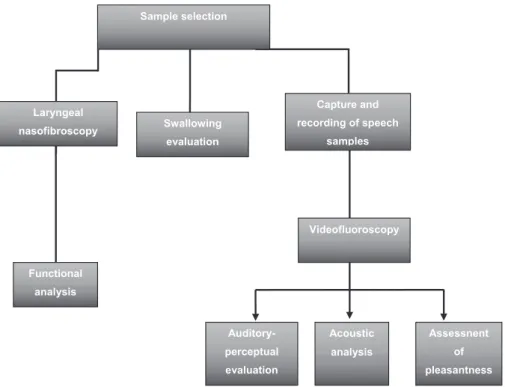
The following procedures were performed (►Fig. 1). 1. Recording of a laryngeal image for the functional assessment of the arytenoid cartilage:laryngoscopy was performed with a Karl Storz (Tuttlingen, Germany) 70° nasofibrolaryngoscope; a Toshiba (Minato, Tokyo, Japan) IK-M41A CCD micro-camera; a Sony (Minato, Tokyo, Japan) SLV-60HFBR VHS DVD media recorder; a Sony KV-1311 CR video monitor; and a ML-8 microphone (Le son, São Paulo, Brazil). The patients remained seated in a chair appropriate for an ear, nose, and throat examination, with the cervical region bent slightly forward. The nasofibrolaryngoscope was introduced through the wider nasal cavity without the use of topical anesthesia to avoid interference with pharyngeal and laryngeal sensitivity. The patients were instructed to breath in regularly, produce a sustained /é/ vowel, and then perform the pre-established tasks in the Brazilian Portuguese version of the consensus auditory-perceptual evaluation of voice (CAPE-V) protocol:14the emission of vowels, sentences and spontaneous speech. The functional conditions of the aryte-noid cartilage were consensually assessed by the physician and a speech therapist with extensive experience. This assessment was confirmed by both professionals after the end of the test based on digital image records.
The following parameters were considered for the classifi -cation of the conserved arytenoids: normal–the arytenoids exhibited no anatomical abnormalities and conserved their mobility and rotation; abnormal–edema, displacement and/ or overturning in each conserved arytenoid, regardless of an association with reduced mobility and/orfixation.
provided in a spoon using a 50% dilution of 100% barium sulfate (Bariogel, Cristália, Rio de Janeiro, RJ, Brazil) and 50% food. For the preparation of the liquid-pasty consistency, 4.5 g of thick-ener were used in 100 ml water, and for the pasty consistency, 9 g of thickener were used in 100 ml water. For the liquid consistency, mineral water was used. The focus of thefl uoro-scopic image was bounded: in the anterior region by the lips; in the upper region by the nasal cavity; in the posterior region by the cervical spine; and in the lower region by the bifurcation of the airways and the cervical esophagus. Thefindings were subsequently analyzed by two speech therapists specializing in dysphagia and experienced with videofluoroscopy evalua-tion, and the results were obtained by consensus.
The VF was analyzed through a modification of the Logemann15procedure, considering the presence or absence of the following parameters: penetration, silent penetration, aspiration, silent aspiration, and vallecular and/or hypophar-yngeal stasis. Swallowing was analyzed after a single spon-taneous deglutition or at most after the second, without the use of maneuvers for each food offered.
3. Recording of speech samples: voice samples were recorded with the participants performing again the tasks included in the Brazilian Portuguese version of the CAPE-V protocol.14A desktop computer, the Audacity program, the Edirol UA-101 interface, and a unidirectional condenser AKG-520 headset microphone (AKG, Vienna, Austria) were used. The microphone was positioned 3-5 cm from the patient’s mouth, and at a 45° to 90° angles. During the vocal tasks, the individual sat comfortably with his back straight, and the evaluator was alert to any indication of stress resulting from the test.
4. Auditory-perceptual analysis of voice:the recorded voices were edited, assigned identification numbers, and organized in random order to ensure blinding. They were
then presented to two raters who were speech therapists specializing in voice and experienced in the care of head and neck cancer patients. The raters analyzed the samples inde-pendently, without contact with each other. The speech samples were presented through speakers directly con-nected to the computer. The evaluations followed the CAPE-V protocol.14 Four voices from individuals with no complaints and no vocal changes were also analyzed; these cases composed the sample used to analyze intra-rater reliability.
5. Acoustic analysis:for this analysis, samples of at least three seconds of phonation of the sustained vowel /a/ were used. In the broadband analysis, the formants F3 and F4 were analyzed considering the mean of 2,500 Hz for F3 and 3,500 Hz for F4.16In the narrowband analysis, the harmo-nics-to-noise ratio was extracted. The maximum phonation time was measured in milliseconds with a timer. The mean values for each item (formants F3 and F4, harmonics-to-noise ratio and maximum phonation time) were calculated.
6. Voice pleasantness using the visual analogue scale:
voice pleasantness was analyzed by 22 undergraduate speech therapy students in the first year of the study program; the voice samples were presented in a silent room through speakers connected to the computer. The students could not talk to one another, and the voices were identified only by numbers. The analysis was per-formed quantitatively, and the classification was based on a 100-mm visual analogue scale in which the extreme left represented a very pleasant voice, and the extreme right represented an extremely unpleasant voice. Subsequently, each distribution range was numerically and qualitatively tabulated; scores from 0 mm to 35.5 mm represented a pleasant voice; from 35.6 mm to 50.5 mm, they represented
Sample selection
Laryngeal
nasofibroscopy Swallowing evaluation
Functional
analysis
Auditory-perceptual
evaluation
Acoustic
analysis
Assessnent
of
pleasantness Videofluoroscopy
Capture and
recording of speech
samples
a slightly pleasant voice; from 50.6 mm to 90.5 mm, they represented an unpleasant voice; and from 90.6 mm to 100 mm, the scores represented an extremely unpleasant voice (adapted from Yamasaki et al17). The mean pleasant-ness score of the voice was calculated.
Analysis of the Results
A descriptive analysis of the evaluated parameters was performed. Fisher test was used to compare the following variables: adjuvant therapy (radiotherapy and/or che-motherapy); and penetration, aspiration and stasis versus number (one or two) and functionality (normal or abnormal) of the remaining arytenoids. The significance level was set at 5%. When the incidence was low, the distribution was described in absolute numbers.
The intra-rater reliability and inter-rater agreement were analyzed by calculating the Kappa coefficient.
Results
A total of 12 patients were selected during the study period. However, one patient did not undergo laryngeal nasofi bro-scopy due to disease recurrence, and was excluded at the end. The sample consisted of 11 men, all undergoing reconstruc-tion with CHEP, and aged between 60 to 80 years old (mean¼68.2 years; standard deviation¼6.19 years).
Most of the patients had: tumors in stages T2 and T3; a 30-day interval from the surgery to the assessment; cervical lymph node dissection and the absence of a tracheostomy tube at the time of assessment; and moderate-to-severe dysphonia accompanied by roughness, breathiness, strain and loudness deviation (►Table 1).
Adjuvant treatment was prescribed to three patients, two due to positive N and one due to a positive margin.
The three patients who still had the tracheostomy tube in place at the time of the assessment exhibited postoperative complications (laryngeal stenosis), and two underwent an adjuvant treatment.
At the time of data collection, three patients had been discharged from speech therapy; the remaining patients were undergoing speech therapy to improve their voice patterns. All of the participants were consuming an oral diet, and were classified as levels 5 to 7 on the American Speech-Language Hearing Association National (ASHA) Out-come Measurement System (NOMS) Swallowing Rating Scale,18which was considered safe deglutition for the pur-poses of the present study. The VF was performed one month after the reintroduction of the oral diet.
Eight patients had two preserved arytenoids; of these patients, six presented abnormal arytenoid functionality. Three patients had only one preserved arytenoid, and two of these patients exhibited some type of abnormality (►Table 2).
One patient exhibited epiglottic edema.
The VF detected silent penetration in the 5 patients with penetration (45.5%); of these, 3 exhibited aspiration (27.3%), which was silent in 1 case. Vallecular and hypopharyngeal stasis was detected in 4 cases (36.4%).
Penetration was proportionally more frequent in the patients who underwent arytenoidectomy, and 2 of the 3 patients who had undergone the procedure exhibited aspira-tion. Among the cases in which both arytenoids were pre-served, aspiration was observed in one case in which both arytenoids were functionally abnormal (►Table 3).
The overall score for pleasantness of the voice was 71.3, which corresponds to an unpleasant voice. The mean number of sessions was 12.3. The patients with one preserved
Table 1 Sample characterization with case distribution according to tumor TNM stage, interval between surgery and assessment, adjuvant radiotherapy and/or chemotherapy, cervical lymph node dissection, tracheostomy at the time of the assessment and voice aspects in absolute numbers and percentages
N (%)
Stage
T1 3 (27.2)
T2 3 (27.2)
T3 5 (45.5)
N0 8 (72.8)
N1 3 (27.2)
M0 11 (100)
Interval between surgery and assessment
Up to 30 days 7 (63.6)
Up to 60 days 2 (18.1)
Up to 90 days 2 (18.1)
Adjuvant therapy
Radio- and chemotherapy 1 (9.1)
Radiotherapy 3 (27.3)
Cervical lymph node dissection
Yes 8 (72.7)
No 3 (27.2)
Tracheostomy tube present at the time of the assessment
Yes 3 (27.2)
No 8 (72.8)
Total 11 (100)
Voice aspects by CAPE-V Mean
Overall vocal deviation grade 61.5
Roughness 64.5
Breathiness 66.5
Strain 77.0
Pitch deviation 55.0
Loudness deviation 77.0
F3 (Hz) 3.124
F4 (Hz) 4.141
Harmonics-to-noise ratio (dB) 2.6
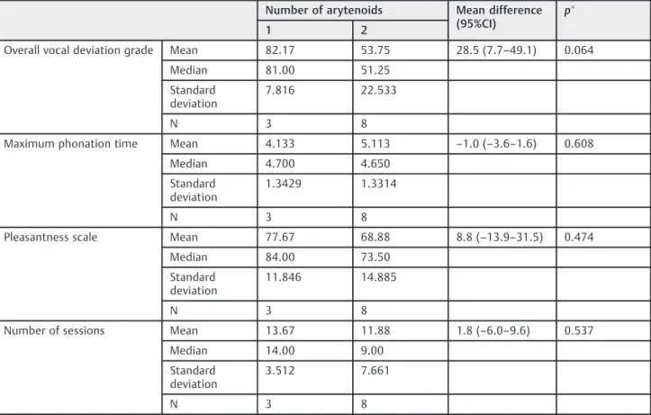
arytenoid exhibited a higher grade of voice deviation com-pared with the rest of the sample. The parametersmaximum phonation time (MPT), voice pleasantness and number of sessionsexhibited more favorable results when both aryte-noids were preserved (►Table 4).
A comparison considering the number of preserved aryte-noids and arytenoid functionality showed that the mean values for vocal deviation and MPT were less satisfactory among the patients with functional abnormalities (►Table 5).
Similar results were obtained when the full sample was classified according to the functionality of the remaining arytenoids. The mean overall vocal deviation grade, MPT, pleasantness scale and number of sessions were more satis-factory in the patients without arytenoid functional abnorm-ality (►Table 6).
Comparisons of the variables adjuvant therapy (radio-therapy and/or chemo(radio-therapy), penetration, aspiration and stasis versus number of preserved arytenoids (one or two) and functionality (normal or abnormal) of the preserved arytenoids indicated no significant difference (p>0.05). The
remaining variables could not be statistically compared due to their low incidence.
The intra-rater reliability and inter-rater agreement among the speech therapists and lay examiners were moderate.
Discussion
Because supracricoid laryngectomy has restricted indica-tions, there are few reports of its outcomes in the literature, and the related case series are limited. Consequently, new studies that increase the knowledge regarding arytenoid preservation are valuable. Other aspects that justify the performance of new studies related to this surgical proce-dure are the heterogeneity of the assessments and the lack of guidelines indicating the risk and prognostic factors that are likely to optimize voice and deglutition outcomes.6,19
The sample consisted of male patients over 50 years old who had undergone supracricoid laryngectomy with recon-struction using CHEP, and the majority were classified as T2/ T3 in accordance with the case series reported in the
Table 2 Distribution of remaining arytenoid functionality in absolute numbers
No
abnormality
Edema Displacement Edema and displacement
Total
One arytenoid
Preserved mobility with rotation 1 1 3
Fixed 1
Two arytenoids
Preserved mobility with rotation 2 1 1 8
Onefixed 1 1
Bothfixed 2
Total 11
Table 3 Distribution of the sample regarding the number and functionality of the preserved arytenoid cartilages according to the presence of penetration, silent penetration, aspiration and silent aspiration on videofluoroscopy
Number of preserved arytenoids
1 2
Functionality
Normal (n¼1) Abnormal (n¼2) p Normal (n¼2) Abnormal (n¼6) p
Silent penetration – 0.464
present 1 2 1 1
absent 0 0 1 5
Aspiration – –
present 1 1 0 1
absent 0 1 2 5
Silent aspiration – –
present 1 0 0 0
absent 0 2 2 6
literature.3,5,6,11,20,21Schindler et al6published an extensive review of supracricoid laryngectomy procedures in which 28% of 25 articles had samples smaller than 20 cases. Many studies do not focus specifically on supracricoid laryngec-tomies, comparing them with other types of surgical procedures.6,22,23
Adjuvant therapy was prescribed for patients with more advanced stages of the disease or positive margins. So et al12 reported indications for radiotherapy in 10 out of 15 patients
who underwent supracricoid laryngectomy; there was no difference in the functional outcomes between the patients who underwent radiotherapy and those who did not. The use of radio- and chemotherapy was not associated with the number of preserved arytenoids or arytenoid functionality. There was no difference in the mean age between the patients who underwent adjuvant therapy and those who did not.
Other studies suggested an assessment of the laryngeal configuration using laryngoscopy and ensuring that the
Table 4 Distribution of overall vocal deviation grade, maximum phonation time, pleasantness scale and number of sessions per number of preserved arytenoids
Number of arytenoids Mean difference (95%CI)
p
1 2
Overall vocal deviation grade Mean 82.17 53.75 28.5 (7.7–49.1) 0.064
Median 81.00 51.25
Standard deviation
7.816 22.533
N 3 8
Maximum phonation time Mean 4.133 5.113 –1.0 (–3.6–1.6) 0.608
Median 4.700 4.650
Standard deviation
1.3429 1.3314
N 3 8
Pleasantness scale Mean 77.67 68.88 8.8 (–13.9–31.5) 0.474
Median 84.00 73.50
Standard deviation
11.846 14.885
N 3 8
Number of sessions Mean 13.67 11.88 1.8 (–6.0–9.6) 0.537
Median 14.00 9.00
Standard deviation
3.512 7.661
N 3 8
Abbreviation: 95%CI, 95% confidence interval. Note:statistically significant, Wilcoxon test.
Table 5 Sample distribution of the overall vocal grade deviation, maximum phonation time and pleasantness scale according to the number and functionality of the preserved arytenoid cartilages
Number of preserved arytenoids
1 2
Functionality
Normal (n¼1)
Abnormal (n¼2)
Md (95%CI)
p Normal (n¼2)
Abnormal (n¼6)
Md (95%CI)
p
Overall vocal deviation grade
81 83 –2.0 (n.a.) – 50.5 55 –4.3 (n.a.) 0.615
Maximum phonation time
5.1 3.6 1.5 (n.a.) – 5.9 4.8 1.1 (–7.0–9.3) 0.311
Pleasantness scale 64 84 –20.5 (n.a.) – 71 68 3.5 (–15.0–22.1) 1.000
analysis was performed by professionals with extensive experience.12,13,24,25
Regarding the characteristics of the neoglottis and its interference with voice production and deglutition func-tions, the literature has shown an association between these functions and the type of reconstruction; furthermore, pub-lished studies have analyzed the effects of the presence of one or two arytenoids on supracricoid laryngectomy out-comes. In a previous study, Nemr et al11observed 22 cases (7 CHP and 15 CHEP) and found that the glottis was the primary site associated with moderate dysphagia, and that the supra-glottis was primarily associated with severe dysphagia. The progression of dysphagia was better than that of dysphonia; in addition, the degree of dysphagia and dysphonia was not associated with the presence of one or two arytenoids. Park et al26 compared 84 patients with 2 preserved arytenoids with 32 patients who had 1 preserved arytenoid, and observed a better functional outcome in those without arytenoidectomy. Schindler et al6 called attention to the fact that this parameter is still controversial in the literature, which reinforces the need for further studies on this subject. In the present study, the presence of one versus two arytenoids was not a determinant of deglutition performance in the assessed individuals. However, a case-by-case analysis showed penetration and aspiration in the three patients with
one preserved arytenoid, including the one patient whose remaining arytenoid was functionally normal. Another rele-vantfinding is that the cartilages were abnormal in six of the eight patients with two preserved arytenoids. The two cases without arytenoid functional abnormality exhibited silent penetration, and one of them also exhibited stasis. It should be noted that all of the participants had undergone speech therapy and were in a safe deglutition condition.18
Video-fluoroscopy enables the visualization of the aspiration of small amounts of food, which are often not identifiable on clinical assessments or with video endoscopy. Some studies have suggested the routine performance of VF to assess deglutition in this population of patients, regardless of whether they have undergone arytenoidectomy or not.9,20In the present study, VF was performed one month after the oral diet was reintro-duced, a time interval considered adequate for the instru-mental assessment of deglutition conditions.
Zacharek et al,27in a study with 10 cases of supracricoid laryngectomy (4 CHP and 6 CHEP), highlighted the fact that the 3 patients who had undergone VF exhibited significant aspiration; however, they did not develop postoperative pneumonia, and they made satisfactory progress with the reintroduction of an oral diet. These authors described data suggesting the importance of the mobility of the arytenoid cartilages, the action of the base of the tongue, and the
Table 6 Distribution of overall vocal deviation grade, maximum phonation time, pleasantness scale and number of sessions according to arytenoid function
Arytenoid abnormality
No Yes Md (95%CI) p
Overall vocal deviation grade
Mean 60.67 61.81 –1.1 (–63.3–61.1) 1.000
Median 75.00 66.50
Standard deviation
30.172 22.723
N 3 8
Maximum phonation time
Mean 5.667 4.538 1.2 (–1.3–3.6) 0.151
Median 5.100 4.500
Standard deviation
1.2503 1.3201
N 3 8
Pleasantness scale Mean 69.00 72.13 –3.1 (–17.9–11.7) 0.413
Median 69.00 77.00
Standard deviation
5.000 16.600
N 3 8
Number of sessions Mean 13.33 12.00 1.3 (–10.9–13.6) 0.837
Median 17.00 9.50
Standard deviation
6.351 7.151
N 3 8
remaining supraglottic tissue for a competent neoglottic sphincter with consequent vibration during phonation, and airway protection during swallowing.
This detailing of the functional conditions of the pre-served arytenoids can help clinicians understand prognostic factors and determine the therapeutic techniques that will hasten the reintroduction of an oral diet and promote an improvement in thefinal vocal quality.
There is a consensus in the literature that the voice is the most severely impaired function after supracricoid laryn-gectomy.6 This fact led specialists to seek alternatives to minimize the negative impact on communication. Allegra et al13detected a better voice pattern in a group of patients that underwent modified supracricoid laryngectomy invol-ving the use of the sternohyoid muscle compared with a group treated using the conventional technique.
In the auditory-perceptual analysis of voice, strain and loudness deviation were considered severe, which corrobo-rates the reports in the literature that patients who underwent subtotal laryngectomy maintain marked dysphonia.7,11,23 Relative to the overall vocal deviation grade, the qualitative analysis showed that dysphonia was moderate among the participants who had not undergone arytenoidectomy, and that it was severe among those with a single preserved arytenoid. The difference in the overall voice deviation grade according to the functionality of the remaining arytenoid, in absolute numbers, was practically the same.
Another notable issue related to the auditory-perceptual analysis of voice is that most of the reviewed articles used the grade, roughness, breathiness, asthenia, and strain (GRBAS) scale.3,22,23,28,29In patients with severe voice changes result-ing from the surgical procedure discussed here, the CAPE-V is considered the most adequate instrument for this type of analysis because, in addition to considering all the aspects included in the GRBAS scale, it comprises the perception of pitch and loudness (which exhibited severe abnormalities in the investigated sample). In addition, the CAPE-V with 100-mm visual analogue scale enables a much more detailed assessment of vocal deviation; for instance, it can establish whether a moderate degree of dysphonia is closer to the mild or the severe degrees. Furthermore, it enables better mon-itoring of therapy progression with the perception of discreet improvements.30
The mean maximum phonation time remained below normal limits due to inefficient glottal closure; the mean value found was similar to that reported in a previous study.12 Higher mean values were obtained in a study that assessed 24 patients with a mean interval of 34.5 months between the surgery and the assessment.3 As most participants in the present study were still undergoing speech therapy at the time of data collection, and considering that the interval from the surgery to the assessment was pf up to 3 months, a course of speech therapy longer than the average undergone by the present study’s sample (12.3 sessions) might help to improve some voice parameters, especially MPT. It should be noted that in absolute terms, the MPT was longer among the individuals with two preserved arytenoids. The same was true for the group with functionally normal arytenoids.
In addition, the possible coexistence of chronic lung diseases in these patients with respiratory limitations cannot be dismissed. The presence of lung disease may contribute to a reduced MPT, in addition to the limitations expected from age and neoglottic space. Therefore, the results of aerody-namic measures may contribute to a greater understanding of the phonation mechanism of these individuals and the limits of speech therapy. These factors further corroborate the loudness deviations found in the analyzed sample.
Regarding the short-term acoustic analysis, the narrow bandfilter provides relevant data regarding the presence and amount of harmonics and the harmonics-to-noise ratio. The mean harmonics-to-noise ratios were below the reference value due to the lower degree of glottal closure, which favors vocal production with fewer harmonics and more noise. This
finding is in agreement with the literature.3,31
The broadbandfilter enabled an analysis of the formants. The third formant (F3) is influenced by cavities formed behind and in front of the constriction of the tongue, and the fourth formant (F4) is influenced in part by the length of the vocal tract and in part by the dimensions of the laryngeal tube and the surrounding area. Considering that the frequency of the formants is influenced by the length of the vocal tract (the longer the vocal tract, the lower the frequencies of the for-mants),16we chose to investigate F3 and F4 as a function of the shortening of the vocal tract and the neoglottis.
The F3 and F4 means were above the reference values, confirming the hypothesis that the length of the vocal tract and the neoglottis influence the formant frequencies. The refer-ence values used in this study were extracted from the Swedish language, as reference values for Brazilian Portuguese corre-sponding to the study’s population of elderly men were not found. Even when the mean F3 for young men in Brazilian Portuguese (2,638 Hz, with a standard deviation of 120 Hz)32 was considered, the values exceeded this mean. Considering that the elderly larynx is lower and thus all formants are lower in frequency compared with young adults, the values obtained in this study support the elevation of the neolarynx and pharynx constriction as a function of the neoglottis.
No studies that measured formants in partial or subtotal laryngectomy patients were found. This measurement may be relevant for the comparative assessment of individuals without voice abnormalities, and to monitor the progression of the treatment.
Formant measurement has been scarcely addressed in studies on head and neck cancer. Timon et al33investigated the impact of thyroidectomy on the vocal tract based on F1 and F2, and suggested that the patients who underwent thyroid surgery presented a significant reduction in vocal tract stabi-lity compared with the controls. They further noted that the aforementioned measurements offer a potential instrument to investigate the functional implications of any thyroid surgery-related disturbance of pharyngeal innervations.
vocal pattern resulting from supracricoid laryngectomy and, consequently, the young adults’ perceptions of these patients’voices as unpleasant.
Naudo et al10 evaluated phonation as good or satisfac-tory in 97% of supracricoid laryngectomy patients at the 12th postoperative month. Dworkin et al24 performed an auditory-perceptual evaluation of the voice in 10 indivi-duals undergoing supracricoid laryngectomy, and found a mean of 2.8 on a 5-point scale in which 1 represented poor vocal quality, and 5, excellent vocal quality. The authors called attention to the differences between CHP and CHEP, mentioning that preservation of the epiglottis can facilitate the rehabilitation process by providing a stable opposing anatomical surface against which the mobile arytenoid(s) can establish neoglottic closure. Notably, none of these studies included assessments performed by inexperienced examiners; in contrast, the present study included fi rst-year speech therapy undergraduate students as examiners. In the analyzed sample, both the overall mean voice pleasantness and the numerical differences between the groups according to the number of arytenoids and aryte-noid functionality were within the range rated as unplea-sant. It should be noted that most of the participants were still undergoing speech therapy to improve their vocal pattern.
This finding suggests the need to reflect upon how the general public might perceive these voices, a factor that can negatively affect the patients’quality of life, especially during conversations with strangers, on the phone, or in noisy environments.
The students’ perceptions of the patients’ voices as unpleasant plus the functional conditions presented in this study reinforce the need to deepen the knowledge regarding supracricoid laryngectomy.
The findings of the present analysis shed light on two aspects that require more thorough study. Firstly, the thera-peutic tests to select the vocal techniques that are most likely to optimize the vocal pattern within the shortest possible course of speech therapy should be studied; and secondly, ongoing research regarding the impact of the number and functionality of the remaining arytenoids should be per-formed with larger samples to establish prognostic factors for satisfactory voice and deglutition.
Some limitations of the present study should be noted. The small sample size may account for the lack of statistical evidence related to the number and functionality of the remaining arytenoids. Multicenter studies might provide more robust results, and, consequently, guide the formula-tion of new paradigms for voice and deglutiformula-tion after supra-cricoid laryngectomy.
Three patients who still had the tracheostomy tube in place at the time of the assessment exhibited postoperative complications, and two underwent adjuvant treatment. The presence of these patients in the studied sample should be considered a potent confounding factor, since the data of their evaluations could be more altered, which is not clear when observing the data individually, and, due to the small sample size, it cannot be further analyzed.
One of the contributions of this study is that it alerts specialists to the need to pay attention to the condition of the remaining arytenoids and to the auditory-perceptual and acoustic aspects of the voice, including acoustic measure-ments of formant frequency. A multi-professional assess-ment at the onset of speech therapy that considers the aspects emphasized in the present study will help specialists formulate strategies to obtain more favorable outcomes in terms of voice quality, with consequent improvement of the patients’quality of life. Similarly, it is necessary to manage deglutition using VF following the clinical assessment of safe deglutition.
Conclusion
In the present sample of elderly men who underwent CHEP, both arytenoids were preserved and exhibited functional abnormalities in most cases. Videofluoroscopy performed one month after the reintroduction of an exclusive oral diet enabled the management of dysphagia, which proved to be persistent in a large portion of the analyzed sample that exhibited silent penetration, aspiration of small amounts of food, and/or vallecular and hypopharyngeal stases.
The participants exhibited moderate-to-severe dysphonia accompanied by roughness, breathiness, strain, and loudness deviation. The maximum phonation time and harmonics-to-noise ratio were below the reference range, while the mean value of the third and fourth formants were above the reference range. The participants’ voices were rated as unpleasant by young adults.
The number and functionality of the preserved aryte-noids were not found to be prognostic factors for favorable deglutition efficiency outcomes. However, the qualitative analysis showed that the preservation of both aryte-noids and the absence of functional abnormalities were associated with more satisfactory voice and deglutition patterns.
References
1 Piquet JJ, Desaulty A, Decroix G. Crico-hyoido-epiglotto-pexy. Surgical technic and functional results. Ann Otolaryngol Chir Cervicofac 1974;91(12):681–686
2 Pressman JJ. Submucosal compartmentation of the larynx. Ann Otol Rhinol Laryngol 1956;65(03):766–771
3 Saito K, Araki K, Ogawa K, Shiotani A. Laryngeal function after supracricoid laryngectomy. Otolaryngol Head Neck Surg 2009; 140(04):487–492
4 Gonçalves AJ, Bertelli AA, Malavasi TR, Kikuchi W, Rodrigues AN, Menezes MB. Results after supracricoid horizontal partial laryn-gectomy. Auris Nasus Larynx 2010;37(01):84–88
5 Nakayama M, Miyamoto S, Seino Y, Okamoto T, Kano K. Neoglottal revisions after supracricoid laryngectomy with cricohyoidoepi-glottopexy. Jpn J Clin Oncol 2016;46(07):642–645
6 Schindler A, Pizzorni N, Mozzanica F, et al. Functional outcomes after supracricoid laryngectomy: what do we not know and what do we need to know? Eur Arch Otorhinolaryngol 2016;273(11): 3459–3475
8 Kruk-Zagajewska A, Szmeja Z, Szyfter W, Wójtowicz J, Citowicki W, Wierzbicka M. [Swallowing disorders in patients after the laryngeal cancer surgery]. Otolaryngol Pol 1995;49(01):15–22 9 Lewin JS, Hutcheson KA, Barringer DA, et al. Functional analysis of
swallowing outcomes after supracricoid partial laryngectomy. Head Neck 2008;30(05):559–566
10 Naudo P, Laccourreye O, Weinstein G, Jouffre V, Laccourreye H, Brasnu D. Complications and functional outcome after supracri-coid partial laryngectomy with cricohyoidoepiglottopexy. Otolar-yngol Head Neck Surg 1998;118(01):124–129
11 Nemr NK, Carvalho MB, Kohle J, Leite GCAL, Rapoport A, Szeliga RMS. Estudo funcional da voz e da deglutição na laringectomia supracricóide. Rev Bras Otorrinolaringol 2007;73(02):151–155 12 So YK, Yun YS, Baek CH, Jeong HS, Son YI. Speech outcome of
supracricoid partial laryngectomy: comparison with total laryn-gectomy and anatomic considerations. Otolaryngol Head Neck Surg 2009;141(06):770–775
13 Allegra E, Lombardo N, La Boria A, et al. Quality of voice evaluation in patients treated by supracricoid laryngectomy and modified supracricoid laryngectomy. Otolaryngol Head Neck Surg 2011; 145(05):789–795
14 Behlau M. Consensus Auditory- Perceptual Evaluation of Voice (CAPE-V), ASHA 2004. Refletindo sobre o novo/New reflexions. Rev Soc Bras Fonoaudiol 2004;9(03):187–189
15 Logemann JA. Evaluation and treatment of swallowing disorders. 2nd ed. Texas: Pro ED; 1998
16 Sundberg J. Ciência da voz: fatos sobre a voz na fala e no canto. Trad. Salomão GL. São Paulo: Editora da Universidade de São Paulo; 2015
17 Yamasaki R, Madazio G, Leão SHS, Padovani M, Azevedo R, Behlau M. Auditory-perceptual evaluation of normal and dys-phonic voices using the voice deviation scale. J Voice 2017;31 (01):67–71
18 American Speech-Language Hearing Association National Out-come Measurement System (NOMS). Adults speech-language pathology training manual. Rockville, Md: American Speech-Language Hearing Association; 1998
19 Marioni G, Marchese-Ragona R, Ottaviano G, Staffieri A. Supra-cricoid laryngectomy: is it time to define guidelines to evaluate functional results? a review. Am J Otolaryngol 2004;25(02): 98–104
20 Prado PRP, Dias FL, Santos IC, Freitas E, Ferreira LP. Avaliação videofluoroscópica no pós-operatório tardio de pacientes
sub-metidos à laringectomia supracricoidea com cricohioidoepiglo-topexia. Rev Bras Cir Cabeça Pescoço 2012;41(03):124–127 21 Ozturk K, Akyildiz S, Gode S, et al. Post-surgical and oncologic
outcomes of supracricoid partial laryngectomy: a single-institu-tion report of ninety cases. ORL J Otorhinolaryngol Relat Spec 2016;78(02):86–93
22 Torrejano G, Guimarães I. Voice quality after supracricoid laryn-gectomy and total larynlaryn-gectomy with insertion of voice prosthe-sis. J Voice 2009;23(02):240–246
23 Crosetti E, Garofalo P, Bosio C, et al. How the operated larynx ages. Acta Otorhinolaryngol Ital 2014;34(01):19–28
24 Dworkin JP, Meleca RJ, Zacharek MA, et al. Voice and deglutition functions after the supracricoid and total laryngectomy proce-dures for advanced stage laryngeal carcinoma. Otolaryngol Head Neck Surg 2003;129(04):311–320
25 Makeieff M, Giovanni A, Guerrier B. Laryngostroboscopic evalua-tion after supracricoid partial laryngectomy. J Voice 2007;21(04): 508–515
26 Park JO, Joo YH, Cho KJ, Kim NG, Kim MS. Functional and oncologic results of extended supracricoid partial laryngectomy. Arch Oto-laryngol Head Neck Surg 2011;137(11):1124–1129
27 Zacharek MA, Pasha R, Meleca RJ, et al. Functional outcomes after supracricoid laryngectomy. Laryngoscope 2001;111(09):1558–1564 28 Schindler A, Favero E, Nudo S, Albera R, Schindler O, Cavalot AL. Long-term voice and swallowing modifications after supracricoid laryngectomy: objective, subjective, and self-assessment data. Am J Otolaryngol 2006;27(06):378–383
29 Bron L, Pasche P, Brossard E, Monnier P, Schweizer V. Functional analysis after supracricoid partial laryngectomy with cricohyoi-doepiglottopexy. Laryngoscope 2002;112(7 Pt 1):1289–1293 30 Nemr K, Simões-Zenari M, Cordeiro GF, et al. GRBAS and Cape-V
scales: high reliability and consensus when applied at different times. J Voice 2012;26(06):812.e17–812.e22
31 Portas JG, Queija DdosS, Arine LP, et al. Voice and swallowing disorders: functional results and quality of life following supra-cricoid laryngectomy with cricohyoidoepiglottopexy. Ear Nose Throat J 2009;88(10):E23–E30
32 Svicero MAF. Caracterização acústica e de imagens de ultrassono-grafia das vogais orais do Português Brasileiro [dissertação]. São Paulo: Pontifícia Universidade Católica de São Paulo; 2012 33 Timon CI, Hirani SP, Epstein R, Rafferty MA. Investigation of the