online | memorias.ioc.fiocruz.br
Subcellular localisation of FLAG tagged enzymes of the dynamic
protein S-palmitoylation cycle of
Trypanosoma cruzi
epimastigotes
Cassiano Martin Batista1, Felipe Saad1, Stephane Pini Costa Ceccoti1, Iriane Eger2, Maurilio José Soares1/+
1Fundação Oswaldo Cruz-Fiocruz, Instituto Carlos Chagas, Laboratório de Biologia Celular, Curitiba, PR, Brasil 2Universidade Estadual de Ponta Grossa, Departamento de Biologia Geral, Ponta Grossa, PR, Brasil
Dynamic S-palmitoylation of proteins is the addition of palmitic acid by zDHHC palmitoyl transferases (PATs) and depalmitoylation by palmitoyl protein thioesterases (PPTs). A putative PAT (TcPAT1) has been previously identified in Trypanosoma cruzi,theetiological agent of Chagas disease. Here we analyse other 14 putative TcPATs and 2 PPTs in the parasite genome. T. cruzi cell lines expressing TcPATs and TcPPTs plus a FLAG tag at the C terminus were produced for most enzymes, with positive detection by indirect immunofluorescence. Overexpressed TcPATs were mostly found as single spots at the parasite anterior end, while the TcPPTs were dispersed throughout the parasite body.
Key words: dynamic S-palmitoylation - Trypanosoma cruzi - protein expression
doi: 10.1590/0074-02760180086
Financial support: CNPq, CAPES and Fiocruz. + Corresponding author: [email protected] Received 16 February 2018
Accepted 2 May 2018
Dynamic protein S-palmitoylation concerns the addition of palmitate to cysteines of the modified pro-tein by zDHHC palmitoyl transferases (PATs) through thioester linkages and depalmitoylation by palmitoyl protein thioesterases (PPTs) (Conibear and Davis 2010). Protein S-palmitoylation cycles promote the insertion of target proteins into membranes, regulating their locali-sation and function (Linder and Deschenes 2003).
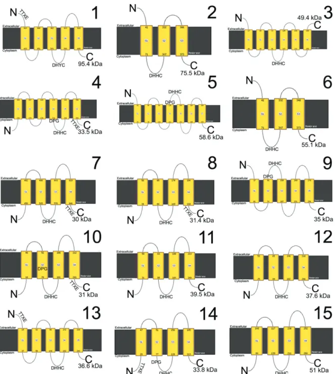
PATs are key transmembrane enzymes, with cyste-ine rich domains in the DHHC motif (CRD-DHHC do-main), in addition to DPG and TTxE structural domains (Greaves and Chamberlain 2011). PATs are involved in diverse biological processes in several organisms, such as Homo sapiens cancer (Ducker et al. 2004) and neurologi-cal diseases (Young et al. 2012, Cho and Park 2016), yeast endocytosis (Feng and Davis 2000), Cryptococcus neo-formans virulence (Santiago-Tirado et al. 2015), Giardia lamblia encystation (Merino et al. 2014) and invasion in Apicomplexa (Frénal et al. 2013). TbPAT7 is responsible for flagellar localisation of calflagin in the trypanosoma-tid protozoan Trypanosoma brucei (Emmer et al. 2009).
PPTs belong to the serine hydrolases family, are less abundant in number than PATs and are characterised by the presence of a serine active site for hydrolysis of the substrate, being able to cleave amide, ester and thioester bounds (Long and Cravatt 2011).
Goldston et al. (2014) identified, by in silico search, 15 PATs in the Trypanosoma cruzi genome, as opposed to 12 in T. brucei and 20 in Leishmania major. However, up to now only one PAT has been characterised in T. cruzi, the etiological agent of Chagas disease: TcHIP, or TcPAT1 (Batista et al. 2013). TcPAT1 is a 95.4 kDa Golgi protein
expressed in different developmental stages of the parasite, with a modified DHYC motif (Batista et al. 2013). Such modified motif is functional in the homologue Akr1p en-zyme of Saccharomyces cerevisae (Roth et al. 2002).
Cassiano Martin Batista et al. 2|6
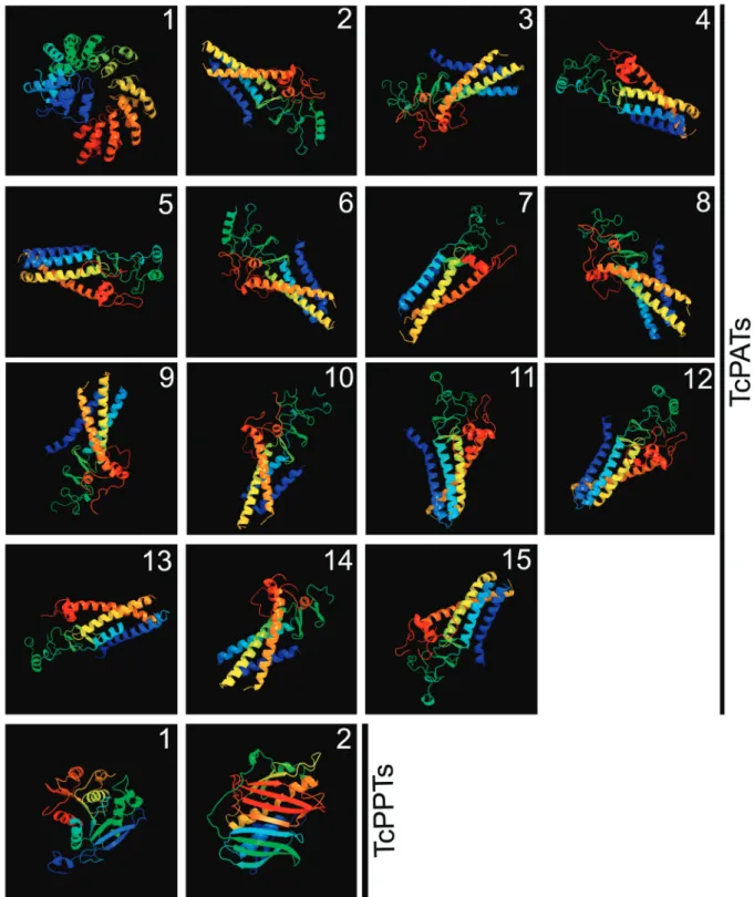
All TcPATs showed similar predicted 3D models, except for TcPAT1 (larger and with ankyrin repeats). TcPPTs 1 and 2 were very different from each other. All 3D models had 100% confidence (Fig. 2).
Aiming to produce transfectant cell lines of T. cruzi epimastigotes expressing TcPATs plus a FLAG tag at the C terminus (FLAGC tagged TcPAT), the genes were amplified using specific primers (Table I) with recombi-nation sites for the Gateway cloning platform (Thermo
Fischer Scientific, Waltham, MA, USA) by using the entry plasmid vector pDONR 221 and the destination T. cruzi vector pTcGWFLAGC (Batista et al. 2010, Kuger-atski et al. 2015). All genes were cloned, except TcPAT6 and TcPAT1 (already characterised). Three-day-old T. cruzi epimastigotes were transfected with a Gene Pulser XCell BIORAD electroporator (BIORAD Inc., Hercules, CA, USA), selected with 500 µg.mL-1 G418 and main-tained with 250 µg.mL-1 of the same antibiotic, as
Fig. 2: predicted 3D models of Trypanosoma cruzi PATs and PPTs. The software Phyre2 was used. All 3D models had 100% confidence.
ously described (Batista et al. 2010). Twelve resistant cell lines could be selected, with the exception of TcPAT4.
For subcellular localisation by indirect immuno-fluorescence assays (IFA), T. cruzi transfectants were washed twice in PBS, fixed for 10 min with 4% parafor-maldehyde, adhered to 0.1% poly-L-lysine coated cover-slips, permeabilised with 0.5% Triton/PBS, and incubat-ed for one hour at 37ºC using a mouse anti-flag antibody
Cassiano Martin Batista et al. 4|6
TABLE I
Primers used for palmitoyl transferase (PAT) isolation from gDNA of Trypanosoma cruzi (clone Dm28c) epimastigotes
PAT/Gene ID F’/R’ (5’-3’)
TcPAT1/* ATGCAGGTGTTTGGCGCTCGGATG
ACGGCGTTCATCTTTCACCT
TcPAT2/ TcCLB.506297.250 ATGCCACAGACTAACAGCACGGAATGG/
GGGTTCTCTGACTTCATGCGC
TcPAT3/ TcCLB.510899.50 ATGGGGCCCATACGCGTTGAAAGAG/
CACCTGCGTGGCACACAACT
TcPAT4/ TcCLB.508479.200 ATGTCAGGTTTCTGGTCTGTTCAGC/
CACCTCTGCTGTTTCAACGACAATAT
TcPAT5/ TcCLB.509029.170 ATGTCCGGAGAGACTTTTGCTTG/
CTCATATTTCATCCTCCGTTCTCCT
TcPAT6/ TcCLB.506177.40 ATGCGGTCATCTATGTTGCTGCTTTT/
TTCCTCCTTCATCTCCTCCTCGCT
TcPAT7/ TcCLB.510687.130 ATGGATGAATCAAACGACGCG/
CACGTCATTCTCAGCGTTTCG
TcPAT8/ TcCLB.511897.19 ATGGGTAAGATTTTTGAAATGGAGGT/
CCGTATCAAATCAACAAGAGTTCTCC
TcPAT9/ TcCLB.509769.33 ATGGATTGCGTGGTAGGTATGCGGAAT/
AACTAGAGCCTCAGTGTTCAACCAC
TcPAT10/ TcCLB.508239.40 ATGATGTCATTGTTATCACGATGGG/
CACAAGGTCGGCGTCATCG
TcPAT11/ TcCLB.511823.50 ATGTCGTTGCTTTGTTGTGATCC/
GTCATATTTGGGTGAAATGGGTG
TcPAT12/ TcCLB.506855.10 ATGGGGTCGTTGATTCCGC/
CACCGGCAACATCACCTCATC
TcPAT13/ TcCLB.510747.18 ATGAATGTACCCACTTCATCCAGTCCGAT/
CACATAAAACTCGGCGTTTTCC
TcPAT14/ TcCLB.511153.60 ATGGAGTCCGTGGAAGTGCTAGT/
TGCGATGATGGGGCTCTTATT
TcPAT15/ TcCLB.509105.20 ATGCGGTGCTGTGGGCG/
CATACCACCAGATCCGGGAAGCGAC
*: Batista et al. (2013); F’: forward primer; R’: reverse primer.
Hoechst 33342 (Sigma-Aldrich St. Louis, MO, USA) and the coverslips were mounted with Prolong Gold antifad-ing agent (Thermo Fischer Scientific, Waltham, MA, USA). The slides were observed in a Nikon Eclipse E600 epifluorescence microscope.
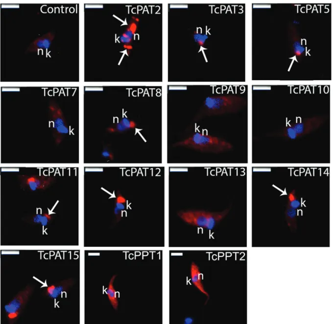
As a result, TcPATs 3, 5, 8, 11, 12, 14 and 15 were located as single dots at the anterior region of the para-site, close to the kinetoplast and the flagellar pocket (Fig. 3). Interestingly, most PATs with four transmem-brane domains (five out of seven) showed this pattern. The positive reaction was frequently found lateral to the kinetoplast, which suggests Golgi, flagellar pocket or contractile vacuole localisation. TcPAT2 labeling appeared as strong dots distributed throughout the cell body, suggestive of localisation in some cytoplasmic or-ganelle (Fig. 3). TcPAT13 presented a stronger labeling at the perinuclear region (Fig. 3). These patterns were expected, since PATs are usually found at the endoplas-mic reticulum, Golgi and plasma membranes (Ohno et al. 2006). No positive reaction was detected for TcPATs 7, 9, 10 (Fig. 3). Transcriptomic data from TritrypDB
in-dicate that TcPATs 7 and 9 are expressed in metacyclic trypomastigotes, but not in epimastigotes. Therefore, gene expression of these two enzymes (and possibly also TcPAT10) can be down-regulated in epimastigotes. In summary, these results indicated that at least nine TcPATs could be overexpressed in T. cruzi epimastigotes.
In order to characterise the TcPPTs, a genomic data search was performed as described above, and two genes were identified (Table II). TcPPT1 is an 843 base pairs gene and the product (30.2 kDa) is homologue to H. sapi-ens acyl-protein thioesterase-1 (APT1) and lysophospho-lipase genes, which are involved in cytosolic and lyso-somal protein depalmitoylation (Long and Cravatt 2011). TcPPT2 is a 951 base pairs gene and the product (35.5 kDa) is homologue to H. sapiens acyl-protein thioester-ase-2 (APT2), involved in cytosolic depalmitoylation (Long and Cravatt 2011). Primers were then designed for isolation and amplification of these genes (Table II).
TABLE II
Identification, in silico analysis and primer design of Trypanosoma cruzi palmitoyl thioesterase (PPT)
PPT/Gene ID BP kDa F’/R’ (5’-3’)
TcCLB.506797.70 (TcPPT1) 843 30.2 ATGATCGGAACGCCGATAGAAAACT/
AGCCTTGGACTCAATCGCCGGCAATACCT
TcCLB.504149.55 (TcPPT2) 951 35.5 ATGCTTCTGCAGGACGTTATTGGAG/
GAGTCTCGATTTGTAGCCCTTTCCTG
BP: number of base pairs; kDa: molecular weight of the predicted protein; F’: forward primer; R’: reverse primer.
Cassiano Martin Batista et al. 6|6
TcPPTs). Resistant cell lines expressing TcPPT1 and TcPPT2 were selected with 500 µg.mL-1 G418. After IFA in the same conditions as described above, both TcPPTs showed strong labeling dispersed through the cell body, suggesting a cytoplasmic localisation (Fig. 3), indicating that T. cruzi epimastigotes overexpressed both TcPPTs, in the expected cytoplasmic localisation.
In conclusion, our data indicate that a dynamic protein S-palmitoylation machinery (nine PATS and two PPTs) could be overexpressed in T. cruzi. Future studies will be crucial to determine the importance of this machinery for the parasite survival. Palmitoylation and depalmitoylation of proteins can play an important role in this parasite, in events as diverse as nutrition, protein traffic, differentia-tion, host-cell interaction and infection establishment.
ACKNOWLEDGEMENTS
To the Program for Technological Development in Tools for Health-PDTIS-FIOCRUZ for use of its facility (Confocal and Electronic Microscopy Platform RPT07C) at the Instituto Carlos Chagas/Fiocruz-PR, Brazil.
AUTHORS’ CONTRIBUTION
CMB planned the experiments, designed the PATs primers,
performed part of the cloning experiment, selected Trypanosoma
cruzi cell lines and wrote the first manuscript draft; FS performed cloning and IFAs; SC made PPTs primer design and cloning; IE helped to plan the experiments and revised the manuscript; MJS conceived the study and edited the final form of the manuscript. All authors read and approved the final manuscript.
REFERENCES
Batista CM, Kalb LC, Moreira CM, Batista GT, Eger I, Soares MJ. Identification and subcellular localization of TcHIP, a putative Golgi zDHHC palmitoyl transferase of Trypanosoma cruzi. Exp Parasitol. 2013(1); 134: 52-60.
Batista M, Marchini FK, Celedon PA, Fragoso SP, Probst CM, Preti H, et al. A high-throughput cloning system for reverse genetics in
Trypanosoma cruzi. BMC Microbiol. 2010; 10: 259.
Brown RW, Sharma AI, Engman DM. Dynamic protein S-palmitoylation mediates parasites life cycle progression and diverse mechanisms of virulence. Crit Rev Biochem Mol Biol. 2017; 52(2): 145-62. Cho E, Park M. Palmitoylation in Alzheimer’s disease and other
neu-rodegenerative diseases. Pharmacol Res. 2016; 111: 133-51. Conibear E, Davis NG. Palmitoylation and depalmitoylation
dynam-ics at a glance. J Cell Sci. 2010; 123(23); 4007-10.
Contreras VT, Araujo-Jorge TC, Bonaldo MC, Thomaz N, Barbosa HS, Meirelles MNSL, et al. Biological aspects of the DM28c clone of Trypanosoma cruzi after metaciclogenesis in chemically defined media. Mem Inst Oswaldo Cruz. 1988; 83(1): 123-33. Ducker CE, Stettler EM, French KJ, Upson JJ, Smith CD. Huntingtin
interacting protein 14 is an oncogenic human protein: palmitoyl acyltransferase. Oncogene. 2004; 23(57): 9230-7.
Emmer BT, Souther C, Toriello KM, Olson CL, Epting CL, Eng-man DM. Identification of a palmiytoyl acyltransferase required for protein sorting to the flagellar membrane. JCell Sci. 2009; 122(6): 867-74.
Feng Y, Davis NG. Akr1p and the type I casein kinases act prior to the ubiquitination step of yeast endocytosis: Akr1p is required for kinase localization to the plasma membrane. Mol Cel Biol. 2000; 20(14): 5350-9.
Frénal K, Tay CL, Mueller C, Bushell ES, Jia Y, Graindorge A, et al. Global analysis of apliconplexan protein S-acyl transferases re-veals an enzyme essential for evasion. Traffic. 2013; 14(8): 895-911. Goldston AM, Sharma AI, Paul KS, Engman DM. Acylation in try-panosomatids: an essential process and potential drug target. Trends Parasitol. 2014; 30(7): 350-60.
Greaves J, Chamberlain LH. DHHC palmitoyl transferases: sub-strates interactions and (patho)physiology. Trends Biochem Sci. 2011; 36(5): 245-53.
Kelley LA, Mazulin S, Yates CM, Was MN, Sternberg MJE. The Phyre2 web portal for protein modeling, prediction and analysis. Nat Protoc. 2015; 10(6): 845-58.
Kugeratski FG, Batista M, Inoue AH, Ramos BD, Krieger MA, Marchini FK. pTcGW plasmid vectors 1.1 version: a versatile tool for Trypanosoma cruzi gene characterisation. Mem Inst Oswaldo Cruz. 2015; 110(5): 687-90.
Linder ME, Deschenes RJ. New insights into the mechanisms of pro-tein palmitoylation. Biochemistry. 2003; 42(15): 4311-7. Lobo S, Greentree WK, Linder ME, Deschenes RJ. Identification of
a Ras palmitoyltransferase in Saccharomyces cerevisiae. J Biol Chem. 2002; 277(43): 41268-73.
Long JZ, Cravatt BF. The metabolic serine hydrolases and their func-tions in mammalian physiology and disease. Chem Rev. 2011; 111(10): 6022-63.
Merino MC, Zamponi N, Vranych CV, Touz MC, Rópolo AS. Identifi-cation of Giardia lamblia DHHC proteins and the role of protein S-palmitoylation in the encystation process. PloS Negl Trop Dis. 2014; 8(7): e2997.
Ohno Y, Kihara A, Sano T, Igarashi Y. Intracellular localization and tissue-specific distribution of human and yeast DHHC cysteine-rich domain-containing proteins. Biochim Biophys Acta. 2006; 1761(4): 474-83.
Roth AF, Feng Y, Chen L, Davis NG. The yeast DHHC cysteine-rich domain protein Akr1p is a palmitoyl transferase. J Cell Biol. 2002; 159(1): 23-8.
Santiago-Tirado FH, Peng T, Yang M, Hang HC, Doering TL. A sin-gle protein S-acyl transferase acts through diverse substrates to determine Cryptococcal morphology, stress tolerance, and patho-genic outcome. PLoS Pathog. 2015; 11(5): e1004908.