Ana Maria Vaz Portugal Silva
Outubro de 2013
Theta and Alpha Neurofeedback for
Age-Related Cognitive Deficits
Dissertação de Mestrado
Mestrado em Ciências da Saúde
Trabalho realizado sob a orientação do:
Doutor Nuno Sérgio Mendes Dias
Escola de Ciências da Saúdeiii
As pessoas têm medo das mudanças.
Eu tenho medo que as coisas nunca mudem.
v
AGRADECIMENTOS
Um obrigada ao Nuno por, há dois anos, me teres aberto as portas a um mundo desconhecido. Mais que isso, um obrigada por, uns tempos depois, teres confiado em mim e teres-me dado a oportunidade de desenvolver este projecto. O teu apoio, a tua calma, mas também a tua teimosia e frontalidade foram peças fundamentais neste ano de trabalho. Por fim, um muito obrigada pelas horas longas a “bater pedra”! no código, no jogo, na estatística, na escrita…
À Daniela: a ti agradeço-te teres partilhado comigo essa pessoa que és. E os meses lindos que passámos no CCA! E o concerto de Ornatos: São coisas / São só coisas…
À Joana, agradeço as conversas filosóficas e a amizade que fomos construindo. Além disso, um obrigada gigante por, quando tive que me ausentar, teres agarrado e tomado conta do trabalho com os participantes!
Ao Ricardo, agradeço toda a ajuda, disponibilidade e empenho, que foram tão importantes na elaboração do protocolo.
Ao “pessoal do CCA”, à Nadine, ao Carlos, à Teresa, à Liliana e ao Pedro, um obrigada por toda a orientação e apoio com os participantes e todas as longas horas a fazer avaliações EEG! Ao Professor Doutor Nuno Sousa, e a todos os NeRDs, agradeço a oportunidade de integrar uma equipa fantástica e entusiástica!
Às minhas coleguinhas: Cláudia, Ana Rita, Mika, Joaninha, agradeço-vos a amizade e a partilha destes dois anos! Sem vocês nada teria sido igual. Especialmente, agradeço a Mika, por tar aqui ao meu lado até ao fim. E à Rita por ser quem é, e me ter ensinado a acordar contente de manhã! Às minhas meninas de sempre: Carol, Funkie, Nocas, Guidinha e Sílvia. Obrigada por estarem aqui sempre e para sempre. À Lia por também ela estar aqui desde sempre e para sempre. À Rita pela amizade e por ser a minha Queen B!
À minha grande família, por apesar de longe, estar sempre atenta. Ao João e a Tânia, obrigada por me “emprestarem” a Madalena e me terem feito tão feliz!
À Teresa, por me perguntar todos os dias se preciso de alguma coisa, e sempre me facilitar a vida! Acima de tudo, obrigada pelo carinho e pela amizade.
vi
Acima de tudo, quero agradecer aos meus pais, ao meu irmão, e ao Zé.
Ao meu pai, pelo entusiamo, pelo apoio, por me aturar todas as manhãs, e pela procura incessante de soluções. À minha mãe, por me escutar todos os dias, pela amizade, pela cumplicidade, e por também ela ter tido um papel activo na realização deste trabalho.
Ao João, porque mesmo não estando cá, estiveste sempre aqui. Não há melhor irmão que tu. A ti, Zé, obrigada pela escuta, pelo apoio, pelos conselhos, pelo companheirismo. Preciso que saibas que, sem ti, não teria sido capaz. Perdoa-me se durante este ano não te pude dar o tempo e atenção que merecias. Mais do que tudo, quero-te agradecer pela tua espera e pelo teu amor. Espero que, quando chegar a altura, consiga desempenhar tão bem este papel como tu.
Por fim, um muito obrigada à USF S. João, por ter tornado este trabalho concretizável!
Agradeço também a cada um dos participantes, por de uma maneira ou de outra, terem sido tão especiais.
vii
ABSTRACT
With the growing life expectancy, the number of elderly people is increasing tremendously worldwide. The progressive decrease of synaptic plasticity and neuronal inter-connectivity, concomitant with neurophysiological and behavioral alterations in the ageing brain, may be delayed by neurorehabilitation. Current approaches used to modify cognitive capabilities are often divided into behavioral training procedures and techniques for direct modulation of neural mechanisms, as neurostimulation or neurofeedback. Neurofeedback, which most of the times is based on electroencephalogram signals, is used to train individuals on learning how to influence their own brain functions based on the online-analysis of the brain activity. However, the potential greater effects of rehabilition through a combined methodology of these two trends are poorly investigated. In the present study, we wanted to examine the effects of a protocol with neurofeedback training interleaved with neurocognitive tasks. It was hypothesized that the combined approach might have a superior impact on cognitive performance, in comparison with a neurofeedback training alone approach. A protocol for neurorehabilitation covering the two proposed methodologies was developed. It supports Alpha and Theta neurofeedback up-training, and can be interleaved with neurocognitive tasks, namely the n-Back Task (the 1-back and the 2-back versions) and the Corsi Block-Tapping Task (either in forward or in backward order). Then, 10 participants from a Health Care Centre from Braga, aged above 55 years-old, were intervened in a twelve-day protocol with either a neurofeedback-combined cognitive protocol or a neurofeedback-single protocol.
In general, the protocol established appear to induce an enhancement of Alpha and Theta activity as an enhancement in working-memory overall state. However, no clear conclusions could be drawn about the real effects of the intervention due to the small sample size and inter-individual differences.
With a forthcoming increase in the number of participants (with more participants already being recruited and intervened) we hope to better address the potential enhancement effects of the combined approach of behavioral training and neurofeedback, as well as understand the possible explanations in the origin of these effects.
ix
RESUMO
Com o aumento da esperança média de vida, a população idosa tem vindo a aumentar exponencialmente no mundo inteiro, e Portugal não é excepção. O decréscimo progressivo da plasticidade sináptica e da inter-conectividade neuronal, em simultâneo com as alterações neurofisiológicas e comportamentais, decorrentes do envelhecimento do cérebro, podem ser atenuados pela neuro-reabilitação. As actuais abordagens utilizadas para estimular as capacidades cognitivas podem ser divididas entre treino comportamental e técnicas de modulação directa de mecanismos neuronais, como a neuroestimulação e o neurofeedback. O neurofeedback, que na maioria dos casos é baseado em sinais de electroencefalograma, é usado para treinar sujeitos no sentido de uma aprendizagem de como influenciar as suas próprias funções cerebrais com base numa análise em tempo real da actividade cerebral. Ainda assim, um possível benefício acrescido da reabilitação através de uma combinação metodológica destas abordagens não tem sido explorado com afinco.
No presente estudo, procurámos examinar os efeitos de um protocolo de treino de neurofeedback intercalado com tarefas neurocognitivas. Partindo da hipótese de que uma abordagem combinada poderia ter um impacto positivo mais significativo no comportamento cognitivo, quando comparada com um treino de neurofeedback em exclusivo, desenvolveu-se um protocolo para neuroreabilitação abrangendo as duas metodologias propostas. O referido protocolo promove um treino de aumento da potência de Alfa e Teta e pode ser intercalado com tarefas neurocognitivas, nomeadamente a tarefa n-Back (as versões 1-back e 2-back) e a tarefa Corsi Block-Tapping (quer directa quer invertida). De seguida, foram recrutados 10 participantes do Centro de Saúde de Braga, com idade superior a 55 anos, que participaram num protocolo de treino de 12 dias, ou com um protocolo neurocognitivo e de neurofeedback combinado, ou com um protocolo simples de neurofeedback.
Em termos gerais, o protocolo estabelecido poderá induzir um aumento da potência de Alfa e Teta, bem como uma melhoria da memória de trabalho. Salienta-se, contudo, que devido à pequena amostra de indivíduos neste trabalho, nenhuma conclusão exacta acerca dos efeitos da intervenção pode ser retirada.
De todo o modo, com o aumento do número de participantes, esperamos estar aptos a responder e a providenciar um entendimento mais conclusivo acerca dos efeitos de uma abordagem combinada de treino comportamental e de neurofeedback, sem descurar as possíveis explicações na base destes efeitos.
xi
TABLE OF CONTENTS
Agradecimentos ... v Abstract ... vii Resumo ... ix Table of contents ... xiAbbreviations list ... xiii
1. Introduction ... 1
1.1. State of the Art ... 3
1.2. Research Objectives ...13
2. Material and Methods ...15
2.1. Participants Characterization ...17
2.2. Electroencephalogram Acquisition ...18
2.3. Rehabilitative Protocol ...19
2.4. Offline Signal Processing ...28
2.5. Statistical Analyses ...30
3. Results ...33
3.1. Performance in the Arrow Flanker Test...35
3.2. Intervention effects on behavior, Theta and Alpha power ...36
3.3. Training Longitudinal Analysis ...43
3.4. Correlations of MMSE score and training effects on the Trail-Making Test ...50
4. Discussion and Conclusions ...51
4.1. Neurocognitive And Neuropsychological Profile Of Participants And Performance On The Arrow Flanker Test ...53
4.2. Effects Of The Cognitive Intervention Protocol ...53
4.3. Neurofeedback Training Procedure ...55
4.4. Protocol Considerations ...56
4.5. Further Investigations ...58
4.6. Concluding Remarks ...59
5. References ...61
xiii
ABBREVIATIONS LIST
ADHD Attention Deficit and Hyperactivity Disorder
BCI Brain-Computer Interface
EEG Electroencephalogram
fm Frontal-midline
Fp Fontral-polar
GDS Geriatric Depression Scale
Hz Hertz
ICA Indepedent Component Anlysis
MMSE Mini-Mental State Examination
ms Milliseconds
NC Neurocognitive
NF Neurofeedback
NFB Neurofeedback training blocks
PEBL Psychology Experiment Building Language
PFC Pre-Frontal Cortex
s Seconds
SEM Standard Error of the Mean
TMT Trail-Making Test
WCST Wisconsin Card Sorting Test
1
3 1.1. STATE OF THE ART
Continued increases in life expectancy implies fundamental changes in population structure representing an exponential increase in the number of elderly people (Lutz, Sanderson, & Scherbov, 2008; Oeppen & Vaupel, 2002). One out of every ten persons is now 60 years old or above, and this number is expected to increase to one out of five in 2050 (DESA United Nations, 2001). In Portugal, in the past 10 years elderly population (> 65 years old) increased from 16% to 19% (Instituto Nacional de Estatística, 2012).
As a consequence of this population ageing, the burden of age-associated disorders, such as Alzheimer and other kinds of dementia, is also exponentially growing, affecting about 50% of all elderly patients, with a high cost to society and a major impact on family and caregivers (Vicioso, 2002). In 2005, Ferri et al. globally estimated that 24 million individuals were living with dementia, and that this number would double in 20 years (Ferri et al., 2005). Impaired cognitive status is perhaps the single most disabling health condition in seniors and a hallmark of dementia. Poor cognitive abilities have been associated with a number of risk factors including decreased physical activity (Laurin, Verreault, Lindsay, MacPherson, & Rockwood, 2001; Yaffe, Barnes, Nevitt, Lui, & Covinsky, 2001), low levels of education (Callahan et al., 1996; Paulo et al., 2011), the lack of social engagement (Paulo et al., 2011), health conditions such as diabetes and hypertension (Kuo et al., 2005), and the presence of certain pathological and genetic traits (McKeith et al., 1996). Also at academic research, these concerns are reflected in the increasing number of articles on cognitive ageing in the last years (R Cabeza, Nyberg, & Park, 2004). In sum, these population changes are likely to have a profound influence on individuals’ lives and society at large.
Therefore, it is of enormous relevance for the actual society to understand how the brain ages and develop strategies to preserve and promote elderly cognitive abilities. The age at which cognitive decline begins still remains subject of much debate (Finch, 2009; Nilsson, Sternäng, Rönnlund, & Nyberg, 2009; Salthouse, 2009). In 2004, there was not any reports on evidence of cognitive losses before the age of 60 (Hedden & Gabrieli, 2004) but in 2012 cognitive decline has been described to be apparent in middle-age (age 45-49) (Singh-Manoux & Kivimaki, 2012). This threshold appears to be very important to behavioral, pharmacological or even neurophysiological interventions designed to delay cognitive ageing trajectories since they seem to have better results if applied when individuals first begin to experience decline. These cognitive intervention approaches are nowadays ever more relevant and attractive and have boosted a market of products
4
aimed at preventing or even reversing the effects of age on cognitive and mental abilities (Rabipour & Raz, 2012).
1.1.1. AGE-RELATED COGNITIVE DECLINE
Ageing is associated with brain structural and physiologic transformations that impair functional abilities. As we age, our physical, physiological, and psychological functions begin to deteriorate, which ends in a progressive loss of capabilities.
Healthy ageing (i.e. elders who are free of overt diseases) has been frequently associated with decreased cognitive performance and alterations in neural features and brain activity. Both cross-sectional and longitudinal studies on cognitive functions in healthy ageing reported a decreased performance on perceptual processing speed (Salthouse, 1996), a reduced capacity of encoding new memories of episodes or facts (Balota, Dolan, & Duchek, 2000), and a deficit in inhibitory processing (Kramer, Humphrey, Larish, & Logan, 1994). Moreover, there are also considerable age-related differences in tasks involving working-memory (WM) (Grady & Craik, 2000; Salthouse, 1994), attention (Connelly, Hasher, & Zacks, 1991; Madden, 1990) and cognitive flexibility (Cepeda, Kramer, & Gonzalez de Sather, 2001; Kramer, Hahn, & Gopher, 1999), all of which categorized in the high level ‘executive’ functions. Working-memory (WM), i.e. the ability of short-term retention of information, while allowing it to be prioritized, modified, utilized and protected from interference, is an essential feature in human cognition and it is typically reduced in older adults.
Alongside with these behavioral alterations, there are some neurophysiological characteristics altered in the ageing brain, as the proportion of neuron and glial cells, the cortical volume, the blood flow and the synaptic density and neuronal inter-connectivity (Rossini, Rossi, Babiloni, & Polich, 2007). Typically, during healthy ageing, all brain regions experience some loss in white matter integrity (O’Sullivan et al., 2001; Raz et al., 2005) and in white and gray matter volume, with the largest changes happening in the frontal cortex (Fjell & Walhovd, 2010; Salat et al., 2004). These ageing brain changes appear to be concomitant with decreased synaptic density (Terry, 2000) and can be accompanied by changes associated with various neurotransmitters’ concentrations, transporters availability and receptors density and with modifications in connections between regions (Hedden & Gabrieli, 2004).
5
Additionally and consistent with behavioral data, reduced brain activity in older adults has been described during a variety of memory tasks (R Cabeza et al., 2004; Grady & Craik, 2000). However, increased brain activity in older adults, compared with that in young adults, have also been reported associated with a better cognitive performance (Roberto Cabeza, Anderson, Locantore, & McIntosh, 2002). It is thought that this process may be explained by an over recruitment of brain activity to compensate for age-related changes in brain structure and function (Grady, 2008).
Indeed, not only behavior and morphology changes with age, but also brain activity patterns, which can be measured by means of electroencephalography (EEG). EEG can reflect different cognitive, sensory or motor processes and may help to explain what happens in the ageing brain.
1.1.2. ELECTROPHYSIOLOGY OF THE AGEING BRAIN
Concerning the alterations in the ageing brain, studies in patients of Alzheimer Disease (Babiloni et al., 2000) and mild cognitive impairment (Rossini et al., 2007) suggested that patients’ temporal and spectral EEG features significantly differ from healthy subjects’.
EEG has emerged as the most important methodology for acquiring brain signals in humans. EEG signals measure bioelectric potentials, recorded from electrodes placed on the scalp, which may echo the spatial-temporally collective activity of large populations of cortical neurons located underneath the sensor position. The signals typically reveal oscillatory activities in specific frequency bands and may reflect neural mechanisms enabling brain communication and cognition (Christoph S Herrmann, Munk, & Engel, 2004). Additionally, EEG is low-cost, robust and potentially mobile, and is its high temporal resolution (usually around few milliseconds) that makes it ideal for cognitive neuroscience of ageing research and for real-time Brain-Computer Interface applications (details in section 1.1.3).
A. EEG and cognition
Several studies have demonstrated a close relationship between increases in theta oscillation (4–8 Hz) and alpha oscillation (8–12 Hz) power and cognitive task performance (Wolfgang Klimesch, 1999; Mitchell, McNaughton, Flanagan, & Kirk, 2008).
Concerning working memory, some reports have shown an increase of theta activity during the encoding and retrieval of information (Karrasch, Laine, Rapinoja, & Krause, 2004; Wolfgang
6
Klimesch, 1996, 1999). Moreover, specifically frontal-midline theta has been associated with focused attention, sustained concentration (Pennekamp, Bösel, Mecklinger, & Ott, 1994), and higher cognitive functions such as increasing working-memory load or cognitive demands (Grunwald et al., 2001; Jensen & Tesche, 2002).
Regarding alpha band, it has been characterized as being reflective of cognitive functioning in general. Alpha has been associated to attention and binding processes (C S Herrmann & Knight, 2001; Wolfgang Klimesch, 1999) and its power increase is closely related to the successful inhibition of irrelevant information (Werkle-Bergner, Freunberger, Sander, Lindenberger, & Klimesch, 2012). Also, alpha power has been shown to increase with working memory load and to be a good predictor of good working memory performance.
Moreover, Beta waves (12–20 Hz) commonly associated with motor functions are also assumed to be involved in the activation of attentional processes (Fan et al., 2007), memory (Hanslmayr, Staudigl, & Fellner, 2012), and language processing (Weiss & Mueller, 2012). Finally, gamma activity (>30 Hz) may play a ‘universal’ role in sensory and cognitive processing (Başar, Başar-Eroğlu, Karakaş, & Schürmann, 2000) and has been linked with fluid intelligence (Jaušovec & Jaušovec, 2005; Stankov et al., 2006) and memory functions (Fell, Fernández, Klaver, Elger, & Fries, 2003; Jensen, Kaiser, & Lachaux, 2007; Sederberg, Kahana, Howard, Donner, & Madsen, 2003).
B. Abnormal EEG and ageing
A challenge in cognition-related EEG field has been to understand the brain mechanisms that might underlie a better or a worse performance in the elders.
It has been clearly established that ageing involves a general decrease of EEG activity (Obrist, 1954), although some evidence have been reported too of an abnormally enhanced theta associated with a greater cognitive impairment in patients with either mild cognitive impairment or dementia (Rossini et al., 2007). Furthermore, alpha-peak frequency, which is the individual’s dominant frequency in the alpha range, has been negatively associated with ageing and Alzheimer’s disease (W Klimesch, Vogt, & Doppelmayr, 1999). The individual's dominant frequency in a given band seems to vary considerably as a function of age, neurological diseases, and brain volume (Wolfgang Klimesch, 1999). Indeed the considerable individual differences in the alpha-peak have led to an individual adjusted frequency procedure with alpha and sometimes adjacent frequencies (Wolfgang Klimesch, Schimke, & Pfurtscheller, 1993).
7
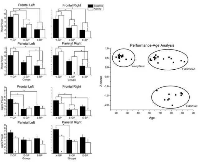
Recently, our group has studied the associations between ageing and cognitive performance and its markers on EEG – Figure 1. It appears that theta power is sensible to age and performance and that alpha power seems to be more related with age than with performance, decreasing in the elders. The study tried to investigate cognitive decline EEG phenotypes by applying to young and elderly subjects the Wisconsin card sorting test (WCST). In this test, cognitive processes like working memory and rule shifting are evaluated. Both theta and alpha activity seem to be lower in elders and poorer performers (Ferreira & Dias, 2012) .
Figure 1. Performance-Age Power Analysis. The Wisconsin Card Sorting Test was applied to young and elder participants, to identify EEG phenotypes of deficits on cognitive performance. Three groups were established: young-good performance (Group 1), elder-young-good performance (Group 2) and elder-bad performance (Group 3). Differences in signal power on theta and alpha rhythms are presented. Adapted from Ferreira & Dias, 2012.
The correlations observed between certain EEG frequency bands and various aspects of cognition led to the conceptualization of neurofeedback (NFB) as an agent of cognitive change. Neurofeedback may be producing effects by enhancing synaptic strength through repeated firing, stimulating neuroplasticity.
8
Neuroplasticity is the capacity of the human brain, even an elder brain, to reorganize neuronal circuits and to produce new synapses throughout life (Eriksson et al., 1998). Plastic changes in the brain are characterized by neural redundancy and plastic remodeling of brain networking and can occur associated with training, practice or learning (Kolb & Whishaw, 1998) whenever task demands diverge from available capacities (Lövdén, Bäckman, Lindenberger, Schaefer, & Schmiedek, 2010).
1.1.3. BRAIN-COMPUTER INTERFACE AND NEUROFEEDBACK
Traditionally BCI applications are based on recordings of brain activity and aim at produce relevant data that assist human functioning. Applications of BCIs are among others ambulatory monitoring, control and communication devices, gaming and entertainment and even safety and security. However noticeable attention has been paid to BCI applications that address the functional recovery of the central nervous system to repair or improve either physical or mental abilities. Although these applications’ context significantly differ from the classic BCIs (Nicolelis, 2001), the NFB-oriented BCI perspective has been suggested as a promising tool to enhance plasticity and able to provide new outcomes for cognitive functional recovery (Daly & Wolpaw, 2008).
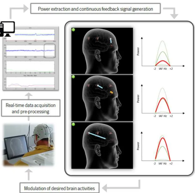
Neurofeedback is a biofeedback technique used to train individuals to control or modify their cortical activity through learned self-regulation (Lecomte, 2011). Within a neurofeedback protocol, individuals receive continuous, real-time visual or auditory feedback over their brain activity patterns so they learn to modulate these signals in the desired direction (Heinrich, Gevensleben, & Strehl, 2007), typically up- or down-regulation of one’s own brain activity.
An EEG neurofeedback protocol involves brain signal acquisition and recording, data pre-processing, feature extraction and generation and presentation of EEG signals to participants, in a way that they can be capable of modulating or altering their brain activity. Linking this feedback signal to a paradigm, for example, a computer game, it is possible for the subject to learn to control his neuronal rhythms. The success during the game dictates the success on rhythms control (Cohen & Evans, 2011).
The mechanism of neurofeedback is considered as an operant conditioning paradigm (Vernon et al., 2003) that might be able to guide neuroplasticity to an induced change in brain activity which may subsequently promote recovery of brain functions.
9 A. Neurofeedback clinical efficacy
Neurofeedback has been used for treating epileptic patients (Sterman & Egner, 2006) and for attention deficit and hyperactivity disorders (ADHD) symptoms relief (Butnik, 2005), but also in the context of addictions (Scott, Kaiser, Othmer, & Sideroff, 2005), depression and anxiety (Hammond, 2005), and chronic pain (Middaugh & Pawlick, 2002).
Without doubt ADHD has been the major focus of relevant clinical investigations being considered nowadays as “efficacious and specific” (Arns, de Ridder, Strehl, Breteler, & Coenen, 2009; Lofthouse, Arnold, & Hurt, 2012). Favoring neurofeedback are its long lasting changes in EEG activity (Gani & Birbaumer, 2008) its effects at the level of network connectivity (Ros et al., 2013); and also its comparability to pharmacologic approaches (Meisel, Servera, Garcia-Banda, Cardo, & Moreno, 2013).
Neurofeedback has been employed more recently to improve the physical or cognitive performance of human beings.
B. Neurofeedback in cognition
Based on the associations between alpha and memory, Bauer tried for the first time to study the effect of 4 NFB training sessions of alpha up-regulation on short-term memory in young adults. The results showed an increase of alpha activity but failed to observe a conclusively effect of NFB on memory performance (Bauer, 1976). Nevertheless, more recent neurofeedback procedures have successfully been used to alter participants' alpha activity and thereby increase cognitive capabilities in mental rotation and memory.
In a study of 18 young adults, Hanslmayr and colleagues investigated raising individual upper-alpha power versus reducing theta power in a single-session NFB training. They observed that 5 participants had learned to increase the power of the upper alpha waves, 6 participants had learned to decrease the power of the theta waves and 4 participants had learned to do both. 3 participants were unsuccessful in controlling either. They showed that upper-alpha stimulation improved spatial rotation accuracy while learned down-regulation of theta was not related to neither the behavioral nor the EEG outcome (Hanslmayr, Sauseng, Doppelmayr, Schabus, & Klimesch, 2005).
After that, other studies have been reporting similar results on alpha power NFB protocols. Zoefel and colleagues explored up-training of individual parietal upper-alpha in five daily sessions versus a non-training control group. Successful training was observed in 11of 14 participants, seen
10
by a linear increase across days both in training and baseline EEG amplitude. They reported a rotation ability and upper-alpha amplitude higher due to neurofeedback training effects (Zoefel, Huster, & Herrmann, 2011). Similarly, in another study five consecutive days of parietal and occipital individual upper-alpha band training were compared to a no-training control, showing a better performance on working-memory in 6 out of 9 participants who were capable of NFB modulation (Escolano, Aguilar, & Minguez, 2011).
Working-memory has also been examined as an outcome measure comparing 16 participants who up-regulate central-midline individual upper-alpha with non-training controls (Nan et al., 2012). A memory enhancement correlated positively with an increase in relative alpha power during training but there was no effects in the post-training resting EEG, which they explained by a much less training density and duration compared to that in Zoefel et al., 2011.
Concerning theta, and based on assumptions that frontal theta has potential as a marker of executive functions and cognitive control, up-training of theta power in frontal-midline regions have been investigated and reported as an effective protocol for cognitive enhancement, specifically in attention and working memory.
Vernon et al. (2003) studied the effects of 8 sessions of NFB training on semantic working-memory and visual attention performance, in a protocol of stimulation of theta waves and inhibition of delta and alpha. The observations did not indicate any changes in EEG activity or in cognitive performance. On the other hand, more recently some studies had explored the trainability of frontal-midline (fm) theta in thirty one participants, who were either up-training theta or in a pseudo feedback protocol. The results showed a significantly enhanced fm-theta power only in the proper neurofeedback training group at the end of the training, as well as during the whole course of sessions (Geppert, Huster, Scharfenort, Mokom, Vosskuhl, et al., 2013; Enriquez-Geppert, Huster, Scharfenort, Mokom, Zimmermann, et al., 2013).
C. Neurofeedback in ageing
Despite several studies showing the effectiveness of neurofeedback on cognitive enhancement in young population, few previous studies have provided evidence showing that NFB can improve cognitive function in the elderly (Angelakis et al., 2007; Becerra et al., 2012; Lecomte, 2011; Wang & Hsieh, 2013).
Similarly to Hanslmayr in 2005, in 2007, Angelakis and colleagues studied six elderly individuals (aged 70–78 years) in contrasting neurofeedback protocols targeting peak alpha
11
frequency (PAF) and alpha amplitude. With thirty one to thirty six sessions they concluded that peak alpha frequency training improved the speed of information processing as well as the resistance to interference; and that training the amplitude of alpha improved memory performance. Additionally, some pronounced effects on the EEG of frontal areas, following peak-alpha training, were described.
The implications of this pilot study were followed by Lecomte that studied the up-regulation of upper-alpha while down-regulating theta in 30 participants aged between 65 and 85 years who were assigned to one of three groups: 4 sessions NFB training protocol, a relaxation control protocol, or a no-intervention protocol. Cognitive improvements occurred in all groups but none of the improvements were associated with neurofeedback learning, although it was achieved by over half of the participants. The limited number of sessions of the study were surely too few with elderly participants (Gruzelier, 2013).
Regarding theta, Becerra and colleagues tried to reduce theta in fourteen participants ageing 60–85 years, who had evidence of abnormally high theta. Thirty 30-min sessions were given over 10–12 weeks, with theta power being successfully reduced, resulting in an improvement in EEG and behavioral measures. However, the control group also showed improved EEG values and memory performance.
In contrast, Wang and Hsieh investigated frontal-midline theta up-training in elders (61–72 years) compared with young students. 8 to 12 sessions over four weeks of theta training resulted in improved attention and working memory performance in ageing adults, accompanied by an increase in theta activity in the resting state. In addition, they reported that younger participants also benefited from the protocol in terms of improving their executive function.
Protocols on gamma and beta neurofeedback have also been reporting improved visual processing (Salari, Büchel, & Rose, 2012), enhanced managing of episodic retrieval (Keizer, Verment, & Hommel, 2010) and improved memory and intelligence in elders (Staufenbiel, Brouwer, Keizer, & van Wouwe, 2013), however in this case without a clear relation with improvements.
To complete, a number of exploratory attempts have been made for preserving cognitive functions in the healthy elderly, with the clear conclusion that age does not exclude neurofeedback ability to regulate brain activity.
12
1.1.4. NEUROREHABILITATION
Neurorehabilitation and mental activity foster the putative promise of enhancing or rehabilitating behavior and brain function (Rabipour & Raz, 2012). Brain training programs are used nowadays to decrease age effects on cognition, by increasing an individual’s baseline level so that age-related declines begin to affect daily-life activities later in life ((Hultsch, 1998; Wilson et al., 2002). Such training can produce changes measured at the behavioral as well as at the neuroanatomical and functional levels. There is evidence on brain training effectiveness and durability (Lustig, Shah, Seidler, & Reuter-Lorenz, 2009) and it can be used to improve cognitive function when exercising attention (Rueda, Rothbart, McCandliss, Saccomanno, & Posner, 2005), working-memory (Klingberg, 2010; Klingberg et al., 2005), or executive and associative functions. In addition, extensive training may modify neural structures and functions by increasing gray matter volumes in specific brain regions (Draganski et al., 2004; Scholz, Klein, Behrens, & Johansen-Berg, 2009).
In healthy elderly populations, brain training delay the natural progression of cognitive decline (Buiza et al., 2009; Park, Kwon, Seo, Lim, & Song, 2009). Indeed, reasoning, memory and speed of process can be trained and a 10 1-h sessions seem to be effective in improving performance on elders in the specific trained abilities (Ball et al., 2002; Jobe et al., 2001). More specifically, training appears to improve memory in healthy ageing individuals (Dunlosky, Kubat-Silman, & Hertzog, 2003; Rebok, Carlson, & Langbaum, 2007) and even in individuals with mild cognitive impairment (Belleville et al., 2011) or mild-to-moderate Alzheimer’s (Zanetti et al., 1997).
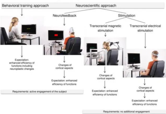
Even if brain training may improve specific cognitive abilities in health ageing, this type of intervention does not appear to improve overall cognitive function and also showed poor results on transferability of tasks for real-life activities (Lustig et al., 2009). Therefore, combination with other approaches are suggested in order to reach higher levels of rehabilitation (Lustig et al., 2009). As we have seen there are now a set of approaches used to alter cognitive capacities, summarized in Figure 2 – one is based on behavioral training procedures and the others on the up- or down-modulation of neural mechanisms by neurofeedback or neurostimulation (directly stimulation of specific brain regions via electrodes that are mounted on the scalp or via a magnetic field delivered by a coil) (Enriquez-Geppert, Huster, & Herrmann, 2013). It has been shown that both neurostimulation and neurofeedback affect the amplitude of cognitive related EEG oscillations (Demos, 2005; Egner, Strawson, & Gruzelier, 2002; Enriquez-Geppert, Huster, Scharfenort, Mokom, Zimmermann, et al., 2013; Hanslmayr et al., 2005; Zaehle, Rach, & Herrmann, 2010;
13
Zoefel et al., 2011). The combination of approaches may result in the cumulative effects of both, leading to higher benefits. The simultaneous cognitive training and neurostimulation protocol has already started to be investigated in improving numerical and response inhibition abilities (Cohen Kadosh, Soskic, Iuculano, Kanai, & Walsh, 2010; Ditye, Jacobson, Walsh, & Lavidor, 2012) with potential greater effects.
Figure 2. Training approaches for cognitive rehabilitation. Behavioral/neurocognitive training, neurofeedback and neurostimulation can be used nowadays to enhance cognitive capacities (Enriquez-Geppert, Huster, & Herrmann, 2013).
1.2. RESEARCH OBJECTIVES
Considering there are few studies in the literature investigating the combined effects on cognitive enhancement in the elderly, and none of them involve neurofeedback technique, the aim of the present study was to examine the effects of a protocol with NFB training interleaved with classic neurocognitive tasks. The central hypothesis is that the combined approach might have a greater impact on cognitive performance, in comparison with a neurofeedback training alone approach.
14
First, based on evidences that both frontal-midline theta and alpha activity are potential effective parameters for cognitive enhancing in elderly population, and based on proven effects of working-memory training in delaying cognitive ageing decline, a combined protocol that comprised both approaches was developed (Portugal, Ferreira, Reis, Pinho, & Dias, 2013) – Annexes 1.
Then, a twelve-days cognitive intervention protocol for memory training was design, combining neurofeedback training (Alpha and Theta power enhancement) with common neurocognitive tasks (working-memory), and validated on 10 Portuguese participants (> 55 years-old), whom were submitted to one of the two cognitive intervention approaches:
o Experimental Group 1: Neurofeedback (NF) training single-methodology;
o Experimental Group 2: Neurofeedback training and Neurocognitive training (NF+NC) combined-methodology.
Considering that this work is included in a broader project which will also recruit subjects for other experimental groups aiming at controlling both neurofeedback and neurocognitive interventions, the scientific goals of this report are to:
Identify NF+NC intervention effects on EEG power spectrum, working-memory and cognitive flexibility performances, as a potentiation technique for NF.
Assess dynamic changes on EEG power spectrum and behavioral measures across training sessions for both experimental groups.
Ultimately we wanted to evaluate, and compare, the alterations induced by both rehabilitation intervention protocols on subjects’ cognitive performances. In addition, this study may support investigations concerning theta and alpha enhancement as a promising parameter in neurofeedback for cognitive enhancement.
15
17 2.1. PARTICIPANTS CHARACTERIZATION
For this project 10 participants (4 males and 6 females) were recruited from a Health Care Centre from Braga and are community-dwelling individuals living in the Minho Region of Portugal. Only participants without any diagnosed dementia, cerebrovascular or neurological pathology were invited to the study. The participants did not present a high academic level (mean years of schooling: 5,4 years ± 1,68 years) and were unemployed or retired. At the beginning of the experiment they answered some questions about their educational qualifications, current or previous occupation and prescribed medication.
The cohort was established in accordance with the principles expressed in the Declaration of Helsinki and the work approved by the national ethical committee (Comissão Nacional de Protecção de Dados) and by local ethics review boards.
All participants were right-handed and presented normal or corrected vision. The Edinburgh Handedness Test was used to determine if the subjects were either right-handed or left-handed.
All the participants joined voluntarily the study, after they had the experimental nature and protocol procedure explained to them. At the time, all the participants sign a voluntarily informed consent for the use of the collected data.
The participants’ cognitive and mood profile was assessed previously to the training protocol by a team of trained psychologists and MD students. Briefly, cognitive state was evaluated using a battery of neurocognitive tests to measure general cognition, attention, learning, short-term memory, verbal memory, cognitive flexibility, verbal fluency and processing speed. Additionally, psychological tests were applied for assessing mood, anxiety and stress profile, personality, functional ability, quality of life and memory perception. Most notably, Geriatric Depression Scale (GDS) and the Mini-Mental State Examination (MMSE) were comprised in this battery. Regarding GDS, participants mean score was 14,8 ± 7,4, which characterize this sample as "mildly depressed" (Yesavage et al., 1983). In the MMSE the participants mean was 19,4 ± 2,32.
Participants within each gender were randomly assigned to either the Neurofeedback (NF) training-group (N = 5, mean age of 61,2 ± 4 years, range: 55– 66 years) or the Neurofeedback and Neurocognitive (NF+NC) training-group (N = 5, mean age of 61,8 ± 5,8 years, range: 55– 67 years).
18
At the end of the study, all participants completed a questionnaire about their general opinion of the study.
2.2. ELECTROENCEPHALOGRAM ACQUISITION
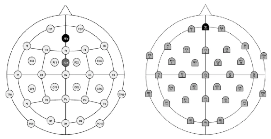
Two systems were used for the electroencephalogram signal acquisition. They are both based on the international 10-20 system (32 channels standard electrode layout – Figure 3, with ground and reference electrode). EEG signals were acquired with either the QuickAmp®, Brain Products, GmbH or the ActiCHamp®, Brain Products, GmbH.
Figure 3: Electrodes layout for the EEG acquisition. Left: electrodes layout of the QuickAmp equipment; Right: electrodes layout of the ActiChamp equipment.
The whole system was constituted by: Ag/AgCl active electrodes, a cap for the placement of the electrodes – actiCAP or EASYCAP (Brain Products, GmbH) – electrolyte gel (to decrease the contact impedance between electrodes and the scalp) and straps to keep the cap in place. Ground was located at forehead and reference was FCz channel when using QuickAmp equipment and Cz when using the ActiCHamp equipment.
For each participant, the same equipment was used for all EEG data acquisitions. During recordings, all participants were instructed to not make any movements beyond the required ones and, when necessary, to answer always with the same hand.
19 2.3. REHABILITATIVE PROTOCOL
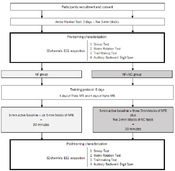
All the participants followed a protocol of 12 days accordingly to the diagram in Figure 4.
Figure 4. Schematic diagram of the design and procedure in the present study protocol.
During the intervention, participants were sited in an illuminated and acclimatized room, distancing 50-80 cm from a 17 inch computer screen, with touch technology. All the moments of the study were conducted in ‘Centro Clínico Académico’ in Hospital de Braga.
2.3.1. ARROW FLANKER TEST
In the first 2 days of the protocol all participants were submitted to an attention test: the Arrow Flanker Test during approximately 15 minutes (5 blocks of 85 trials). This step aimed at guarantee
20
that all participants presented minimum attention level in order to be able to perform the subsequent requested tasks of the intervention.
The Arrow Flanker Test was implemented in the framework used during the training protocol, the BCI++ (details below). It was adapted from the Flanker Task in the PEBL (Psychology Experiment Building Language) Test Battery and it was used for attention domain assessment. In each trial of the test, the participant is presented with a set of arrows. He has to pay attention to the central target arrow and give a directional response – left or right – within a timeout of 1500 milliseconds. The target arrow can be flanked by stimuli in the same direction (congruent stimuli), in the opposite direction (incongruent stimuli), or not be flanked (neutral stimuli) (Figure 5). A fixation cross is presented before every trial. Measures of mean accuracy, mean response time (RT) and conflict (difference in response time between incongruent and congruent stimuli) are reported at the end of the task. Additionally, measures of accuracy and response time for each stimuli condition can be assessed.
Figure 5. Representative image of the Arrow Flanker Test implemented in the protocol. A set of arrows is presented and the participant has to give a directional response to the central arrow (target), in a determined set of time (1500 ms). The type of stiimulus that can flank the target arrow are also represented at the bottom of figure.
Direction responses were given on a modified keyboard, either by pressing a yellow key on the left of the keyboard or a blue one on the right, whether the answer was left or right direction, respectively.
21 2.3.2. PRE AND POST-TRAINING CHARACTERIZATION
At the third and at the last day, EEG signals from 32-channel were acquired from all the participants while performing a battery of cognitive tests. This battery was applied to evaluate mainly working-memory and cognitive flexibility. It comprises the Stroop Test, the Matrix Rotation Test, the Trail-Making Test and the Auditory Backward Digit Span Test. The EEG data was used for further power analysis.
A. EEG recordings
During the characterization moments, OpenVibe was used for acquiring, plotting and recording the EEG signal (Renard et al., 2010). Also, it was used to implement the synchronization between the EEG data and the software of visual stimulation (PEBL) with markers for offline analysis. The four cognitive tests were preceded by a one-minute length eyes-open baseline, where the participants were instructed to minimize blinking and to stare at the center of the computer screen in grey, the more relaxed as possible.
B. Cognitive battery
PEBL (Psychology Experiment Building Language) was used for stimulus presentation. PEBL is a psychology software for designing and running computer-based tests and experiments, which has ready-made paradigms for assessing cognitive and psychological domains (Mueller, 2010). The four tests used in the battery were already implemented on PEBL Test Battery, and were adapted for the cognitive characterization.
1) Stroop Test (Stroop): it is a popular neuropsychological test with different test variants (Lezak, 2004) considered to measure selective attention, cognitive flexibility and processing speed (Lezak, 2004; Strauss, Sherman, & Spreen, 2006). The version applied here consisted of two blocks. On the first one, the participant read random color names (red, green, blue, yellow) printed in colored ink (red, green, blue, yellow) or black ink, ignoring the color of the print – in equal proportions the print color could correspond to the color name (Condition 1), the print color could not match the color name (Condition 2), or the print color could be black (Condition 3). In the second block, the participant had to name the ink color in which the color names were printed
22
and to ignore their verbal content – similarly to the previous block, stimuli color and reading name could match, stimuli color could not match the reading name or stimuli could consist of groups of letters "xxxx" in a given color. 60 stimuli were presented in each block of the test. Every stimulus must be paired "correctly" for the test to proceed, which means participants may make multiple errors before complete a trial. Four labels with the four color names (red, green, blue, yellow) printed in the matching color ink were visible on the bottom of the screen for responses.
2) Matrix Rotation Test: it is a test that evaluates spatial working-memory and mental rotation skills, described in the Unified Tri-Service Cognitive Performance Assessment Battery (UTC-PAB) (Englund et al., 1987). A set of 4 to 4 cell matrices were presented to the participant, each with 4 highlighted yellow cells. The participant was required to compare successive matrices and determine if they had the same pattern, but turned 90 degrees to the left or right, or if they were different matrices, regarding the immediately preceding matrix. Participants decided on the time they needed to memorize the study matrix. There were 10 matrices per condition (same and different). Two labels of response (designated as “same” or “different”) were visible on the bottom of the screen.
3) Trail-making Test (TMT): it is used to measure attention, speed, and cognitive flexibility. There were two parts of the test, both consisting of 26 circles spread over the screen. In part 1, the circles were numbered 1 to 26 consecutively, and the participant had to connect the numbers in ascending order. In part 2, the circles included both numbers (1 – 13) and letters (A – N), and the participant had to connect the circles in an ascending pattern, but alternating between numbers and letters (i.e., 1-A-2-B-3-C, etc.). The trial finished when all the circles had been successfully clicked in the correct order. Two trials were performed for each part of the test.
4) Auditory Backward Digit Span (Backward Digit Span): it is a test for assessing working-memory and consists of a series of trials presenting random digits at the rate of one digit per second. In each trial, the participant had to listen the digits sequence. Then, he was instructed to enter the digits, in the exact inverse order, in a digital keyboard on the screen. The test started with sequence length 3 and had 2 trials at each length. The length increased until the participant failed twice to recollect every digit.
23
All the responses were given using the computer touch screen. The tests were preceded by practicing. For all the participants, tests were applied in the same order.
2.3.3. TRAINING SESSIONS
Depending on the cognitive intervention group, participants were randomly allocated to the neurofeedback training protocol or to the combined neurofeedback and neurocognitive training protocol.
During the 8 sessions of training, participants were submitted to a 30-minute intervention protocol each day, since this training intensity seems to produce effects on behavior and electroencephalogram measures (Ball et al., 2002; Keizer et al., 2010; Ros et al., 2013), according to their experimental group:
• NF training-group – participants performed only neurofeedback training (NFB) – modulation of theta and alpha rhythm. The 30-minute training period consisted of six 5-minutes blocks of a neurofeedback task, preceded by a 3-min baseline measurement.
• NF+NC training-group – participants performed training of neurofeedback (NFB) – modulation of theta and alpha rhythm – and a set of neurocognitive tasks: Corsi Block-Tapping Test and n-Back Test (see details below). The 30-minutes training comprised three 5-minutes blocks of a neurofeedback task, preceded by a min baseline measurement, plus five 3-minutes blocks of neurocognitive tasks.
The participants were asked about motivation and interest for attending the sessions prior to the beginning of each session; and were asked about general concentration and train difficulty at the end of it. Exceptional stress or tiredness was documented for posterior analysis.
The 8-days protocol of NFB enhancement was conducted separately for alpha and theta rhythms, each with a duration of 4 days. The initial frequency to be trained was randomly selected for each participant.
For this part of the intervention protocol it was used a software dedicated to the development and fast prototyping of Brain-Computer Interface systems and pc-driven protocols, the BCI++ platform (Maggi, Parini, Perego, Andreoni, & Milano, 2008). BCI++ is divided in two main interconnected modules: one module oriented for the acquisition and real-time processing of the EEG signals, and a second module responsible for providing visual and auditory stimuli to the
24
participant. Both modules have customized blocks for the development and implementation of C++ or Matlab based algorithms and paradigms.
During the training protocol, EEG signals were acquired continuously, sampled at 500 Hz, from the Fp1, Fp2, Fz and Pz channels. Alpha or theta feedback was calculated from the Fz channel and Fp1 and Fp2 channels were used for detection of ocular movements.
A. Neurofeedback training procedure
For NFB training, EEG data was processed in real-time by a custom algorithm developed in C++ and Matlab® (Matlab® Engine).
Every 200 ms, data was filtered and baseline corrected, and Fast Fourier-Transforms (FFT) were computed, using a spectrum estimation function of the Chronux matlab toolbox (Andrews et al., 2008), for calculation of theta or alpha power. The spectrum measurement was based on 1 s data windows (containing the last 200 ms of data and 800 ms of outdated data), which provides the participants with a smooth appearance of the visual feedback and avoids large shifts in feedback.
At first, a baseline measurement of 3 min was recorded, during which the measurement of power spectrum was recorded. At the end, the amplitude of theta and alpha rhythms was calculated in all artifact-free data windows. An average of all power spectrum estimations was used for detection of the individual alpha peak frequency (IAPF). Then, theta and alpha frequency bands were defined from 4 Hz to IAPF– 3 Hz and from IAPF– 2 Hz to IAPF + 2 Hz, respectively. The baseline power, calculated as the mean power of all segments, was then used as a participant-specific reference for the feedback training.
During the training blocks, feedback was given as the ratio of the power amplitude measured at Fz channel to the baseline power amplitudes, updated for each training session. Feedback was updated 5 times per second.
The neurofeedback training task, where visual feedback was provided to the participant about his own brain rhythms, was design with a therapeutic approach in order to keep participants motivated and stimulate them to improve. So, a virtual scenario is presented to the participant, as seen in Figure 6, with a human head, three neuronal cells and a fire. During NFB blocks, the feedback was given by means of a blue bar (symbolizing water) that comes out of the first neuronal cell and must reach the fire. The length of the bar indicated the power amplitude in relation to the baseline measurements.
25
Depending on the current power amplitude, the bar length was displayed as longer, closer to the fire, whenever the power amplitude was increased and the bar turned shorter, further from the fire when the power amplitude was attenuated, in comparison to the baseline power measurement. In the game paradigm, the three neuronal cells indicated the three levels of the game. The length scale covered 95% of the amplitude range measured during the baseline. At the first level, values below the 2,5 percentile were displayed with no bar and full-bright fire, while values above the 55 percentile were displayed with a full length bar and total extinction of the fire. A level update
Figure 6. Representative images of the Neurofeedback task implemented in the protocol. An online power analysis of the EEG is computed for Fz, updated every 200 ms. Results of this analysis are visually presented as a blue bar (symbolizing water) whose length, depending on the current power amplitude, was displayed as longer, closer to the fire, whenever the power amplitude was increased and the bar turned shorter, further from the fire when the power amplitude was attenuated, in comparison to the individual baseline power measurement. The three levels of the game are displayed. Red lines represent current power amplitude and green lines represent the minimum, medium and maximum reference values – 2,5 percentil, 55 percentil and 97,5 percentil.
26
happened when the mean value for the last 10 seconds was above the 40 percentile. At the second level, values below the 2,5 percentile were again displayed with no bar and full-bright fire, while values above the 75 percentile were displayed with a full length bar and total extinction of the fire. At this moment, a level update could happen when the mean value for the last 10 seconds was above the 60 percentile or below the 40 percentile. If the mean value was below 40 percentil, the participant went back to the first level scenario. If the mean value was above the 60 percentil, the participant went to the third level. At this level, values below the 2,5 percentile were displayed with no bar and full-bright fire, while values above the 97,5 percentile were displayed with a full length bar and total extinction of the fire.. The mean value for level adaptation was monitored after 12 seconds in the same scenario level, and afterwards was updated with the same frequency as the feedback.
To assure the comparability of the baseline measurements and the NFB blocks, the virtual scenario of the first level was presented during recording of baseline, with the blue bar randomly changing its length. In order to maintain the subject cognitively active, they were asked to count the number of times the bar approached the fire (Enriquez-Geppert, Huster, Scharfenort, Mokom, Zimmermann, et al., 2013; Zoefel et al., 2011).
Because artifact contaminated feedback signals may influence the neurofeedback learning outcome and since their power unfolds in frequency bands often used for neurofeedback training (Huster, Mokom, Enriquez-Geppert, & Herrmann, 2013), as with Theta, we discard from feedback data windows showing contamination of ocular artifacts, like eye blinks, detected whenever the signal amplitude of the Fp1 or Fp2–Fp1 exceeded an adjustable threshold. In this case, the feedback was suppressed and the length of the bar did not change.
A set of strategies which earlier studies reported as successful were given to the participants. In the first sessions they could test these strategies and find the most effective strategy. The strategies listed were separated in positive (like love, family or friendship), negative (death, diseases or conflicts) or neutral (like visual attention to the bar, mental operations and calculus or breathing). Participants were informed that they could use these strategies favoring an increase of the bar and thus the fire extinction. Also, they were instructed to be concentrated and keep pursuing this goal as much as possible. At the end of each block of training, the strategy used and its effects were reported. Additionally, average measures of theta and alpha power in each block were kept for posterior analyses.
27 B. Neurocognitive tasks
For the neurocognitive practice, two working-memory neurocognitive tests were implemented: the Corsi Block-Tapping Test, adapted from PEBL Test Battery, and the n-Back Test. Responses to the tasks were given using the touch screen.
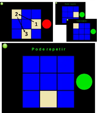
1) Corsi Block-Tapping Test (forward and backward): is a classic test to assess visual-spatial short-term working memory (Corsi, 1973). The player has to track a sequence of up to nine identical spatially separated blocks being highlighted and reproduce it, either in forward or backward order - Figure 7. The sequence starts with length two (two blocks highlighted), but when the player repeats correctly the sequence, the length is increased in the next trial. On the other hand when the participant repeats wrongly, the length decreases. The task finishes when the participant performs 15 trials. At the end, measures of block memory Span, number of corrected trials, mean length of the sequence and a combined score of memory block span and corrected trials were reported.
Figure 7. Representative image of the Corsi Block-Tapping Task implemented in the protocol. There are 9 blue blocks and a sequence of them is presented to the player, who has to reproduce it. When the player reproduces correctly, the number of the next sequence is increased (up to nine), and when it reproduces it incorrectly the number is decreased.
28
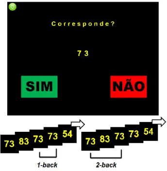
2) n-Back Test (1-back and 2-back): is a commonly accepted task to measure cognitive performance in working memory domain. The participant is presented successively with digits and has to indicate whether or not the current digit matches the one n instances before (1-back – the previous digit; 2-back – the digit that appeared before the previous digit) – Figure 8. In the 1-back task, which was originally introduced by Kirchner (1958), the participant only has to evaluate if the current digit is the same as the previous one. In the 2-back task, a variation proposed by Jaeggi et al. (2003), the participant has to remember and compare the current digit to the digit that appeared prior to the previous one (Jaeggi et al., 2003; Kirchner, 1958). Measures of accuracy and reaction time, for every type of stimuli (one-back, two-back, or random) were reported at the end. Each block had 65 stimuli.
Figure 8. Representative image of the 2-Back Task implemented in the protocol. A digit is consecutively appearing in the screen, and the player has to warn if it matches the digit presented two times earlier in the game.
2.4. OFFLINE SIGNAL PROCESSING
Analyzer 2®, Brain Products, GmbH and Matlab, The MathWorks, Natrick, USA were used for offline EEG processing and power analysis.
At first, all raw signals were filtered with a notch filter to reject the 50 Hz band, and a bandpass filter from 0,3 to 100 Hz.
29
2.4.1. 32-CHANNELS EEGCHARACTERIZATION
An algorithm for correction of ocular artifacts based on independent component analysis (ICA) (Hyvärinen & Oja, 2000) was applied, and segments with motor artifacts were removed by rejecting segments of the signal based on threshold criteria of signal amplitude , difference between the maximum and minimum in a time envelope and signal gradient. The adequate thresholds were adjusted for every session and every task.
In order to increase the independence of the signals on neighbouring electrode locations, the current source density (CSD) method was applied on the data. In this way, the channels generated did not have any specifications with respect to the channel reference (Makeig, Jung, Bell, Ghahremani, & Sejnowski, 1997).
From the 32 electrodes recorded, only Fz, F4, F3, FC2, FC1, FC6 and FC5 were used to power spectrum estimation. The last 6 channels were selected to observe hemispheric lateralization.
At the end, EEG signals were segmented. The signal of every cognitive test was separated in Baseline (EEG acquired during resting state) and Activity (EEG acquired during task performance), and these were divided in segments of 1 s length with 0,8 s overlap. A baseline correction was applied in each segment.
2.4.2. EEGRECORDINGS OF TRAINING SESSIONS
Segments with ocular and motor artifacts were removed by rejecting segments of the signal based on threshold criteria of signal amplitude , difference between the maximum and minimum in a time envelope and signal gradient. The adequate thresholds were adjusted for every session and block. Only the Fz channel were used to calculate the power spectrum.
For the neurocognitive blocks, the EEG data was separated in Baseline and Activity.
Then, all EEG signals were divided in segments of 1 s length with 0,8 s overlap. A baseline correction was applied in each segment.
30
2.4.3. POWER ANALYSIS
The signal power was calculated through the Fourier transform (Sanei & Chambers, 2007) in the Analyzer software. The average of the calculus of the Fourier transform in all the segments was exported to Matlab.
In Matlab, the signal power was analysed for each frequency band: theta (4-8 Hz) and alpha (8-13 Hz). The limits for each frequency bands were adjusted accordingly to the individual alpha peak frequency (IAPF). The IAPF was calculated as the frequency with the highest peak in the alpha band range (8-13 Hz). For each of the characterization moments, the IAPF measurement was based on the average of the four baselines performed. In the case of the training sessions, IAPF detection was based on the start baseline of each day. Theta and alpha were defined as the frequency bands from 4 Hz to IAPF – 3 Hz and IAPF – 2 Hz to IAPF + 2 Hz, respectively. Then, power for each individual frequency band was extracted.
2.5. STATISTICAL ANALYSES
Considering the low number of available subjects and not assuming the normal distribution of the population, non-parametric tests were used for statistical analyses. For comparisons between groups and conditions the Kruskal-Wallis ANOVA was used in order to assess if there were any significant differences (considered for p-values below 0.05). For testing positive or negative effects of the intervention (testing if the median is greater or lesser than 0) the one-sample Wilcoxon signed-rank test (significance was considered for p-values below 0.05) was used. Spearman´s Rank Correlation Coefficient Test was performed to observe statistical dependence between EEG and behavioural measures. The statistical analyses were performed using OriginLab.
2.5.1. EVOLUTION OF ALPHA AND THETA POWER ACROSS SESSIONS
Dynamical changes in power due to NFB training were identified analyzing variations in power measurements throughout the 8 days. Thus, for each participant, the power amplitudes in the theta and alpha band were extracted and averaged across blocks, for each training session. Power was extracted for baseline EEG as well.
31
In order to analyze the training effects on the EEG amplitudes, it was examined separately the four days corresponding to theta NFB and the four days corresponding to alpha NFB. To investigate the relationship between the amplitudes in the above mentioned EEG bands and session number a regression line was fitted for each subject.
As previous studies have reported, a subset of subjects does not respond to NFB training (Enriquez-Geppert, Huster, Scharfenort, Mokom, Zimmermann, et al., 2013; Hanslmayr et al., 2005; Zoefel et al., 2011). Thus, statistics was performed only considering responders. Training results were inspected and a participant was classified as responder if the gradient of training was positive, and as non-responders if this gradient was null or negative.
Gradients were used to test differences between groups (NF vs. NF+NC). Also, differences between starting the training with alpha NFB and starting with theta NFB were examined.
With regard to the mental strategy analysis, strategies employed in each block were collected and the most frequently used were reported for each participant.
In the same way as with the power in NFB, performance in the neurocognitive tasks were assessed. Measures of performance along the 8 days were extracted and a regression line was fitted for each participant of the NF+NC group. Gradients were used to investigate if there was an improvement over the days in the cognitive performance measures.
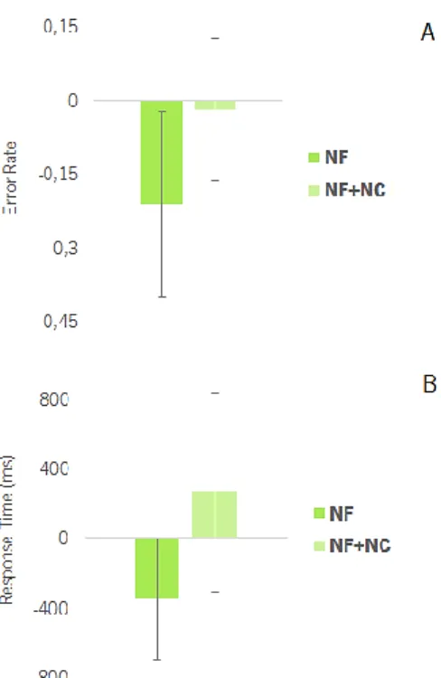
2.5.2. TRAINING EFFECTS ON BEHAVIOR AND ALPHA THETA POWER
The cognitive battery of four tests and the EEG recordings while performing them were used for assessment of training effects.
For each participant, Fz-power of theta and alpha frequencies was extracted for the first and final characterization moments. Baseline and activity was discriminated for each of the four tests. For the cognitive evaluation, different measures were collected from the first and last tests applications. In the case of Stroop, the measures adopted were the error rate and the mean response time for condition 2 of the second block. A measure of interference between conditions 1 and 2 was also taken into account. For the Matrix Rotation test, the mean accuracy, study time and response time were extracted. Regarding the TMT, measures of accuracy and mean response time concerning only the part 2 of the test were registered. In the Digit Span, a combined measure was calculated based on the memory span – the last digit that was successfully recollected – and the number of correct trials.
32
At the end, differences between the pre-training moment and the post-training moment were calculated in order to evaluate and compare the alterations induced by both intervention protocols on participants’ behavioral performance and neurophysiologic state.
Outlier observations were detected if a value was two standard deviations away from the mean.
2.5.3. BRAIN LATERALIZATION
For each participant, power of theta and alpha bands was extracted in F4, F3, FC2, FC1, FC6 and FC5 channels. To observe hemispheric lateralization power ratios of F4 to F3, FC2 to FC1, and FC6 to FC5 were calculated both in the pre- and post-characterization, discriminating baseline and activity for each of the four tests. Differences between the pre-training moment and the post-training moment were also calculated.
33
35
This section presents the results obtained after data analysis and their statistical testing. We begin by verifying that participants in both experimental groups (NF and NF+NC) had no significant differences in age, education and score obtained in both the Geriatric Depression Scale and the Mini Mental State Examination.
3.1. PERFORMANCE IN THE ARROW FLANKER TEST
During the first two days of the intervention protocol participants had to perform 5 3-min blocks of the Arrow Flanker Test, which is an attentional test. Based on researcher observations, it was clear that all participants understood the instructions that were given to them, they answered appropriately and kept engaged in the task. Most importantly, individual reports of the task were analyzed and all participant had very few number of errors in each block (maximum 6 errors in 85 stimulus).
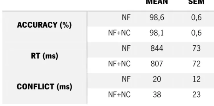
Accuracy, response time (RT), and conflict (difference in time response between incongruent and congruent stimulus) are presented for both groups in Table 1. There were no significant differences between the groups for any of the measures, which suggests there were no performance differences between the NF and the NF+NC group in the Arrow Flanker Test.
Table 1. Performance measures in the Arrow Flanker Test. Mean and standard error of the mean (SEM) are
discriminated for accuracy, response time and conflict (difference between incongruent and congruent stimulus).
MEAN SEM ACCURACY (%) NF 98,6 0,6 NF+NC 98,1 0,6 RT (ms) NF 844 73 NF+NC 807 72 CONFLICT (ms) NF 20 12 NF+NC 38 23
Additionally, all participants presented a mean accuracy in the Arrow Flanker Test greater than 97,5 % (Z = 2,344 and p-value = 0,0068). Additionally, mean response time was lower than 1000 milliseconds (Z = -2,548 and p-value = 0,0029).