Dissertação de Doutoramento
Characterization of Molybdenum and
Tungsten Formate Dehydrogenases from
Sulfate Reducing Bacteria.
Cristiano de Sousa Mota
Cristiano de Sousa Mota
Characterization of Molybdenum and
Tungsten Formate Dehydrogenases from
Sulfate Reducing Bacteria.
Lisboa, 2011
- Nº DE ARQUIVO
AKNOWLEDGEMENTS
First of all, I would like to express my gratitude to my advisor Prof. Isabel Moura and to Prof. José Moura, for accepting me to carry out my Ph.D. work in their research group, for all the trust in my work and ability to get the results and for all the help in key questions of my Ph.D.
Maria Gabriela Rivas, thank you for all the help, all the teaching, all the patience, all the persistence since the very first beginning. Thanks to you, I’m today a best student and a best scientist (I want to think that!).
Pablo Javier Gonzalez, for all the useful discussions, for all help in the headaches with EPR theory and practice, for all the information about Gaussian v03 and ORCA 2.8 and, for all the fruitfulness brainstorms with the software PES v09 and v10, thank you.
To the latter two, I would like to express my recognition for all the work and contribution in the achievement of this thesis.
Carlos Brondino, Nuno Cerqueira, Alain Dolla, Özlen Ferruh Erdem, Marie-Claire Durand, Edward Reijerse and Wolfgang Lubitz. The collaborators that contributed with experimental data, data discussion and, help to write and discuss manuscripts. Thanks for your useful knowledge.
To Bioin and Bioprot groups thanks for your friendship, you teach me a lot about research and lab work, it was a pleasure work with you every single day.
Thanks to Fundação para a Ciência e Tecnologia for funding (Grant SFRH/BD/32478/2006).
To my family, the most important people in my life, thank you for all the support in good and bad moments.
RESUMO
As enzimas de Mo e W têm funções vitais na catálise de passos fundamentais do metabolismo do carbono, azoto e enxofre. Estes metais em condições fisiológicas são activos redox, variando entre os estados de oxidação IV, V e VI.
Esta tese reporta sobre desidrogenases de formato (Fdhs) que contêm Mo e W, estas enzimas pertencem à família das DMSO reductases das enzimas mononucleares de Mo e W. No centro activo destas enzimas o ião metálico é coordenado por quatro átomos de enxofre pertencentes a duas moléculas de piranopterina, um selénio de uma seleno-cisteína (SeCys) e um enxofre inorgânico. Estas enzimas contêm também nas suas estruturas outros cofactores redox como centros [4Fe-4S] e em alguns casos, grupos hemos.
No Capítulo I é feita uma introdução geral ao âmbito das enzimas de Mo e W, enquanto uma visão mais aprofundada no conhecimento actual das Fdhs de Mo e W é apresentada no Capítulo II.
O Capítulo III descreve os estudos aplicados para compreender os efeitos da suplementação com Mo e W, no meio de cultura, nos níveis de expressão e propriedades bioquímicas das Fdhs isoladas de D. alaskensis. Duas Fdhs diferentes são expressas,
uma que pode conter no centro activo tanto Mo como W (Mo/W-Fdh), e outra que apenas pode incorporar W (W-Fdh). Ambas as enzimas foram purificadas de células cultivadas em meio suplementado com Mo, enquanto em meio suplementado com alta concentração de W apenas a W-Fdh pode ser isolada. Os genes que codificam para ambas as enzimas foram identificados e os níveis de expressão nas condições optimizadas foram avaliados por RT-PCR. Os resultados mostraram que os genes que codificam a Mo/W-Fdh (Mo/W-fdh) foram fortemente reprimidos na presença de W e
ligeiramente sobrexpressos na presença de Mo. Contudo, a expressão dos genes W-fdh
são menos susceptíveis tanto à suplementação com Mo como com W. Foi ainda observado que tanto a presença de Mo como de W induz a repressão dos genes envolvidos no transporte de Mo e W. As características das Fdhs e a especificidade para incorporar tanto Mo como W são discutidas neste capítulo.
presente no centro activo. O mecanismo catalítico foi simulado usando ferramentas de DFT e correlacionado com as propriedades cinéticas experimentais tanto de Fdhs isoladas de três espécies de Desulfovibrio como de outros exemplos reportados na
literatura. Os nossos estudos indicaram que a substituição do Mo por W no centro activo não afectava a velocidade de reacção. A função de alguns aminoácidos conservados perto do centro metálico é também discutida.
O Capítulo V foca os tipos de inibição que nitrato, azida e cianeto produzem nas Fdhs. Foram determinadas as constantes cinéticas de três Fdhs similares, de espécies
Desulfovibrio, na presença destes inibidores. O nitrato inibe a oxidação de formato de
um modo competitivo enquanto a azida é um forte inibidor e exibe um tipo de inibição misto. O cianeto inibe a Fdh de Mo de D. desulfuricans, demonstrando um tipo de
inibição misto, mas não tem qualquer efeito nas Fdhs de W isoladas de D. gigas e D.
alaskensis. Esta diferença observada entre as enzimas de Mo e W é discutida baseada
nas propriedades do metal do centro activo, nos aminoácidos na vizinhança do centro e nas subunidades que compõem as enzimas.
No Capítulo VI foram aplicadas ferramentas de DFT de forma a tentar entender os dados espectroscópicos disponíveis na literatura. Foram propostos modelos estruturais do centro de Mo com o objectivo de clarificar as propriedades espectroscópicas dos sinais de EPR de Mo(V) observados na Fdh-H de E. coli e Fdh de D. desulfuricans. A
importância destes modelos do centro activo no mecanismo catalítico das Fdhs é também proposto.
ABSTRACT
Mo and W-containing enzymes have crucial physiological roles in catalysis of key steps in the carbon, nitrogen and sulfur metabolism. These metals are redox-active under physiological conditions ranging between oxidation states VI, V and IV.
This thesis deals with Mo and W containing formate dehydrogenases (Fdhs), which belong to the DMSO reductase family of mononuclear Mo and W enzymes. The metal ion at the active site is coordinated by four sulfur atoms from two pyranopterin molecules, a Se from a SeCys and an inorganic sulfur atom. These enzymes also harbor in their structures other redox cofactors such as [4Fe-4S] centers and in some cases heme groups.
A general introduction to the field of Mo- and W-enzymes is described in Chapter I whereas a deeper overview of the current knowledge regarding Mo- and W-Fdhs is summarized in Chapter II.
Chapter III describes studies aimed to understand the effects of Mo and W supplementation on the gene expression levels and biochemical properties of the Fdhs isolated from D. alaskensis. Two different Fdhs can be expressed, one that can harbor
either Mo or W in the active site, and a second one that only incorporates W. Both enzymes were purified from the cells cultured in medium supplemented with Mo, whereas only the W-Fdh can be isolated from cells cultured in medium supplemented with high W concentrations. The genes coding both enzymes were identified and the expression levels under the conditions tested were evaluated by RT-PCR. The results showed that the genes encoding the Mo/W-Fdh (Mo/W-fdh) were strongly
down-regulated in the presence of W and slightly up-down-regulated in the presence of Mo. However, the W-fdh genes expression was less susceptible to either Mo or W
different Desulfovibrio species and some other examples reported in the literature. Our
studies indicate that the substitution of Mo for W in the active site does not affect the turnover rates. Also, the role of some conserved aminoacids located near to the metal site is discussed.
The Chapter V focuses on the determination of the type of inhibition that nitrate, azide and cyanide have on Fdhs. The kinetics constants of three similar Fdhs from
Desulfovibrio species in presence of such inhibitors were determined. Nitrate inhibits
the formate oxidation in a competitive way while azide is a strong inhibitor showing a mixed inhibition type. Cyanide can inhibit the Mo-Fdh from D. desulfuricans in a
mixed way but has no effect on W-Fdhs isolated from D. gigas and D. alaskensis. This
difference observed between the Mo and W enzymes is discussed based on the properties of the active site metal, aminoacids in the neighborhood of the active site, and the subunit composition of the enzymes.
In Chapter VI, DFT tools have been used to explain the spectroscopic data available in the literature. Structural models of the Mo-site were proposed in order to clarify the spectroscopic properties of both the formate and azide Mo(V) EPR signals observed in
Ec Fdh-H and Dd Fdh. The relevance of these active site models in the catalytic
mechanism of Fdhs is discussed.
ABBREVIATIONS
3D Three dimensional
Å Angstron
A Value of the hyperfine coupling
A. thaliana Arabidopsis thaliana
a.u. Atomic unit
A280 nm Absorbance at 280 nm
A400 nm Absorbance at 400 nm
Aa Aromateleum aromaticum
Aba3 Protein involved in Moco sulfurilation in plants ABC transporter Adenosine triphosphate binding cassete transporter
ADP Adenosine diphosphate
AMP Adenosine monophosphate
AOR Aldehyde ferredoxin oxidoreductase
AOX Aldehyde oxidase
Arg Arginine
Asn Asparagine
Asp Aspartate
ATCC American type culture collection
ATP Adenosine triphosphate
β-ME Beta mercaptoethanol
BV Benzyl viologen
c.a. From the latin "circa" that means around
cDNA Complementary DNA
CMP Cytidine monophosphate
Cn Cupriavidus necator
Cnx1E/G Protein involved in Moco biosynthesis from plants Cnx2/3/5/6/7 Proteins involved in Moco biosynthesis from plants CO dehydrogenase Carbon monoxide dehydrogenase
CODH Carbon monoxide dehydrogenase
CPCM Conductor-like polarizable continuum model cPMP Cyclic pyranopterin monophosphate
CW Continuous wave
Cys Cysteine
D. alaskensis or Da Desulfovibrio alaskensis NCIMB 13491
D. desulfuricans G20 Desulfovibrio desulfuricans G20
D. desulfuricans or Dd Desulfovibrio desulfuricans ATCC 27774
D. gigas or Dg Desulfovibrio gigas NCIB 9332
D. vulgaris Desulfovibrio vulgaris Hildenborough
Da Dalton
DE-52 Anion exchange resin based on cellulose with diethylaminoethyl ligands
DFT Density functional theory
DMSO Dimethyl sulfoxide
DMSOr Dimethyl sulfoxide reductase
DNA Deoxyribonucleic acid
dNTPs Deoxynucleotide triphosphates
DTT Dithiothreitol
ε UV-Vis extinction coefficient
E. acidaminophilum Eubacterium acidaminophilum
E. coli or Ec Escherichia coli
E.C.C. Enzyme Commission Classes
Ea Activation energy
EBDH Ethylbenzene dehydrogenase
EC Enzyme Classe
EDTA Ethylenediamine tetraacetic acid ENDOR Electron nuclear double resonance
EPR Electron paramagnetic resonance
Er Reaction energy
ES Enzyme-substrate complex
ESEEM Electron spin echo envelope modulation
et al From the Latin "et alli" means and others
Euk Eukaryotic
EXAFS Extended X-ray Absortion Fine Structure
Fdh Formate dehydrogenase
fdh formate dehydrogenase gene
fdhD/E Gene codifying for a Fdh chaperone
Fdh-H Formate dehydrogenase-H from E. coli
Fdh-N Fotmate dehydrogenase N from E. coli
Fdhs Formate dehydrogenases
FeMoco Molybdenum iron cofactor
g g-factor
Geph-E/G Proteins involved in Moco biosynthesis from human
Gln Glutamine
GMP Guanosine monophosphate
GTP Guanosine triphosphate
hfc Hyperfine coupling
His Histidine
HPLC High-performance liquid chromatography HYSCORE Hyperfine sub-level correlation
ICP-AES Inductively coupled plasma atomic emission spectroscopy
Ile Isoleucine
IRC Internal reaction coordinate
kcal kiloCalories
kcat Catalysis constant
kDa kiloDalton
Kic Competitive inhibition constant Kiuc Uncompetitive inhibition constant
KM Michaelis-Menten constant
KPB Potassium phosphate buffer
Leu Leucine
M. barkeri Methanosarcina barkeri
M. formicium Methanobacterium formicium
M. thermoautotrophicum Methanobacterium thermoautotrophicum
M. wofei Methanobacterium wolfei
MCD Molybdopterin cytidine dinucleotide
Met Metionine
Mg-ATP Magnesium-binding adenosine triphosphate MGD Molybdopterin guanosine dinucleotide
Mo/W-Fdh Fdh incorporating molybdenum or tungsten
MoaA Protein involved in Moco biosynthesis from E. coli moaABCDE Operon involved in Moco biosynthesis
MoaB/C/D/E Proteins involved in Moco biosynthesis from E. coli
MobA/B Proteins involved in guanosine/cytidine monophosphate attachement to the Moco in E. coli
mobAB Operon involved in Moco biosynthesis
Moco Molybdenum cofactor
MocoCD Human Moco deficiency
MOCOS Protein involved in Moco sulfurilation in human
Mocs1 Human MoaA homologue
MOCS1A/B or 2A/B or 3A Proteins involved in Moco biosynthesis from human
mod Operon that codifies to molybdenum ABC transporter
modA Gene codifying to the periplasmic molybdenum-binding
protein from themolybdenum ABC transporter ModA Molybdenum ABC transporter, periplasmic
molybdenum-binding protein
modB Gene cofying to the permease protein from molybdenum
ABC transporter
ModB Molybdenum ABC transporter, permease protein
modC Gene codifying to the ATP-binding protein from
molybdenum ABC transporter
ModC Molybdenum ABC transporter, ATP-binding protein ModE Protein involved in Moco biosynthesis regulation
moeA Gene that express the protein involved in
molybdenum/tungsten incorporation
MoeA Protein involved in molybdenum/tungsten incorporation by the cofactor
moeAB Operon involved in Moco biosynthesis
MoeB Protein involved in Moco biosynthesis from E. coli
mogA Gene involved in Moco biosynthesis
MogA Protein involved in Moco biosynthesis from E. coli
MPT Metal-binding pterin
MPT synthase Metal-binding pterin synthase MPT-AMP Adenylylated metal-binding pterin
MW Molecular weight
Nar Dissimilatory nitrate reductase
NarGH/I Nitrate reductase GH / nitrate reductase GHI NCIB National collection of industrial bacteria
NCIMB National collection of industrial, marine and food bacteria
NR Nitrate reductase
O. carboxidovorans Oligotropha carboxidovorans
OD Optical density
P. aerophilum Pyrobaculum aerophilum
P. denitrificans Paracoccus denitrificans
PCR Polymerase chain reaction
PDB Protein data bank
P. furiosus or Pf Pyrococcus furiosus
PGD Pyranopterin guanine dinucleotide
Pi Inorganic phosphate
pI Isoelectric point
ppm Part per million
psi Pound-force per square inch
qRT-PCR Quantitative real-time PCR
R. capsulatus Rhodobacter capsulatus
REST Relative expression solftware tool
RMSD Root mean square deviation
RNA Ribonucleic acid
RNAse Ribonuclease
ROS Reactive oxygen species
SDH Sulfite dehydrogenase
SDS-PAGE Sodium dodecyl sulfate polyacrylamide gel electrophoresis
SECIS element Selenocysteine insertion element
SeCys Selenocysteine
selA/B/C/D Genes that express the protein involved in
selenocysteine insertion
SelB Selenocysteine tRNA-specific elongation factor
Ser Serine
Si Inorganic sulfur
SO Sulfite oxidase
sp. Species
SRB Sulfate reducing bacteria
T. weissflogii Thalassiosira weissflogii
TMAO Trimethylamine N-oxide
tup Operon codifying to the molybdenum ABC transporter
tupA Gene codifying to the periplasmic tungsten uptake
protein from the Tungsten-specific ABC transporter TupA Tungsten-specific ABC transporter, periplasmic
tungsten uptake protein
tupB Gene codifying to the permease protein from the
Tungsten-specific ABC transporter
TupB Tungsten-specific ABC transporter, permease protein
tupC Gene codifying to the ATP-binding protein from
theTungsten-specific ABC transporter
TupC Tungsten-specific ABC transporter, ATP-binding protein
U Enzyme unit - amount of the enzyme that catalyzes the conversion of 1 mmol of substrate per minute
UGA codon Stop codon
UV-Vis Ultraviolet-visible
Val Valine
Wco Tungsten cofactor
W-fdh Dde_0716-07178 genes from D. desulfuricans G20
W-Fdh Fdh incorporating tungsten
WO42- Tungstate
WtpA Tungsten-specific ABC transporter, periplasmic tungsten uptake protein
WtpB Tungsten-specific ABC transporter, permease protein Wtpc Tungsten-specific ABC transporter, ATP-binding
protein
XANES X-ray Absortion near edge structure
XAS X-ray absortion spectroscopy
XDH Xanthine dehydrogenase
XdhC Protein involved in Moco sulfurilation in R. capsulatus
TABLE OF CONTENTS
Page
AKNOWLEDGEMENTS I
RESUMO III
ABSTRACT V
ABBREVIATIONS VII
TABLE OF CONTENTS XIII
FIGURES INDEX XVII
Chapter I:
General introduction
1I.1 Metals in proteins 2
I.2 Cell biology of molybdenum and tungsten 4
I.2.1 Cellular uptake 4
I.2.2 Biosynthesis of the cofactor 5
I.2.3 Molybdenum cofactor deficiency 8
I.3 Molybdenum and tungsten enzymes 8
I.3.1 Categories and general structures of mononuclear Mo/W
containing enzymes 9
I.3.1.1 The xanthine oxidase family 11 I.3.1.2 The sulfite oxidase family 12 I.3.1.3 The DMSO reductase family 12
I.4 Subject and objectives of this thesis 13
I.5 References 14
Chapter II:
Biology and structure of Fdhs
19II.1 Introduction 20
II.2 Fdh: an overview of the biochemical and crystallographic
characteristics 22
II.2.1 Fdh-H from E. coli 22
II.2.2 Fdh-N from E. coli 24
II.2.3 Fdh from D. gigas 25
II.2.4 Fdh from D. desulfuricans 26
Chapter III:
Influence of Mo and W in the expression of Fdh
from D. alaskensis
31III.1 Summary 32
III.2 Introduction 32
III.3 Experimental procedures 34
III.3.1 Bacterial strain, culture media, and growth conditions. 34 III.3.2 Soluble extract preparation and activity tests. 34
III.3.3 Enzymes purification. 36
III.3.4 Protein and metal content quantification. 37 III.3.5 Molecular mass and cofactor content determination. 37 III.3.6 Determination of the N-terminal sequence of Fdhs from
D. alaskensis. 37
III.3.7 Quantitive real-time PCR (qRT-PCR). 38
III.4 Results 40
III.4.1 Carbon source and optimal metal concentration for Fdh
production. 40
III.4.2 Protein purification, identification and biochemical
characterization. 42
III.4.3 Gene expression analysis. 45
III.5 Discussion 48
III.6 References 53
Chapter IV:
The mechanism of formate oxidation by Fdh
59IV.1 Summary 60
IV.2 Introduction 60
IV.3 Experimental procedures 64
IV.3.1 Bacterial strain, culture media, and growth conditions. 64
IV.3.2 Enzymes purification. 64
IV.3.3 Enzyme Kinetic Assays. 64
IV.3.4 Theoretical calculations. 65
IV.3.4A The configuration of the active site. 65
IV.3.4B Methods used. 65
IV.4 Results 67
IV.4.2 DFT calculations. 69 IV.4.2.1 Structure and oxidation state of the active site. 69 IV.4.2.2 Formation of enzyme-substrate complex:
selenium-sulfur shift. 71
IV.4.2.3 Formation of selenide anion and formate
oxidation. 71
IV.4.2.4 Carbon dioxide release. 77
IV.4.2.5 Regeneration of the active site to the initial
state. 79
IV.5 Discussion 82
IV.6 References 87
Chapter V:
Inhibition studies on Fdhs
91V.1 Summary 92
V.2 Introduction 92
V.3 Experimental procedures 94
V.3.1 Bacterial strain, culture media, and growth conditions. 94
V.3.2 Enzymes purification. 94
V.3.3 Fdhs sequences alignments and three-dimensional
structures alignments. 94
V.3.4 Enzyme inactivation test. 94
V.3.5 Enzyme kinetic assays. 95
V.3.6 Analysis of initial rate data. 95
V.4 Results 96
V.4.1 Inhibition studies 96
V.4.1.1 Inhibition by nitrate. 96
V.4.1.2 Inhibition by azide. 101
V.4.1.3 Inhibition by cyanide. 106
V.4.2 Homology between α and β subunits of Fdhs. 107
V.5 Discussion 112
V.6 References 115
Chapter VI:
Theoretical methods revising spectroscopic data
119VI.1 Summary 120
VI.3 Theoretical methods 124
VI.3.1 Structure optimizations 124
VI.3.2 EPR parameters 124
VI.4 Results 125
VI.5 Discussion 130
VI.6 References 132
FIGURES INDEX
Page
Figure I.1| Metals used in catalysis. A, The elements used as cofactors by enzymes are shown in green. The height of each column represents the proportion of all enzymes with known structures using the respective metal. A single enzyme use cadmium (carbonic anhydrase from T. weissflogii [6]). B, The
proportion of proteins using the indicated metals that occur in each of the six enzyme classes: oxidoreductases (EC 1), blue; transferases (EC 2), yellow; hydrolases (EC 3), purple; lyases(EC 4), pink; isomerases (EC 5), green; ligases (EC 6), grey. EC, Enzyme Commission. Figures were taken from reference [3]. 3
Figure I.2| Biosynthesis of the Pyranopterin-based molybdenum cofactors.
Schematic overview of the molybdenum cofactor biosynthesis adapted from reference[12] and based on data derived from studies in E. coli, plants and
humans. Abbreviations of the intermediates are written in bold on the left side of their structures, and the enzymes are on the right side of the arrows. The fifth step present only in prokaryotes. For MPT and MPT-AMP, the dithiolate sulfurs are bonded to a Cu, because in the crystal structure of plant Cnx1G was found a
copper ion in that place. 6
Figure I.3| Active-site structures of the different families molybdo- and tungsto-pterins containing enzymes. XO/XDH: xanthine oxidase/xanthine dehydrogenase; CODH: carbon monoxide dehydrogenase; NDH: Nicotinate
dehydrogenase; SO: sulfite oxidase; SDH: sulfite dehydrogenase; Euk-NR:
eukaryotic nitrate reductase; Dg Fdh: Desulfovibrio gigas formate
dehydrogenase; Ec Fdh-H: Escherichia coli formate dehydrogenase H; Dd
NapA: Desulfovibrio desulfuricans ATCC 27774 periplasmatic nitrate reductase
A; Cn NapAB: Cupriavidus necator periplasmatic nitrate reductase AB; Aa
EBDH: Aromateleum aromaticum ethylbenzene dehydrogenase; Ec NarGH/I: Escherichia coli nitrate reductase GH/nitrate reductase GHI; Pf AOR: Pyrococcus furiosus aldehyde ferrodoxin:oxidoredutase. 10
Figure II.1| NAD-dependent Fdh active site. Crystal structure of NAD-dependent formate dehydrogenase active site from methylotrophic bacterium
Pseudomonas sp. 101, adapted from [1]. 20
Figure II.2| E. coli formate dehydrogenase H structure [17]. Left: Fdh-H structure at 2.27 Å (PDB code: 2IV2). Right: Redox centers in Fdh-H:
molybdenum active site and [4Fe-4S] center. 22
Figure II.3| Active site of Fdh-H from E. coli. A: Oxidized Fdh-H active site [6]. B: Reduced Fdh-H active site [6]. C: Reduced Fdh-H active site
(reinterpretation) [17]. 23
Figure II.4| E.coli Fdh-N structure[7]. Left: Fdh-N structure at 1.6Å (PDB code:1KQF). Right: Redox centers in Fdh-N: molybdenum active site, [4Fe-4S]
Figure II.5| D. gigas Fdh structure[9]. Left: Fdh structure at 1.8Å (PDB code:1H0H). Right: Redox centers in Fdh: tungsten active site and [4Fe-4S]
centers. 26
Figure III.1| In gel Fdh activity test of D. alaskensis cells grown in medium C [17] with different carbon sources. Cells cultured until the end of the exponential phase (OD600=0.5-0.6) in medium C [33] with different carbon sources, harvested and subjected to the procedure described in Material and Methods section to obtain the soluble fraction. The amount of total proteins loaded in each lane was 10 µg. Panel (A) shows the effect of the partial replacement of lactate by formate and/or acetate, and panel (B) the optimization of formate concentration for maximal in-gel specific Fdh activity. Concentrations of lactate, acetate and formate in the culture medium for each growth condition are indicated below the respective lanes. 40
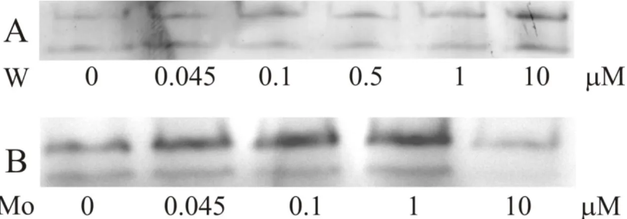
Figure III.2|Influence of W and Mo on the Fdh activity of soluble extract of D. alaskensis. The soluble extracts were obtained as described in the Material and Methods section from cells grown until the end of the exponential phase (OD600=0.5-0.6) in modified medium C supplemented with different concentrations of Na2WO4.2H2O (A) or Na2MoO4.2H2O (B). Total amount of proteins loaded in each lane in tungstate and molybdate supplementation
conditions was 10 µg and 45 µg, respectively. 41
Figure III.3| UV-visible spectrum and electrophoresis gels. UV-visible spectrum of W-Fdh (black line) and Mo/W-Fdh (gray line) isolated from D. alaskensis cells. Inset: (A) SDS-PAGE of W-Fdh (lane 1, 2 µg) and molecular
weight markers (lane 2); (B) SDS-PAGE of molecular weight markers (line 1) and Mo/W-Fdh (lane 2, 1 µg); (C) in-gel activity of the as-purified W-Fdh (lane
1, 2 µg) and Mo/W-Fdh (lane 2, 9 µg). 43
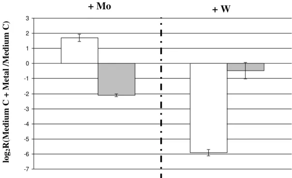
Figure III.4| Influence of metal in Fdh expression. Relative gene expression of
Mo/W-fdh and w-fdh depending on metal supplementation. Ratio of transcripts
abundance of Mo/W-fdhA (Dde_3513) ( ) or W-fdhA (Dde_0717) ( )
from D. alaskensis cultured in modified medium C supplemented with either
1µM molybdate or 10µM tungstate versus culture in modified medium C without
metal supplementation.
46
Figure III.5| Influence of metal in transporters and MoeA expression.
Relative expression of modB (Dde_3519) and tupB (Dde_0233) genes encoding
proteins responsible for Mo and W transport and moeA1 (Dde_0230) and moeA2
(Dde_3228)genes putatively involved in the selective incorporation of Mo or W
in the cofactor [25,30] as determined by using quantitative real-time PCR. The abundance of transcripts was determined from D. alaskensis cells cultured until
the end of the exponential phase (OD600=0.5-0.6) in modified medium C supplemented with either 1 µM Mo ( ) or 10 µM W ( ) and in modified medium C without metal addition.
Figure III.6 Organization of the fdh genes, elements putatively involved in molybdenum transport and metal cofactor biosynthesis and incorporation present in the fdh genes surrounding area. (A) fdh1E and fdh1D genes encode
for proteins probably involved in Fdh formation, MobA gene codes for a protein
that catalyzes the conversion of MPT to MGD, moaA gene codes for a protein
that catalyzes the conversion of GTP into cyclic pyranopterin monoPhosphate (precursor Z), W-fdhA and W-fdhB genes encoding the alpha and beta subunits of
the W-Fdh, respectively. Dde_0719 gene encodes a protein homologous to
MobB which binds GTP and has weak GTPase activity [40]. (B) Mo/W-fdhA and Mo/W-fdhB genes encoding the alpha and beta subunits of the Mo/W-Fdh. The modABC genes encode the molybdate uptake transport system. The modE
encodes a protein that regulates the modABC operon expression [31,41,42]. (C) Dde_0230 gene encodes a homologous MoeA protein involved in Mo/W
insertion into the MPT. Dde_0232-Dde_0234 genes codify for an ABC-type
tungstate transport system (TupA, TupB, and TupC) similar to the modABC gene
cluster. (D) fdh3A and fdh3B genes encoding a third putative periplasmic Fdh.
49
Figure IV.1|E. coli Fdh-H active site in the presence of nitrite. Fdh structure
at 2.9Å (PDB code:1FDI) [6]. 61
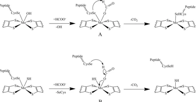
Figure IV.2| Reaction mechanisms proposed for the oxidation of formate to carbon dioxide by Fdhs. (A) Mechanism proposed by Boyington et al where
the SeCys remains bonded to the Mo atom during the catalysis [6]. (B) Mechanism proposed by Raaijamkers et al where the SeCys bond is replaced by
a formate molecule in the first step of the reaction [17]. 62
Figure IV.3| Fdh model. Model used to study the catalytic mechanism of formate dehydrogenase (frozen atoms are marked with the F letter and the truncation of the residues marked with a white asterisk). 65
Figure IV.4| Enzyme activation. Kinetic timescan of the reduction of benzyl viologen monitored at 555 nm catalyzed by Mo-dependent Fdh from D. desulfuricans. Red: with activation, Fdh (35 nM) was incubated with 10 mM of
sodium formate in 60 mM Tris-HCl buffer pH 8.0 and 133 mM β-mercaptoetanol at 37 ºC. The reaction was started adding 7.5 mM benzyl viologen. Black: without activation, Fdh (35 nM) was incubated with 7.5 mM benzyl viologen in 60 mM Tris-HCl buffer pH 8.0 and 133 mM β-mercaptoetanol at 37 ºC. The reaction was started adding 10 mM sodium formate. All assays were performed in Argon atmosphere in stoppered quartz
cell. 67
Figure IV.5| Michaelis-Menten behavior from Fdh. Michaelis-Menten plots of the steady-state kinetic assays performed in the three Fdh enzymes studied. Black and red lines are the fitting of the experimental values obtained with 1HCOO-Na (squares) and 2HCOO-Na (circles) as substrates. 68
Figure IV.7| Enzyme activation. General overview of the energies involved in the activation of the enzyme (the total charge and multiplicity of the system are
between brackets). 72
Figure IV.8| Formation of the ES complex. General overview of the energies involved in the formation of the ES complex (the total charge and multiplicity of
the system is between brackets). 73
Figure IV.9| SeCys dissociation. General overview of the energies involved in the dissociation of the SeCys (the total charge and multiplicity of the system is
between brackets). 75
Figure IV.10| Proton transfer. General overview of the energies involved in the Proton transfer (the total charge and multiplicity of the system are between
brackets). 76
Figure IV.11| Active site after proton transfer. Product obtained after finishing the proton transfer from the formate to the SeCys140. 77
Figure IV.12| Carbon dioxide release. The mechanism of the carbon dioxide release prior (top) and after (bottom) the electron transfer (the total charge and multiciplity of the system is enclosed in brackets). 78
Figure IV.13| Active site regeneration. Regeneration of the active site to the initial oxidized state (the total charge and multiplicity of the system are between
brackets). 80
Figure IV.14| Turnover in presence of formate. Formate binding to an intermediate species produced after oxidation of the active site (the total charge and multiplicity of the system are between brackets). 81
Figure IV.15| Globlal scheme of formate oxidation by Fdh. Global view of the catalytic mechanism of formate dehydrogenase. 83
Figure IV.16| Mo- and W-dependent Fdhs active site-core. Close-up view of the active site of Mo- and W-dependent formate dehydrogenases. The H-Si distance is written in red and the relevant aminoacids in black. Atom color code: white, hydrogen; yellow, sulfur; dark yellow, selenium; cyan, molybdenum. 86
Figure V.1| Comparison of the Nap and Fdh active site-core. Superposition of W-Fdh from D. gigas (red) and NapA (blue) from D. desulfuricans (PDBs:
1H0H and 2JIM). 93
Figure V.2| Normalized initial rates vs. nitrate concentration. (■): Fdh from
D. desulfuricans (35 nM); (▲): Fdh from D. gigas (35 nM); (●): Fdh from D. alaskensis (10 nM). Conditions used as described in section V.3.5, 10 mM of
Figure V.3| Inhibition type characterization of Fdh from D. desulfuricans by nitrate. Direct linear plot showing competitive inhibition. Black and red lines correspond to the experiments in absence and presence of 20 mM of nitrate, respectively; Inhibition constant (Kic) determined by Rivas et al [16]. 97
Figure V.4 Inhibition type characterization of Dg Fdh by nitrate. (a) Direct linear plot showing competitive inhibition. Black and red lines correspond to the experiments in absence and presence of 10 mM of nitrate, respectively; (b) Dixon plot used to calculate Kic. (c) Parallel pattern of Cornish-Bownden plot showing competitive inhibition. Formate concentrations used: (■) 500 µM, (▲)
250 µM, (●) 100 µM and (♦) 50 µM. 98
Figure V.5| Inhibition type characterization of Fdh from D. alaskensis by nitrate. (a) Direct linear plot showing competitive inhibition. Black and red lines correspond to the experiments in the presence or absence of 5 mM of nitrate, respectively; (b) Dixon plot used to calculate Kic. (c) Parallel pattern of Cornish-Bownden plot showing competitive inhibition. Formate concentrations used: (■) 250 µM, (▲) 50 µM, (●) 20 µM and (♦) 12.5 µM. 99
Figure V.6| Normalized initial rates vs. azide concentration. (■): Fdh from D. desulfuricans (35 nM); (▲): Fdh from D. gigas (35nM); (●): Fdh from D. alaskensis (10 nM). Conditions used as described in section V.3.5, 10 mM of
formate was used. 101
Figure V.7| Characterization of the inhibition type by azide for the Dd Fdh. (a) Direct linear plot showing mixed inhibition. Black and red lines correspond to the experiments in absence and presence of 0.5 mM of azide, respectively; (b) Inhibition constants (Kic and Kiuc) determined by Rivas et al [2,16]. 102
Figure V.8| Characterization of the inhibition type by azide for the Dg Fdh. (a) Direct linear plot showing mixed inhibition. Black and red lines correspond to the experiments in absence and presence of 10 mM of azide, respectively; (b) Dixon plot used to calculate Kic. (c) Cornish-Bownden plot used to calculate Kiuc. Formate concentrations used: (■) 500 µM, (▲) 250 µM, (●) 100 µM and
(♦) 50 µM. 103
Figure V.9| Characterization of the inhibition type by azide for the Da Fdh. (a) Direct linear plot showing mixed inhibition. Black and red lines correspond to the experiments in absence and presence of 0.5 mM of azide, respectively; (b) Dixon plot used to calculate Kic. (c) Cornish-Bownden plot used to calculate Kiuc. Formate concentrations used: (■) 250 µM, (▲) 50 µM, (●) 25 µM and (♦)
15 µM. 104
Figure V.10| Normalized initial rates vs. cyanide concentration. (■): Fdh from D. desulfuricans(35 nM); (▲): Fdh from D. gigas (35 nM); (●): Fdh from D. alaskensis (35 nM). Conditions used as described in section V.3.5, 10 mM of
Figure V.11| Characterization of the inhibition type by cyanide for the Dd Fdh. Direct linear plot showing mixed inhibition. Black and red lines correspond to the experiments in absence and presence of 2.5 mM of cyanide, respectively; Inhibition constants (Kic and Kiuc) determined by Rivas et al [2,16]. 107
Figure V.12| Aminoacids alignment of Fdhs α subunits. FdhDg: α subunit from D. gigas; FdhDa: α subunit from D. alaskensis; FdhDd: α subunit from D. desulfuricans; light grey: twin arginine motif; light yellow: conserved SeCys;
light blue: conserved residues probably involved in enzymatic catalysis; ▼: signal peptide cleavage site; ·: cysteines motifs involved in the coordination of [4Fe-4S] clusters; (*): identity; (:): strongly similar; and (.) weekly similar. 110
Figure V.13| Aminoacids alignment of Fdhs β subunits. FdhDg: β subunit from D. gigas; FdhDa: β subunit from D. alaskensis; FdhDd: β subunit from D. desulfuricans; ●, ●, and ●: cysteine motifs involved in the FeSII, FeSIII and
FeSIV cluster binding; (*): identity; (:): strongly similar; and (.) weekly similar. 111
Figure V.14| Fdhs active sites overlapped. Fdhs active sites with aminoacids from neighborhood from D. gigas (black), D. alaskensis (green) and D. desulfuricans (red). The overlay was done using the structure of Fdh from D.
gigas [19] as support. 115
Figure VI.1| Schematic representations of the several Mo ion coordination sphere proposed by several authors based on different experimental evidences. X-ray crystallography: (A), (B), (D) and (E). DFT calculations: (C).
XAS spectroscopy: (F). 121
Figure VI.2| CW-EPR spectra obtained in Dd Fdh samples (200 µM) in presence or absence of the competitive inhibitor azide. A) Formate-reduced
Dd Fdh incubated with 3 mM sodium azide in H2O-buffer. B) As A) but in D2 O-buffer. C) Formate-reduced Dd Fdh in H2O-buffer. D) As C) but in D2O-buffer. Measurement conditions: Microwave frequency, 9.65 GHz; Modulation frequency, 100 kHz, Modulation amplitude, 5Gpp; Microwave power, 2 mW;
Temperature, 100 K. 123
Figure VI.3| 3D view of the structural model used to predict EPR parameters of the formate Mo(V) species. Predicted g-values and hfc constants
of the exchangeable proton bound to the –SH group are written next to the
model. 128
Figure VI.4: 3D view of the structural model used to predict EPR parameters of the azide (2.094) Mo(V) species. Predicted g-values and hfc
Chapter I
I.1 Metals in proteins
Proteins are the most versatile macromolecules in living systems and have crucial functions in essentially all biological processes. They function as catalysts, transport and store other molecules, provide mechanical support and immune protection, generate movement, transmit nerve impulses, and control cell growth and differentiation. The entire set of proteins of a living cell is called the proteome. Although the genetic information coding for the proteome is stored in the form of DNA within the genome, is the proteome that determines which and at which levels proteins should be expressed under given conditions in a given cell type [1].
It has been estimated that about one third of all proteins bind one or more metal ions as prosthetic groups. A systematic bioinformatics study of 1,371 enzymes with known three-dimensional structures showed that 47% require metals from which 41% contain metals at their catalytic centers [2]. Metalloenzymes occur in all six Enzyme Commission Classes (E.C.C.), accounting for 44% of oxidoreductases, 40% of tranferases, 39% of hydrolases, 36% of lyases, 36% of isomerases and 59% of ligases. Figure I.1A shows the frequency of metals in proteins structures. Magnesium is the most prevalent metal in metalloenzymes being often involved in loose partnerships with phosphate-containing substrates such as ATP. Since this metal is sometimes interchangeable with manganese, the frequency of Mn in vivo may be overestimated [3].
solutions and thus would be more available in anaerobic and highly reducing environment [4].
Figure I.1| Metals used in catalysis. A, The elements used as cofactors by enzymes are shown in green. The height of each column represents the proportion of all enzymes with known structures using the respective metal. A single enzyme use cadmium (carbonic anhydrase from
T. weissflogii [6]). B, The proportion of proteins using the indicated metals that occur in each of
the six enzyme classes: oxidoreductases (EC 1), blue; transferases (EC 2), yellow; hydrolases (EC 3), purple; lyases(EC 4), pink; isomerases (EC 5), green; ligases (EC 6), grey. EC, Enzyme
Commission. Figures were taken from reference [3]. A
Both molybdenum and tungsten are redox-active under physiological conditions (ranging between oxidation states VI, V and IV) and this chemical versatility make them useful in biological systems [4]. The physiological roles of enzymes containing these metals are fundamental and include the catalysis of key steps in carbon, nitrogen and sulfur metabolism [7].
I.2 Cell biology of molybdenum and tungsten
Molybdenum and tungsten are widely distributed in biology. Molybdenum-containing enzymes are present in bacteria, fungi, algae, plants and animals [8,9], whereas the majority of the tungsten-containing enzymes purified to date are found in anaerobic organisms of Archaea and Bacteria [5].
On the basis of cofactor composition and catalytic function, molybdenum and tungsten dependent enzymes can be grouped into two categories: bacterial nitrogenases containing an FeMoco in the active site [10], and pterin-based molybdenum/tungsten enzymes [4,7].
Molybdenum and tungsten are bioavailable as molybdate (MoO42-) and tungstate (WO42-) oxoanions, respectively. Once these oxoanions enters inside the cell (see section I.2.1), they are incorporated into metal cofactors by complex biosynthetic machineries (see section I.2.2).
I.2.1 Cellular uptake
The cellular transport systems for oxoanions like tungstate, molybdate and sulfate have been studied and described for many organisms. In particular, the uptake mechanisms of these oxoanions have been deeply characterized in E. coli [11]. All these transport
systems are members of the adenosine triphosphate (ATP) binding cassette (ABC) transporter group. According to the currently accepted model for cellular Mo uptake, molybdate is captured by ModA, a periplasmic molybdate-binding protein that transfer the metal oxo-ion to ModB, which is a transmembrane channel. At the cytoplasmic side ATP is hydrolyzed to ADP and Pi in ModC, which generates the energy necessary to complete the transport of molybdate into the cell cytoplasm. Additional proteins encoded by the mod operon have functions in intracellular molybdate binding and
of the metal. ModA is also able to bind tungstate with a similar affinity as to molybdate [12,13]. For tungsten, two ABC-type transporters are known: TupABC and WtpABC [13]. The TupABC system is specific for tungstate and was identified in E.
acidaminophilum [14]. More recently, the structurally different WtpABC transporter
was reported in the hyperthermophilic archaeon P. furiosus [15].
I.2.2 Biosynthesis of the cofactor
Molybdenum and tungsten association with enzymes occur in a similar cofactor, which consist of one or two tricyclic pterin moieties usually referred as molybdopterin, tungstopterin or pyranopterin. The pathway of Mo-cofactor (Moco) biosynthesis has been extensively studied in prokaryotes (E. coli) as well as eukaryotes (Arabidopsis
thaliana and Homo sapiens) and appears to be highly conserved [16]. The pathway of
W-cofactor (Wco) biosynthesis is thought to be similar to the pathway of Moco biosynthesis, at least up to the step of the metal insertion. The support of this hypothesis is that homologues of almost all genes that have an assigned function in the Moco biosynthetic pathway are also present in the genomes of organisms that incorporates W instead of Mo in the enzymes active sites.
The first model of Moco synthesis was based on E. coli data [17]. Three operons and
one gene have been identified to be involved in the Moco biosynthesis in this organism:
moaABCDE, mobAB, moeAB and mogA, respectively. Among the ten proteins involved
in the biosynthesis of Moco, eight have an assigned function and two remain to be demonstrated. Figure I.2 schematizes the Moco biosynthesis pathway including the function of each one of this proteins.
Figure I.2| Biosynthesis of the Pyranopterin-based molybdenum cofactors. Schematic overview of the molybdenum cofactor biosynthesis adapted from reference[12] and based on
data derived from studies in E. coli, plants and humans. Abbreviations of the intermediates are
written in bold on the right side of their structures, and the enzymes are on the left side of the arrows. The fifth step present only in prokaryotes. For MPT and MPT-AMP, the dithiolate sulfurs are bonded to a Cu, because in the crystal structure of plant Cnx1G was found a copper
ion in that place.
The next steps involve the coordination of the metal atom (Mo or W) to the dithiolene sulfurs replacing the Cu atom present in the MPT. The proteins MoeA and MogA (Cnx1E and Cnx1G for plant; Geph-E and Geph-G for human) catalyze these steps, and very recently also MoaB was found to be involved in this stage of the cofactor synthesis
+ SAM MOCS1A Cnx2 MOCS1B Cnx3 + GTP/CTP +Mg2+ MoaA MoaC MobA + ATP +Mg2+
+ MoO4
2-+Mg2+ Geph-G Cn1G Geph-E Cnx1E MoeA MogA or MoaB + ATP +Mg2+
+ S
2-OH AMP SH SH MOCS3 Cnx5 MOCS2B
Cnx7 MOCS2ACnx6 MoeB
[20]. The proteins MogA and MoaB catalyse the activation of MPT by adenylylation with Mg-ATP [21,22]. The trimeric MogA proteins are commonly found in bacteria and eukaryotes, whereas the hexameric MoaB proteins are mostly found in archaea and in some bacteria though believed to be specific for the biosynthesis of the tungsten cofactor [20]. Subsequently, MoeA is thought to bind the adenylylated MPT (MPT-AMP) produced after reaction with MogA or MoaB and in the presence of molybdate and/or tungstate the MPT-AMP complex is hydrolyzed. At this step, molybdenum or tungsten displaces the Cu ion coordinated to the dithiolene sulfurs, and the AMP is released. Because Moco is deeply buried within the holoenzymes, it needs to be incorporated before the completion of folding and oligomerization of enzyme subunits domains in the presence of chaperones [12,13].
As a final maturation step (only in prokaryotes), guanosine monophosphate (GMP) or cytidine monophosphate (CMP) can be attached (phosphoester condensation from GTP and CTP) to the MoCo, forming molybdopterin guanosine/cytidine dinucleotide (MGD/MCD) cofactor. This reaction is catalyzed by MobA and MobB [23]. For some prokaryotes enzymes the coupling of a second pyranopterin dinucleotide cofactor can be required, leading to the formation of the Mo/W-bis-pyranopterin dinucleotide cofactor.
The pathway that lead to the formation of this type of cofactors still remains unclear and needs to be more studied [13].
Eukaryotic molybdenum hydroxilases (e.g. xanthine oxidase, aldehyde oxidase) harbor the Moco in the mature protein, but require the addition of a terminal sulfido ligand to the molybdenum to get enzymatic activity. This step is catalyzed by Moco sulfurilase (Aba3 in plants and MOCOS in humans) [12,24]. Among prokaryotes, no Moco sulfurilases homologues to eukaryotic have been found. So far, only one exception was reported and corresponds to the XdhC which is a specific chaperone coded by the R.
capsulatus genome found to perform Moco sulfurilation of xanthine dehydrogenase
I.2.3 Molybdenum cofactor deficiency
Moco biosynthesis is a relevant target of study because many steps of the synthesis still remain unclear and the specificity for Mo or W is not yet understood. The Moco biosynthesis is an important metabolic pathway in cells, for example, human Moco deficiency (MocoCD) results in complete loss of sulfite oxidase, xanthine oxidase and aldehyde oxidase activity. This disease is characterized by progressive neurological damage, leading to early childhood death in most cases. Symptoms are mainly caused by deficiency of sulfite oxidase protecting the organs (in particular the brain) from elevated concentrations of toxic sulfite [12]. The biochemical explanation behind this is that sulfite oxidase catalyzes the last step in the oxidative degradation of sulfur-containing amino acids and lipids. Localized in the intermenbrane space of mitochondria, this enzyme catalyzes the oxidation of sulfite to sulfate. Under conditions of Moco or sulfite oxidase deficiency, sulfite accumulates in the plasma and serum, crosses the blood-brain barrier and rapidly triggers neuronal death [12].
An animal model, in which the gene encoding Mocs1 (a MoaA homologue) was deleted, has been used to study the Moco cofactor deficiency [26]. Homozygous mice displayed a severe phenotype that reflects all biochemical characteristics of human Moco-deficient patients. They failed to thrive, and died within the first 12 days of life. Mice treated with cPMP purified from E. coli [27], gained weight and reached the
adulthood and fertility like their wild-type analogues. When the delivery of cPMP by injections stops, Mocs-1-knockout mice die within 10-14 days. These promising results are already being applied in clinical assays in humans [12].
I.3 Molybdenum and tungsten enzymes
In contrast to the multinuclear iron-molybdenum clusters found in bacterial nitrogenases [10] and copper-molybdenum sites found in carbon monoxide dehydrogenase from O.
carboxidovorans [28], the active sites of all other well-characterized molybdenum- and
reaction coupled to electron-transfer between substrate and other cofactors such as iron-sulfur centers, hemes or flavins.
I.3.1 Categories and general features of mononuclear Mo/W containing enzymes
Based on X-ray structural data, primary sequence alignments, spectroscopic and biochemical characteristics, molybdenum- and tungsten-containing enzymes can be grouped in four broad families (figure I.3) [7]:
1) The xanthine oxidase family: can harbor one MCD or MPT that provides the only attach of the Mo ion to the protein. The Mo coordination sphere can be completed by oxygen (oxo, hydroxo) and sulfur (sulfido) ligands.
2) The sulfite oxidase family (and eukaryotic nitrate reductases): can harbor one MPT that together with a sulfur atom from a cysteine sidechain attach the Mo ion to the protein. The coordination sphere is completed by two oxygenic species (oxo, hydroxo).
3) The DMSO reductase family: can bind two MGD molecules that provide four sulfur ligands to the Mo/W ion arising from the two dithiolene groups. In addition, the Mo/W can bind oxygen, sulfur or selenium from aminoacid sidechains (O-Ser, O-Asp, S-Cys, Se-Cys). The Mo coordination sphere is completed with oxygen (oxo, hydroxo) or sulfur (sulfido) atoms.
4) The tungsten aldehyde oxidoreductase family: can bind two MPT molecules that in a similar arrangement to the DMSO reductase family. The W coordination sphere is completed by two oxygen atoms (oxo, hydroxo). This family of proteins includes a few enzymes within the aldehyde ferrodoxin oxidoreductase (AOR) from P. furiosus
Figure I.3| Active-site structures of the different families molybdo- and tungsto-pterins containing enzymes. XO/XDH: xanthine oxidase/xanthine dehydrogenase; AOR: aldehyde oxidoreductase; CODH: carbon monoxide dehydrogenase; NDH: Nicotinate dehydrogenase;
SO: sulfite oxidase; SDH: sulfite dehydrogenase; Euk-NR: eukaryotic nitrate reductase; Dg
Fdh: Desulfovibrio gigas formate dehydrogenase; Ec Fdh-H: Escherichia coli formate
dehydrogenase H; Dd NapA: Desulfovibrio desulfuricans ATCC 27774periplasmatic nitrate
reductase A; Cn NapAB: Cupriavidus necator periplasmatic nitrate reductase AB; Aa EBDH:
Aromateleum aromaticum ethylbenzene dehydrogenase; Ec NarGH/I: Escherichia coli nitrate
reductase GH/nitrate reductase GHI; Pf AOR: Pyrococcus furiosus aldehyde
I.3.1.1 The xanthine oxidase family
Enzymes of the xanthine oxidase family are the best characterized mononuclear Mo-containing enzymes. With a few exceptions, they catalyze the oxidative hydroxylation of a diverse range of aldehydes and aromatic heterocycles in reactions that involve the cleavage of a C-H and the formation of a C-O bond:
RH + H2O → ROH + 2H+ + 2e
-Members of this family can be found distributed within eukaryotes and prokaryotes. Xanthine dehydrogenase (XDH) [30] is the key enzyme in the catabolism of purines, catalyzing the conversion of hypoxanthine to xanthine, and xanthine to uric acid. XDH can be converted to xanthine oxidase (XO) by proteolysis and cysteines oxidation. XO also catalyzes the reactions of purine catabolism but is not NADH-dependent and reactive oxygen species (ROS) are formed as by-products of the reaction [31]. The mammalian aldehyde oxidase is also an important enzyme involved in metabolism of several aldehyde compounds and the biotransformation of xenobiotics [32].
Enzymes belonging to the XO family have the Mo-MPT cofactor without any covalent attachment to the polypeptide chain. They are organized either as homodimers, with several redox-active cofactors placed within a single subunit, or as multi-subunit enzymes. In the case of a single subunit structure (mammalian xanthine oxidase and aldehyde oxidase (AOX)), two [2Fe-2S] centers are located in the N-terminal domain (~20kDa), followed by the flavin domain (~40kDa) and finally the C-terminal domain (90kDa) that hold the Moco. The prokaryotic aldehyde oxidoreductases isolated from sulfate reducers like D. gigas and D. desulfuricans [33,34] are very similar to the XO
and AOX but they lack the flavin domain.
In multi-subunit structures (e.g. CO dehydrogenase from Oligotropha carboxidorovans
[28] and quinoline oxidoreductase from Pseudomonas putida [35]), the redox centers
I.3.1.2 The sulfite oxidase family
The members of this family include not only sulfite oxidases from plants, animals and bacteria but also the eukaryotic nitrate reductases [36]. These assimilatory nitrate reductases catalyze the first and rate-limiting step of nitrate assimilation in plants, algae and fungi, and are completely different from the bacterial nitrate reductases that belong to the DMSO reductase family.
Three different sulfite-oxidizing subfamilies are known: sulfite oxidase (SO) in animals, SO in plants, and sulfite dehydrogenase (SDH) in bacteria. The animal SO catalyzes the last reaction in the oxidative degradation of the sulfur containing amino acids cysteine and methionine and is critical in transforming the toxic sulfite into the more soluble sulfate anion that can be excreted. Similarly, the plant SO is responsible for detoxifying excess sulfite produced during sulfur assimilation. In bacteria, e.g. Sarkeya novella [37],
SDH has an important role oxidizing the sulfite formed during dissimilatory oxidation of reduced sulfur compounds.
In contrast to the XO family, the polypeptide chain coordinates directly to the Mo ion by a cisteinyl residue. The animal SO are homodimeric and bind Moco and one b-type
heme in different domains. The plants SO are also homodimeric, harbor the Moco domain but lacks the heme domain. The most representative bacterial enzyme is heterodimeric periplasmic sulfite dehydrogenase (SorAB) which contains a Moco and one c-type heme in and subunits, respectively [37]. Some of these enzymes occur as
monomers or dimers and lack the heme groups [38].
I.3.1.3 The DMSO reductase family
With some exceptions, members of the DMSOr family catalyze the transfer of an oxygen atom to or from the substrate. The exceptions are the Fdhs, which catalyze the oxidation of formate to carbon dioxide by cleaving a C-H bond. In addition, the acetylene hydratase from Pelobacter acetylenicus [49] catalyzes a non-redox reaction,
that is, the hydration of acetylene to acetaldehyde. This process is part of an anaerobic degradation pathway of unsaturated hydrocarbons.
The enzymes from DMSOr family present high degree of similarity in the overall polypeptide fold of crystal structures. However, there are some variations in the active site, not only at the metal coordination sphere, but also with the surrounding amino acids residues. These slight but important differences at the catalytic sites of the different enzymes can explain the remarkable diversity of functions performed by enzymes of the DMSOr family. The amino acid that coordinates the molybdenum or tungsten atom differs among the several enzymes of the family: cysteine in the periplasmic nitrate reductase and acetylene hydratase, selenocysteine in Fdh, aspartate in the membrane-bound respiratory nitrate reductases and ethylbenzene dehydrogenase, and a serine in DMSO reductase and TMAO reductase. Only in the arsenite oxidase the Mo ion has no ligands from the polypeptide chain.
To date, it is known that only Fdhs [50] and DMSO reductases [51] are capable of incorporating either molybdenum or tungsten at their active sites.
I.4 Subject and objectives of this thesis
This thesis deals with formate dehydrogenases isolated from sulfate reducing bacteria (SRB), namely Desulfovibrio species. These organisms can use formate, the product of
the reaction catalyzed by pyruvate formate lyase, as carbon source and electron donor [52]. The protons released in formate oxidation by Fdh will contribute for a proton gradient and subsequently energy production.
The active site of Fdh comprises a metal ion (Mo or W) coordinated to five sulfur atoms and a selenium. The chemistry of transition metals like Mo and W in biological systems is not yet well understood. Also, the preference of some Fdhs to incorporate Mo or W to perform the catalysis is a phenomenon that lies in the shade for microbiologists.
this work: How substrate binds to the active site? What is the role of the Se in the catalytic mechanism? How is the catalytic mechanism of formate oxidation? Why different active sites structures are proposed in the literature for the same Mo species? Why some Fdhs prefer incorporate Mo and other W in the active site?
The studies described in this work covers a large set of techniques:
- Real Time PCR, purification and biochemical characterization methods were used to obtain information about the influence of Mo and W on the Fdh and metal transport systems expression.
- Kinetics studies and DFT calculations were used to characterize the enzyme by determining the kinetic parameters and to understand the reaction mechanism.
- DFT calculations were also used to reconcile the available structural and spectroscopic data obtained in Mo and W containing Fdhs.
I.5 References
1 Berg, J. M., Tymoczko, J. L. & Stryer, L. Biochemistry. 5th Edition edn, (W. H. FREEMAN AND COMPANY, 2002).
2 Andreini, C., Bertini, I., Cavallaro, G., Holliday, G. L. & Thornton, J. M. Metal ions in biological catalysis: from enzyme databases to general principles. J Biol Inorg Chem 13, 1205-1218, (2008).
3 Waldron, K. J., Rutherford, J. C., Ford, D. & Robinson, N. J. Metalloproteins and metal sensing. Nature 460, 823-830, (2009).
4 Hille, R. Molybdenum and tungsten in biology. Trends Biochem Sci 27, 360-367, (2002).
5 Johnson, M. K., Rees, D. C. & Adams, M. W. Tungstoenzymes. Chem Rev 96, 2817-2840, (1996).
6 Lane, T. W., Saito, M. A., George, G. N., Pickering, I. J., Prince, R. C. & Morel, F. M. M. Biochemistry A cadmium enzyme from a marine diatom. Nature 435, 42-42, (2005).
7 Romao, M. J. Molybdenum and tungsten enzymes: a crystallographic and mechanistic overview. Dalton Trans, 4053-4068, (2009).
9 Williams, R. J. P. & da Silva, J. J. R. F. The involvement of molybdenum in life. Biochem Bioph Res Co 292, 293-299, (2002).
10 Burgess, B. K. & Lowe, D. J. Mechanism of Molybdenum Nitrogenase. Chem Rev 96, 2983-3012, (1996).
11 Rech, S., Deppenmeier, U. & Gunsalus, R. P. Regulation of the molybdate transport operon, modABCD, of Escherichia coli in response to molybdate
availability. J Bacteriol 177, 1023-1029, (1995).
12 Schwarz, G., Mendel, R. R. & Ribbe, M. W. Molybdenum cofactors, enzymes and pathways. Nature 460, 839-847, (2009).
13 Bevers, L. E., Hagedoorn, P. L. & Hagen, W. R. The bioinorganic chemistry of tungsten. Coordin Chem Rev 253, 269-290, (2009).
14 Makdessi, K., Andreesen, J. R. & Pich, A. Tungstate Uptake by a highly specific ABC transporter in Eubacterium acidaminophilum. J Biol Chem 276,
24557-24564, (2001).
15 Bevers, L. E., Hagedoorn, P. L., Krijger, G. C. & Hagen, W. R. Tungsten transport protein A (WtpA) in Pyrococcus furiosus: the first member of a new
class of tungstate and molybdate transporters. J Bacteriol 188, 6498-6505, (2006).
16 Schwarz, G. Molybdenum cofactor biosynthesis and deficiency. Cell Mol Life Sci 62, 2792-2810, (2005).
17 Rajagopalan, K. V. & Johnson, J. L. The pterin molybdenum cofactors. J Biol Chem 267, 10199-10202, (1992).
18 Wuebbens, M. M. & Rajagopalan, K. V. Investigation of the early steps of molybdopterin biosynthesis in Escherichia coli through the use of in vivo
labeling studies. J Biol Chem 270, 1082-1087, (1995).
19 Schwarz, G. & Mendel, R. R. Molybdenum cofactor biosynthesis and molybdenum enzymes. Annu Rev Plant Biol 57, 623-647, (2006).
20 Bevers, L. E., Hagedoorn, P. L., Santamaria-Araujo, J. A., Magalon, A., Hagen, W. R. & Schwarz, G. Function of MoaB proteins in the biosynthesis of the molybdenum and tungsten cofactors. Biochemistry 47, 949-956, (2008).
22 Nichols, J. D. & Rajagopalan, K. V. In vitro molybdenum ligation to molybdopterin using purified components. J Biol Chem 280, 7817-7822, (2005). 23 Palmer, T., Goodfellow, I. P., Sockett, R. E., McEwan, A. G. & Boxer, D. H. Characterisation of the mob locus from Rhodobacter sphaeroides required for
molybdenum cofactor biosynthesis. Biochim Biophys Acta 1395, 135-140, (1998).
24 Bittner, F., Oreb, M. & Mendel, R. R. ABA3 is a molybdenum cofactor sulfurase required for activation of aldehyde oxidase and xanthine dehydrogenase in Arabidopsis thaliana. J Biol Chem 276, 40381-40384, (2001).
25 Neumann, M., Schulte, M., Junemann, N., Stocklein, W. & Leimkuhler, S.
Rhodobacter capsulatus XdhC is involved in molybdenum cofactor binding and
insertion into xanthine dehydrogenase. J Biol Chem 281, 15701-15708, (2006). 26 Lee, H. J., Adham, I. M., Schwarz, G., Kneussel, M., Sass, J. O., Engel, W. &
Reiss, J. Molybdenum cofactor-deficient mice resemble the phenotype of human patients. Hum Mol Genet 11, 3309-3317, (2002).
27 Schwarz, G., Santamaria-Araujo, J. A., Wolf, S., Lee, H. J., Adham, I. M., Grone, H. J., Schwegler, H., Sass, J. O., Otte, T., Hanzelmann, P., Mendel, R. R., Engel, W. & Reiss, J. Rescue of lethal molybdenum cofactor deficiency by a biosynthetic precursor from Escherichia coli. Hum Mol Genet 13, 1249-1255,
(2004).
28 Dobbek, H., Gremer, L., Kiefersauer, R., Huber, R. & Meyer, O. Catalysis at a dinuclear [CuSMo(==O)OH] cluster in a CO dehydrogenase resolved at 1.1Å resolution. Proc Natl Acad Sci U S A 99, 15971-15976, (2002).
29 Chan, M. K., Mukund, S., Kletzin, A., Adams, M. W. & Rees, D. C. Structure of a hyperthermophilic tungstopterin enzyme, aldehyde ferredoxin oxidoreductase. Science 267, 1463-1469, (1995).
30 Truglio, J. J., Theis, K., Leimkuhler, S., Rappa, R., Rajagopalan, K. V. & Kisker, C. Crystal structures of the active and alloxanthine-inhibited forms of xanthine dehydrogenase from Rhodobacter capsulatus. Structure 10, 115-125,
(2002).
32 Garattini, E., Mendel, R., Romao, M. J., Wright, R. & Terao, M. Mammalian molybdo-flavoenzymes, an expanding family of proteins: structure, genetics, regulation, function and pathophysiology. Biochem J 372, 15-32, (2003).
33 Brondino, C. D., Romao, M. J., Moura, I. & Moura, J. J. Molybdenum and tungsten enzymes: the xanthine oxidase family. Curr Opin Chem Biol 10, 109-114, (2006).
34 Brondino, C. D., Rivas, M. G., Romao, M. J., Moura, J. J. & Moura, I. Structural and electron paramagnetic resonance (EPR) studies of mononuclear molybdenum enzymes from sulfate-reducing bacteria. Acc Chem Res 39, 788-796, (2006).
35 Hanzelmann, P., Dobbek, H., Gremer, L., Huber, R. & Meyer, O. The effect of intracellular molybdenum in Hydrogenophaga pseudoflava on the
crystallographic structure of the seleno-molybdo-iron-sulfur flavoenzyme carbon monoxide dehydrogenase. J Mol Biol 301, 1221-1235, (2000).
36 Feng, C., Tollin, G. & Enemark, J. H. Sulfite oxidizing enzymes. Biochim Biophys Acta 1774, 527-539, (2007).
37 Kappler, U. & Bailey, S. Molecular basis of intramolecular electron transfer in sulfite-oxidizing enzymes is revealed by high resolution structure of a heterodimeric complex of the catalytic molybdopterin subunit and a c-type cytochrome subunit. J Biol Chem 280, 24999-25007, (2005).
38 Kappler, U. Bacterial sulfite-oxidizing enzymes. Biochim Biophys Acta, (2010). 39 Moura, J. J., Brondino, C. D., Trincao, J. & Romao, M. J. Mo and W bis-MGD
enzymes: nitrate reductases and formate dehydrogenases. J Biol Inorg Chem 9, 791-799, (2004).
40 Bertero, M. G., Rothery, R. A., Palak, M., Hou, C., Lim, D., Blasco, F., Weiner, J. H. & Strynadka, N. C. Insights into the respiratory electron transfer pathway from the structure of nitrate reductase A. Nat Struct Biol 10, 681-687, (2003). 41 Jormakka, M., Richardson, D., Byrne, B. & Iwata, S. Architecture of NarGH
reveals a structural classification of Mo-bisMGD enzymes. Structure 12, 95-104, (2004).
42 Kloer, D. P., Hagel, C., Heider, J. & Schulz, G. E. Crystal structure of ethylbenzene dehydrogenase from Aromatoleum aromaticum. Structure 14,
![Figure I.2| Biosynthesis of the Pyranopterin-based molybdenum cofactors. Schematic overview of the molybdenum cofactor biosynthesis adapted from reference[12] and based on data derived from studies in E](https://thumb-eu.123doks.com/thumbv2/123dok_br/16494272.733494/34.892.138.734.110.839/biosynthesis-pyranopterin-molybdenum-cofactors-schematic-molybdenum-biosynthesis-reference.webp)
![Figure II.3| Active site of Fdh-H from E. coli. A: Oxidized Fdh-H active site [6]. B: Reduced Fdh-H active site [6]](https://thumb-eu.123doks.com/thumbv2/123dok_br/16494272.733494/51.892.282.607.108.1075/figure-active-site-fdh-oxidized-active-reduced-active.webp)
![Figure II.4| E.coli Fdh-N structure[7]. Left: Fdh-N structure at 1.6Å (PDB code:1KQF)](https://thumb-eu.123doks.com/thumbv2/123dok_br/16494272.733494/53.892.135.737.110.608/figure-coli-fdh-structure-left-fdh-structure-pdb.webp)
![Figure II.5| D. gigas Fdh structure[9]. Left: Fdh structure at 1.8Å (PDB code:1H0H). Right:](https://thumb-eu.123doks.com/thumbv2/123dok_br/16494272.733494/54.892.135.767.112.588/figure-gigas-fdh-structure-left-fdh-structure-right.webp)
![Figure III.1| In gel Fdh activity test of D. alaskensis cells grown in medium C [16] with different carbon sources](https://thumb-eu.123doks.com/thumbv2/123dok_br/16494272.733494/68.892.131.799.552.882/figure-activity-alaskensis-cells-medium-different-carbon-sources.webp)

![Figure IV.1|E. coli Fdh-H active site in the presence of nitrite. Fdh structure at 2.9Å (PDB code:1FDI) [1]](https://thumb-eu.123doks.com/thumbv2/123dok_br/16494272.733494/89.892.278.611.119.471/figure-coli-fdh-active-site-presence-nitrite-structure.webp)