Anaerobic bacteria: an investigation
of metabolic important enzymes
A novel type of oxygen reductase and
enzymes of the tetrapyrrole biosynthesis
Susana André Lima Lobo
Dissertation presented to obtain the Ph.D. degree in Biochemistry at the Instituto de Tecnologia Química e Biológica, Universidade Nova de Lisboa
Supervisor
:
Dr. Lígia M. Saraiva
Co
-
supervisor
:
Prof. Miguel Teixeira
Opponents
:
Prof. Martin Warren
Prof. Ilda Sanches
Dr. Carlos Salgueiro
From left to right: José Martinho Simões, Ilda Sanches, Carlos Salgueiro, Martin Warren, Susana Lobo, Lígia Saraiva, Ana Melo, Célia Romão, Miguel Teixeira
8th of July 2009
Second Edition, July 2009
Molecular Genetics of Microbial Resistance Laboratory Instituto de Tecnologia Química e Biológica
Universidade Nova de Lisboa Av. da República (EAN) 2780-157 Oeiras
I would like to express my gratitude to the people that contributed to this work and beyond:
To my supervisor, Dr. Lígia Saraiva, whose presence, determination and friendship were a constant factor. For believing in me and in my work, for the 24h availability whenever I needed to ask a question or communicate the result of an experiment that just could not wait! For always seeing the bright positive side of things, when I was only seeing the dark ones. For giving me the opportunity to evolve as a scientist (and as a person) in this infinite scientific world.
To my co-supervisor, Prof. Miguel Teixeira, for all his support and fruitful
discussions, for spending so much time in my computer playing with Matlab, and for always paying attention when I started talking and talking and talking about
Desulfovibrio genomes.
To Prof. Martin Warren, for all his collaboration and welcome into his lab, and to all the people in Prof. Martin Warren’s lab, especially to Amanda Brindley, for the help in the tetrapyrrole work, to Evelyne Deery, for her genial genetic tips and to Susanne Schroeder, for her friendly welcome into her home and for all she has done to integrate me when I was in the UK.
Claudia Almeida, for being the excellent person she is, for her patience when I was still learning how to put LA in a Petri dish without solidifying the medium before getting into the plate, for her support and belief in me, and most of all, for her huge friendship.
Célia Romão, who helped me in the anaerobic (and aerobic) protein purification quest, for the 1500000 protein assays, for the structure of the most pinkish protein, which I had the delight to see in 3D in the computer using special glasses, without having to switch my eyes into 3D mode!
Ana Melo that helped me when I started to give my first steps in science (when I was still finishing my fifth year of Biology!).
To my group, the Molecular Genetics of Microbial Resistance, for making the lab such a dynamic and nice place to work. To Lígia Nobre, Vera Gonçalves and Marta Justino, for their friendship and for the “conferences” and ”brain stormings” downstairs or in the lab desk surrounding areas, and Filipa Tavares, Joana Baptista and Mafalda Figueiredo, the three “baby girls” in the lab that were always updating me with things from the “future” (e.g. Hi5), for their friendship, good mood, and for saying things like: “Hemos de porfirinas”. I will never forget this haem description! I
would like also to thank our former group members, Vera Mónica, Sofia, Nuno and Nuno Félix, for the good moments.
Pereira, Smilja Todorovic and to Andreia Veríssimo, Filipa Sousa and João Rodrigues, who were my travel companions the first time I went to a congress outside of Portugal.
João Carita, for the endless Desulfovibrio growths (with and without oxygen!), for the chats and for the help when I had to look into the microscope.
To all the members (and former members) of our neighbour group, the Microbial Biochemistry group, especially to Sofia Venceslau, for the hydrogen bubbling in gels and for all the great chats! And also to Isabel Pacheco, for the welcoming into her lab to use the anaerobic chamber.
To Ricardo Louro for the collaboration on the NMR spectroscopy.
To Manuela Regalla for the N-terminal sequencing and HPLC analysis.
To everyone in the 3rd floor of ITQB, for the friendly environment.
Desulfovibrio for being such an interesting and good cell model which allowed me to study so many fascinating proteins!
To all my friends; especially to Andreia (who is the most friendly, strong and
determined person I know), André and João, for saying: “oh no, there she goes
again….and the lab talking begins!” and for all the fantastic and treasured off-lab moments! A special thanks goes also to Tânia, Ana Raquel, Raquel, Lena, Leonor, Anna and Lara.
To Luísa, Tó, Manel, Luís and Catarina, for their friendship and support.
To my godson Rodrigo and to my godparents, Tó and Isabel, for their friendship and for being part of my life since June 1981!
To António, for his love, patience, unconditional support, for being my best friend and a huge part of my life.
To my family, for all their constant curiosity in my work and in science, for always seeing the artistic and beautiful side of science (even if it was just a dry SDS-PAGE),
for always asking me: “How are your proteins going?” and “Are your bacteria being
nice to you?”, for listening to me every time I went into a lab mode (“bla bla bla, protein bla bla bla, bacteria bla bla bla”), for their support, encouragement and belief in me. Without them I would not be where I am today.
Fundação para a Ciência e Tecnologia is acknowledged for financial support, by awarding a PhD grant (PhD Grant SFRH/BD/19813/2004).
This dissertation is based on original publications, listed by
chronological order:
1. Lobo, S.A., Melo, A.M., Carita, J.N., Teixeira, M., Saraiva, L.M. (2007) “The anaerobe Desulfovibrio desulfuricans ATCC 27774 grows at nearly
atmospheric oxygen levels” FEBS Letters, 581(3):433-436.
2. Lobo, S.A., Brindley, A.A., Romão, C.V., Leech, H.K., Warren, M.J.,
Saraiva, L.M. (2008) “Two distinct roles for two functional cobaltochelatases
(CbiK) in Desulfovibrio vulgaris Hildenborough” Biochemistry, 47(21):5851
-5857.
3. Lobo, S.A., Almeida, C.C., Carita, J.N., Teixeira, M., Saraiva, L.M. (2008)
“The haem-copper oxygen reductase of Desulfovibrio vulgaris contains a
dihaem cytochrome c in subunit II” Biochimica et Biophysica Acta,
1777(12):1528-1534.
4. Lobo, S.A., Brindley, A.A., Warren, M.J., Saraiva, L.M. (2009) “Functional
characterization of the early steps of tetrapyrrole biosynthesis and modification in Desulfovibrio vulgaris Hildenborough” Biochemical Journal,
Desulfovibrio desulfuricans was the first species of a sulphate
-reducing bacterium to be isolated, in 1895. Since that time, many questions were raised in the scientific community regarding the metabolic and ecological aspects of these bacteria. At present, there is
still a myriad of open questions remaining to be answered to enlarge our knowledge of the metabolic pathways operative in these bacteria that
have implications in the sulfur cycle, in biocorrosion, namely in sewers and in oil and gas systems, and in bioremediation of several toxic metals. The work presented in this dissertation aimed at contributing
with new insights of enzymes involved in two different metabolic systems on Desulfovibrio species, namely enzymes that play a role in the response to oxidative stress and that are involved in the haem biosynthetic pathway.
Although for many years Desulfovibrio species have been considered
strict anaerobes, several Desulfovibrio strains are found in
environments where they are exposed to oxygen. In fact, these
organisms contain several systems that enable the survival under oxidative conditions, but no aerobic growth was until now reported. In this dissertation it was shown, for the first time, that a Desulfovibrio species, D. desulfuricans ATCC 27774, has the ability to sustain growth when exposed to nearly atmospheric oxygen concentrations. Our studies suggest that D. desulfuricans ATCC 27774 utilizes oxygen for growth maintaining at the same time the nitrate dissimilatory pathway
metabolism operative. Furthermore, enzymatic studies performed with protein fractions of D. desulfuricans ATCC 27774 cells exposed to 18 % oxygen, showed that proteins involved in scavenge of reactive oxygen
contain genes coding for proteins that perform the reduction of oxygen to
water, including the haem-copper membrane-bound oxygen reductases.
A comprehensive amino acid sequence analysis of the haem-copper
oxygen reductases encoded by the majority of Desulfovibrio sp. genomes,
predicts the existence of a novel A2-type oxygen reductase, ccaa3, with
an extra haem c binding motif (CxxCH) in subunit II. The biochemical
characterization of a truncated form of D. vulgaris Hildenborough ccaa3
subunit II confirmed the binding of the two c-type haems, with a His
-Met haem-iron coordination. Furthermore, we showed that the D.
vulgaris cytochrome c553 donates electrons to the ccaa3 oxygen reductase,
revealing the first possible physiological electron donor to the haem
-copper oxygen reductase in these organisms.
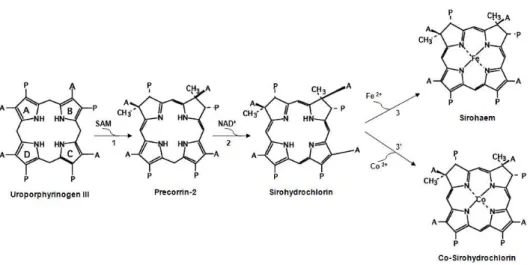
With the objective of clarifying the tetrapyrrole biosynthetic pathway in D. vulgaris Hildenborough, we performed the biochemical
study of several enzymes involved in the generation of sirohydrochlorin,
sirohaem and cobalt-sirohydrochlorin from aminolaevulinic acid, namely
porphobilinogen synthase (HemB), porphobilinogen deaminase (HemC),
uroporphyrinogen III synthase (HemD), uroporphyrinogen III
methyltransferase (SUMT), precorrin-2 dehydrogenase (CysGB) and
sirohydrochlorin ferro- and cobalto-chelatases (CysGB and CbiK,
respectively).
These studies showed that D. vulgaris porphobilinogen synthase is a
hexameric zinc-dependent enzyme and that the porphobilinogen
deaminase contains a dipyrromethane-cofactor. The gene for D. vulgaris
uroporphyrinogen III synthase is fused with the gene encoding uroporphyrinogen III methyltransferase. Both activities were demonstrated in the complete protein, and two separate domains of the
protein, generated by molecular genetic techniques, exhibit the
contain only the dehydrogenase activity, being simply capable of synthesising sirohydrochlorin rather than sirohaem. D. vulgaris
contains two CbiK cobaltochelatases (CbiKP and CbiKC) that were shown
in the present study, to perform in vitro cobalt and iron chelation into
sirohydrochlorin. CbiKP has a signal peptide that exports the protein to
the periplasm, whereas CbiKC is located in the cytoplasm. Expression of
CbiKC and a form of CbiKP lacking the signal peptide, from a multicopy
vector, restored the wild type phenotype of an E. coli mutant strain deficient in sirohydrochlorin ferrochelatase activity, showing that they can act in the in vivo synthesis of sirohaem in E. coli.
D. vulgaris CbiKP is a tetrameric protein that contains a b-type
haem, which is so far a unique feature within the chelatase-family of
enzymes. We have demonstrated that haem b of CbiKP is not involved in
the chelatase activity and the three-dimentional structure of CbiKP
revealed that the haem is located in between two monomers and
coordinated by two histidines, namely His96 of each monomer. Co
-crystallisation of CbiKP with cobalt showed three amino acid residues
involved in cobalt binding: His154, Glu184 and His216. Moreover,
comparison of the structure with and without cobalt shows that His154, a strictly conserved amino acid, is the only residue that undergoes a
significant alteration, most probably to allow the coordination of the cobalt ion.
It was also shown that D. vulgaris Hildenborough synthesises
vitamin B12 at the cytoplasm level, being CbiKC the most likely enzyme
to be involved in this process. Although the function of CbiKP remains
unclear, our analysis of the D. vulgaris genome together with biochemical and genetic studies of cobaltochelatases from other
synthase/methyltransferase suggests that uroporphyrinogen III is not released as an intermediate. Therefore, uroporphyrinogen III cannot be
acted upon by the first enzyme of the haem biosynthetic branch of the tetrapyrrole pathway. This event is consistent with a proposal made in the 1990’s of an alternative pathway being operative in Desulfovibrio, in
which precorrin-2 would be the intermediate in the haem synthesis
pathway. However, the enzymes necessary to convert precorrin-2 to
haem are not known, given that Desulfovibrio genomes lack the genes encoding the canonical haem synthesizing enzymes. Since our analysis of the genome of D. vulgaris show the presence of two genes encoding
NirJ and NirD, two proteins involved in the synthesis of haem d1, which
is made via precorrin-2, we proposed that in Desulfovibrio the enzymes
of the d1 biogenesis may be involved in the conversion of precorrin-2,
sirohydrochlorin or sirohaem, into haem. The proposed pathway of haem
Desulfovibrio desulfuricans foi a primeira espécie do grupo das bactérias redutoras de sulfato a ser isolada, em 1895. Desde essa altura, muitas questões sobre o metabolismo e ecologia destas bactérias foram discutidas na comunidade científica. Actualmente, existem ainda
inúmeras questões em aberto que precisam de ser exploradas, de forma a alargar o nosso conhecimento das vias metabólicas operativas nestes
organismos, que têm implicações directas no ciclo do enxofre, biocorrosão em esgotos e na indústria do petróleo e do gás e na bioremediação de vários metais tóxicos. O trabalho apresentado nesta dissertação teve
como objectivo contribuir para o conhecimento das enzimas envolvidas em dois sistemas metabólicos diferentes de Desulfovibrio, nomeadamente enzimas que desempenham funções na resposta ao stress oxidativo e enzimas envolvidas na via biosintética dos hemos.
Apesar das espécies de Desulfovibrio serem consideradas desde há muitos anos como anaeróbias estritas, várias estirpes de Desulfovibrio vivem em habitats expostos a oxigénio. De facto, estes microorganismos
possuem vários sistemas que permitem a sua sobrevivência em condições oxidativas mas o seu crescimento aeróbio nunca foi reportado. Nesta
dissertação é apresentada pela primeira vez uma espécie de Desulfovibrio, D. desulfuricans ATCC 27774, que tem a capacidade de crescer quando exposta a concentrações atmosféricas de oxigénio. Os nossos resultados sugerem que D. desulfuricans ATCC 27774 utiliza oxigénio para crescer, retendo ao mesmo tempo a capacidade de
respiração de nitrato. Estudos realizados em fracções proteicas de células de D. desulfuricans ATCC 27774 submetidas a 18 % de oxigénio, mostram um aumento da expressão de proteínas envolvidas na
estes organismos possuem genes que codificam para proteínas que fazem
a redução do oxigénio a água, incluindo redutases de oxigénio hemo
-cobre. A análise das sequências de aminoácidos das redutases de
oxigénio hemo-cobre, codificadas nos genomas da maioria das espécies de
Desulfovibrio, prevê a existência de uma nova enzima da família A2, a
ccaa3, que contém na sua subunidade II um motivo extra de ligação a um
hemo c (CxxCH). A caracterização bioquímica de uma forma truncada da
subunidade II da enzima ccaa3 de D. vulgaris Hildenborough confirmou a
ligação de dois hemos do tipo c, com uma coordenação His-Met ao ferro
do hemo. Neste trabalho foi ainda mostrado que o citocromo c553 dá
electrões à ccaa3, revelando-se o primeiro possível dador de electrões
fisiológico para a redutase de oxigénio hemo-cobre nestes organismos.
Com o objectivo de elucidar a via biosintética dos tetrapirroles em D.
vulgaris Hildenborough, foi realizado o estudo bioquímico de várias
enzimas envolvidas na síntese de sirohidroclorina, sirohemo e cobalto
-sirohidroclorina, a partir de ácido aminolevulínico, nomeadamente porfobilinogénio sintase (HemB), porfobilinogénio deaminase (HemC),
uroporfirinogénio III sintase (HemD), uroporfirinogénio III
metiltransferase (SUMT), precorrina-2 desidrogenase (CysGB),
sirohidroclorina ferroquelatase (CysGB) e sirohidroclorina cobalto
-quelatase (CbiK).
Estes estudos mostraram que a proteína porfobilinogénio sintase de D. vulgaris é uma enzima hexamérica, cuja actividade é dependente de zinco, e que a porfobilinogénio deaminase contém um dipirrometano como cofactor. O gene que codifica para a uroporfirinogénio III sintase
em D. vulgaris encontra-se fundido com o gene da uroporfirinogénio III
metil-transferase. Ambas as actividades foram demonstradas na
ferroquelatase, codificada no genoma de D. vulgaris, foi demonstrada possuir apenas a actividade de desidrogenase, sendo capaz de sintetizar sirohidroclorina e não sirohemo, como proposto. D. vulgaris tem duas
cobaltoquelatases (CbiKP e CbiKC) e neste estudo foi demonstrado que
ambas fazem a inserção in vitro de cobalto e ferro na sirohidroclorina. A
CbiKP possui um sinal peptídico que a transporta para o periplasma,
enquanto que a proteína CbiKC é citoplasmática. Quando expressas a
partir de um vector de cópia múltipla, numa estirpe mutante de E. coli que é deficiente em actividade de ferrochelatase de sirohidroclorina, as
duas quelatases (CbiKC e uma forma truncada da CbiKP, em que o sinal
peptídico foi removido) são capazes de restabelecer o fenótipo da estirpe
de E. coli selvagem, demonstrando a sua capacidade de participar na
síntese in vivo do sirohemo.
A CbiKP de D. vulgaris é uma proteína tetramérica que contém um
hemo do tipo b, uma característica que é até agora única na família das quelatases. Os nossos estudos demonstraram ainda que o hemo b da
CbiKP não participa na actividade de ferro- ou cobalto-quelatase e a
estrutura tridimensional da CbiKP revelou que o hemo está localizado
entre dois monómeros sendo coordenado por duas histidinas, uma de
cada monómero (His96). A co-cristalização da proteína CbiKP com cobalto
mostrou que existem três aminoácidos envolvidos na ligação ao cobalto:
His154, Glu184 e His216. A comparação da estrutura da CbiKP com e
sem cobalto permitiu verificar que a His154, um aminoácido estritamente conservado, é o único resíduo que sofre uma alteração posicional significativa, provavelmente para permitir a coordenação do
ião cobalto.
Foi também mostrado que D. vulgaris Hildenborough sintetiza
vitamina B12 ao nível do citoplasma, e que muito possivelmente a
em conjunto com estudos bioquímicos e genéticos efectuados com
cobaltoquelatases de outros organismos, sugere o envolvimento da CbiKP
de D. vulgaris no transporte de hemo ou/e ferro.
A presença em D. vulgaris da proteína bi-functional
uroporfirinogénio III sintase/metil-transferase, sugere que o
uroporfirinogénio III não é libertado como intermediário. Desta forma, este composto não pode servir de substrato à primeira enzima que actua
no ramo biossintético do hemo da via metabólica dos tetrapirroles. Este facto é consistente com uma proposta feita nos anos 90, que sugere a existência de uma via alternativa em Desulfovibrio, na qual a
precorrina-2 seria um intermediário na via biosíntetica dos hemos. No
entanto, as enzimas necessárias para converter a precorrina-2 em hemo
não são conhecidas, uma vez que os genomas de Desulfovibrio não codificam para as enzimas clássicas que participam na via biossintética do hemo. Uma vez que a nossa análise do genoma de D. vulgaris mostra a existência de dois genes que codificam para duas proteínas, a NirJ e a
NirD, que estão envolvidas na síntese do hemo d1, que ocorre via a
precorrina-2, nós propusemos que em Desulfovibrio as enzimas da
biogénese do hemo d1 estão envolvidas na conversão da precorrina-2, da
sirohidroclorina ou do sirohemo, em hemo. A via proposta para a
biossíntese dos hemos em Desulfovibrio é discutida no capítulo final
∆ Deletion Å Acetate ADP ALA Angstrom
─CH2COOH
Adenosine diphosphate
δ-Aminoleaevulinic acid
ATCC American type culture collection
ATP Adenosine triphosphate
bp CoA CO CO2 Da DCIP DNA
Base pair Coenzyme A Carbon monoxide Carbon dioxide Dalton
2,6-Dichlorphenolindophenol
Deoxyribonucleic acid
e- Electron
Em
EPR
Redox potential
Electron paramagnetic resonance FAD
Fe-S
FMN
Flavin adenine dinucleotide Iron-sulphur cluster
Flavin mononucleotide
g EPR g-factor
H2 Hydrogen
H+ Proton
HMB Hydroximethylbilane
HPLC IPTG
High performance liquid chromatography Isopropyl-ß-D-thiogalactopyranoside
LB Luria Bertani
M Mb Methyl NAD NADH Molar Mega base pair
─CH3
β-Nicotinamide adenine dinucleotide, oxidized form
β-Nicotinamide adenine dinucleotide, reduced form
NADP NADPH
β-Nicotinamide adenine dinucleotide phosphate, oxidized form
β-Nicotinamide adenine dinucleotide phosphate, reduced form
NMR Nuclear magnetic resonance PBG
PCR Pi Propionate
Porphobilinogen Polymerase chain reaction Inorganic phosphate
─CH2CH2COOH
RNA Ribonucleic acid ROS Reactive oxygen species SAM/SAH
SO42
-SUMT H2S
S-adenosyl-methionine/ S-adenosyl-homocysteine
Sulphate
S-adenosyl-methionine:uroporphyrinogen III methyltransferase
Sulphide/hydrogen sulphide
sp. Species
SRB Sulphate reducing bacteria TMPD
tRNA UV
N,N, N’,N’-Tetramethyl-p-phenylenediamine
Transfer ribonucleic acid Ultraviolet
Vinyl Vis
─CH═CH2
Visible
e.g. exempli gratia, for example
et al. et alia, and other people
Part I - Introduction
Chapter 1 -Desulfovibrio
1.1 - Sulphate Reducing Bacteria ________________________________________________ 5
1.1.1 - An overview on sulphate reducing bacteria ______________________________ 5 1.1.2 - Historical perspective ________________________________________________ 6
1.2 -Desulfovibrio______________________________________________________________ 8 1.2.1 - Energy metabolism of Desulfovibrio ___________________________________ 9
Sulphate reduction _________________________________________________ 10 Nitrate and nitrite reduction ________________________________________ 11 Hydrogen oxidation ________________________________________________ 12 Lactate, pyruvate and formate oxidation _____________________________ 13 Electron carriers and membrane-bound electron
transport complexes _______________________________________________ 14
1.2.2 -Desulfovibrio in environment and bioremediation ________________________ 16 1.2.3 -Desulfovibrio in human health and disease ______________________________ 17
1.3 - References _______________________________________________________________ 18
Chapter 2 -Desulfovibrio and oxygen
2.1 - Sulphate reducing bacteria in oxic habitats ___________________________________ 25
2.2 - Desulfovibrio responses to oxygen ___________________________________________ 25 2.3 - Proteins involved in the response to oxidative stress __________________________ 27
2.3.1 - Detoxification of reactive oxygen species _______________________________ 29
2.3.2 - Oxygen reduction systems ____________________________________________ 30
Cytoplasmic oxygen reduction ________________________________________ 30 Periplasmic oxygen reduction _________________________________________ 30 Membrane-bound oxygen reductases ___________________________________ 31
2.4 - Transcriptomic and proteomic response to oxidative stress ____________________ 34
Historical perspective _______________________________________________________ 43 Nomenclature of tetrapyrroles _______________________________________________ 45 3.2 - How are tetrapyrroles synthesised? __________________________________________ 46
3.2.1 - Overview on the early steps of the pathway _____________________________ 47
Aminolevulinic acid and Porphobilinogen ________________________________ 47 Hydroxymethylbilane __________________________________________________ 49 Uroporphyrinogen III __________________________________________________ 50 3.2.2 - Overview on the late steps of the pathway ______________________________ 51
Sirohaem and vitamin B12 _____________________________________________ 51
Precorrin-2 __________________________________________________________ 53
Sirohydrochlorin _____________________________________________________ 54 Sirohaem and cobalt-sirohydrochlorin __________________________________ 55
Haem _______________________________________________________________ 57 3.3 - Tetrapyrroles of Desulfovibrio _______________________________________________ 59
3.4 - References ________________________________________________________________ 60
Part II -Results
Chapter 1 - The anaerobe Desulfovibrio desulfuricans ATCC 27774 grows at nearly atmospheric oxygen levels
1.1 - Introduction _______________________________________________________________ 71
1.2 - Materials and Methods _____________________________________________________ 71
1.3 - Results ___________________________________________________________________ 73
1.4 - Discussion and conclusion __________________________________________________ 78
1.5 - Acknowledgments _________________________________________________________ 79
2.1 - Introduction _______________________________________________________________ 85
2.2 - Materials and Methods _____________________________________________________ 87
2.3 - Results and discussion _____________________________________________________ 92
2.4 - Conclusion ________________________________________________________________ 101
2.5 - Acknowledgments _________________________________________________________ 101
2.6 - References _______________________________________________________________ 102
Chapter 3 - Functional characterization of the early steps of the tetrapyrrole biosynthetic pathway in Desulfovibrio vulgarisHildenborough
3.1 - Introduction _______________________________________________________________ 107
3.2 - Materials and Methods _____________________________________________________ 110
3.3 - Results and discussion _____________________________________________________ 116
3.4 - Conclusion ________________________________________________________________ 124
3.5 - Acknowledgments _________________________________________________________ 127
3.6 - References _______________________________________________________________ 128
Chapter 4 - Two distinct roles for two functional cobaltochelatases (CbiK) in Desulfovibrio vulgaris Hildenborough
4.1 - Introduction _______________________________________________________________ 135
4.2 - Materials and Methods _____________________________________________________ 136
4.3 - Results ___________________________________________________________________ 141
4.4 - Discussion and conclusion __________________________________________________ 147
4.5 - Acknowledgments _________________________________________________________ 150
4.6 - References _______________________________________________________________ 151
Chapter 5 - Crystal structure of a haem-containing cobaltochelatase (CbiKP) of Desulfovibrio vulgaris Hildenborough
5.1 - Introduction _______________________________________________________________ 157
5.2 - Materials and Methods _____________________________________________________ 158
5.3 - Results ___________________________________________________________________ 161
5.4 - Discussion and conclusion __________________________________________________ 167
Discussion
1 -D. desulfuricans ATCC 27774grows in the presence of oxygen __________________ 179
2 - A new type of haem-copper oxygen reductase __________________________________ 181
3 - Tetrapyrrole biosynthesis in Desulfovibrio _____________________________________ 184
4 - Insights on an alternative haem biosynthetic pathway ___________________________ 190
Chapter 1
Desulfovibrio
1.1 - Sulphate Reducing Bacteria _________________________________________ 5
1.1.1 - An overview on sulphate reducing bacteria _______________________ 5
1.1.2 - Historical perspective ____________________________________________ 6 1.2 - Desulfovibrio _________________________________________________________ 8
1.2.1 - Energy metabolism of Desulfovibrio _____________________________ 9 Sulphate reduction ______________________________________________ 10 Nitrate and nitrite reduction _____________________________________ 11
Hydrogen oxidation ______________________________________________ 12
Lactate, pyruvate and formate oxidation _________________________ 13
Electron carriers and membrane-bound electron
transport complexes _____________________________________________ 14
1.1 - Sulphate Reducing Bacteria
1.1.1 - An overview on sulphate reducing bacteria
Sulphate reducing bacteria (SRB) are a large and heterogeneous group of prokaryotic microorganisms that perform dissimilatory sulphate
reduction, i.e., they are able to couple the anaerobic oxidation of organic
compounds or molecular hydrogen to the reduction of sulphate to hydrogen sulphide in order to obtain energy for growth (1). Since this is a very ancient process, some of the oldest life forms on Earth probably belong to this group of microorganisms (2). The dissimilatory sulphate reduction
product, hydrogen sulphide, has a typical smell and in the presence of iron minerals forms black precipitates of ferrous sulphide, which makes easy the recognition of SRB habitats (1). In fact, SRB are widespread in nature
and mainly found in sulphate-rich anoxic habitats such as soil, marine and
fresh waters and sediments as well as in the oxic-anoxic interfaces of all
these biotopes and in the gut of many animals, including humans. Although named after a single electron acceptor, SRB are metabolic versatile and therefore able to use other organic or inorganic compounds as
terminal electron acceptors, such as nitrate, nitrite, carbon dioxide, iron(III), fumarate, elemental sulphur and other sulphur species (3, 4).
This group of microorganisms has a significant environmental impact
in several aspects: i) they participate in the carbon and sulphur cycle by
recycling sulphur compounds in the degradation of organic matter; ii) SRB
are involved in bioremediation, being able to reduce several toxic metals that constitute an environmental pollution problem with great impact in public health and economy (5). Apart from the beneficial point of view, SRB are a major problem in oil industry due to the production of sulphide, which in high concentrations leads to corrosion of the pipelines and equipments
biodeterioration of paleolithic paintings (6). As inhabitants of the animal and human intestine, SRB have health relevance and some studies led to the hypothesis that SRB may play a role in inflammatory bowel diseases (7).
1.1.2 - Historical perspective
It was in 1895 that the Dutch microbiologist Martinus Beijerinck
(Figure I-1.1) described the first sulphate reducer, Spirillum desulfuricans
(Figure I-1.2) (8). Beijerinck isolated this bacterium from a Dutch city canal
in Delft and described it as the cause of contamination of the city sewages
in the summer due to the production of hydrogen sulphide. Since he observed that Spirillum desulfuricans was difficult to growth in the absence of aerobic bacteria, that were required to consume the oxygen in the culture, the bacterium was classified as strict anaerobe (9, 10).
Figure I-1.1 - Left: Martinus Beijerinck (1851-1931), Professor of microbiology at the Technical
School in Delft (Netherlands) which isolated the first sulphate reducing bacterium. Right:
Beijerinck's Laboratory for Microbiology in Delft.
time, this bacterium was named Vibrio desulfuricans and later was
renamed Desulfovibrio desulfuricans, by Kluyver and van Niel (11). The discovery that sulphate reducers
could oxidize H2 occurred in 1931 and
was between 1950 and 1960 that the
first steps on sulphate-reducer
biochemistry were made, leading to important discoveries such as the
existence of cytochromes, the pigments that were only known to be associated with aerobic respiration,
and the presence of a green enzyme, named desulfoviridin, further
recognized as a sulphite-reductase (1).
Following this period of time, several areas of study emerged allowing a
Figure I-1.2 - Painting of Vibrio desulfuricans (Desulfovibrio desulfuricans) by the sister of Martinus Beijerinck (9).
better understanding of these bacteria. These included the taxonomic classification of several sulphate reducers, the exploration of alternate
electron donors and acceptors for SRB growth, and the biochemical and functional characterization of several enzymes present in these microorganisms.
In the 1960s, the taxonomic classification of SRB was essentially based in cell morphology and ability to form spores. Although SRB are a group of prokaryotes with different types of cell morphology (e.g. cocci, oval, rods, curved (vibrioid) types, cell aggregates and filaments), these bacteria were
first classified in two genera on the basis of morphology: the vibrio-shaped
and nonspore-forming genera Desulfovibrio and the rod-shaped spore
-forming genera Desulfotomaculum (12). This phylogenetic classification
accurate classification of SRB was achieved based on the 16S rRNA sequences of the several strains (13), and the present phylogenetic classification of SRB takes in account several taxonomic markers such as
the cell morphology, the 16S rRNA, the nutritional characteristics, the presence of special pigments and the DNA guanine+cytosine (G+C) content. In addition, recent studies on lateral gene transfer events of gene fragments encoding enzymes of the sulphate respiration pathway (e.g.
dissimilatory adenosine-5'-phosphosulphate (APS) reductase (ApsAB) and
the dissimilatoty sulphide reductase (DsrAB)) between the several species, provided a more restrict evolutionary classification of SRB (14, 15).
Although sulphate reducers have been studied for more than one
century, only in the last few years a better understanding of their life and the metabolic implications in environment and human health has been
possible as a result of studies at the genetic, biochemical and molecular level (see (16) for review). Still, there are many questions that remain to be addressed to fully understand the importance of these organisms.
1.2 - Desulfovibrio
Desulfovibrio, which belongs to the class of δ-proteobacteria is one of
the best studied genus of sulphate reducing bacteria. Several Desulfovibrio species are found in aquatic habitats (marine and fresh waters), soil and
sediments, oil and natural gas wells, sewages and in the gut of animals and humans.
Cells of Desulfovibrio present
Gram-negative staining, curved or
rod morphology and are often motile
(Figure I-1.3). They typically contain high mol % and, in general, contain the sulphite reductase
desulfoviridin and the tetrahaem
Figure I-1.3 - Desulfovibrio desulfuricans
cytochrome c3. The optimal growth temperature of Desulfovibrio strains is
between 30-38 ºC (17, 18).
Advances in genome sequencing have contributed for a better
understanding of the general metabolism of Desulfovibrio species (19).
Presently, five strains of Desulfovibrio have its genome completely
sequenced: D. vulgaris str. Hildenborough (20), D. vulgaris str. PD4, D.
vulgaris str. Myasaki, D. desulfuricans str. G20 and D. desulfuricans str.
ATCC 27774 (21). Genome sequencing of other Desulfovibrio strains are
currently in progress, namely for D. salexigens str. DSM 2638 (21), D.
magneticus str. RS-1 (22) and D. piger str. ATCC 29098 (23). The mol % of
G+C in these Desulfovibrio genomes varies between 47-67 % and the size of
the genomes contains between 3.5-4.2 Mb, being the genome of D.
desulfuricans ATCC 27774 the smallest, with approximately 2.9 Mb.
1.2.1 - Energy metabolism of Desulfovibrio
As previously mentioned, Desulfovibrio sp. gain energy by performing dissimilatory sulphate reduction, a process where sulphate is reduced to
sulphide, coupled with the oxidation of H2 or organic substrates. Besides
sulphate, several other electron acceptors are used, such as sulphite,
thiosulphate, sulphur, nitrate, elemental Fe(III), CO2 and fumarate.
Growth of Desulfovibrio with fumarate as electron acceptor involves a
quinol:fumarate oxidoreductase (QFR) that reduces fumarate to succinate.
The latter can also catalyse the reverse reaction of succinate oxidation
although this reaction is normally catalysed by succinate:quinone
oxidoreductase (SQR) (24). The most common organic substrates utilized by Desulfovibrio are lactate, pyruvate and ethanol. These substrates are
incompletely oxidized to acetate and carbon dioxide due to the apparent
lack of a mechanism for acetyl-coenzyme A (CoA) oxidation. Consequently
members of this genus are called “incomplete oxidizers”. Desulfovibrio sp.
serine) and sugars (fructose) as electron donors. Growth in the absence of
external electron donor is also possible by fermentation of sulphite and thiosulphate to sulphate and sulphide. This process is known as
disproportionation. Fumarate and malate can also be disproportionated to yield succinate (1, 25).
Sulphate reduction
The reduction of sulphate occurs in the cytoplasm near the inner side of the cytoplasmic membrane by an overall reaction that involves eight
electrons and requires ATP (1):
SO42- + ATP + 8H+ + 8e- HS- + AMP + PPi
The energy loss is compensated by proton-gradient coupled to
phosphorylation of ADP to ATP (1). External sulphate is initially
transported across the membrane, to the cytoplasm, by an ion-gradient.
This uptake is achieved either by simultaneous transport of sulphate with protons, in the case of D. desulfuricans (26) or with sodium ions, as in D. salexigens (27). Upon entering the cytoplasm, sulphate, which is
thermodynamically stable and by itself is not a suitable electron acceptor,
has to be activated (3). The activation of sulphate to adenosine 5´
-phosphosulphate (APS) is made at the expense of ATP in a reaction
catalysed by ATP sulfurylase (Figure I-1.4). The next steps of the pathway
are the reduction of APS to bisulphite and adenosine-5´-phosphate (AMP),
catalysed by APS reductase, and the reduction of bisulphite to sulphide, by the enzyme bisulphide reductase, which in Desulfovibrio is known as desulfoviridin (1, 3). The mechanism for the latter reaction is still not
completely clear and two different pathways have been proposed (Figure I
-1.4). In one of the pathways (trithionate pathway), the reduction proceeds
have shown that growth of D. vulgaris on sulphate and H2 was not
impaired when thiosulphate reduction was abolished, thus it was proposed
that under these growth conditions, APS and HSO3- are the only
intermediates (30).
Figure I-1.4 - Proposed pathways of dissimilatory sulphate reduction (adapted from (25) and (17)).
Nitrate and nitrite reduction
D. desulfuricans ATCC 27774 is capable of using nitrite and/or nitrate as electron acceptors. Nitrate is reduced to ammonia in a dissimilatory
pathway that involves the nitrate reductase and nitrite reductase enzymes. The first step is the conversion of nitrate to nitrite, performed by the enzyme nitrate reductase. D. desulfuricans ATCC 27774 has a dissimilatory periplasmic nitrate reductase (Nap) which contains one
molybdenum cofactor in the active site and a [4Fe-4S] cluster. The crystal
structure of D. desulfuricans Nap was the first to be solved for a nitrate reductase (31). The six electron reduction of nitrite to ammonia is
performed by nitrite reductase (NiR) enzymes. Two different types of NiRs
are known: the cytoplasmic sirohaem NiRs, proposed to be involved in the
detoxification of nitrite, and the membrane bound or periplasmic
cytochromes c NiRs, which are involved in the dissimilatory nitrate
pathway. The dissimilatory cytochrome c nitrite reductase of D.
desulfuricans ATCC 27774 is a membrane-bound protein composed by two
side, and the transmembrane tetrahaem subunit, NrfH (32). D. vulgaris Hildenborough is not able to reduce nitrate but it can reduce nitrite to ammonium through a constitutive cytochrome c NiR, in a reaction that does not sustain cell growth (33).
Hydrogen oxidation
Hydrogen can be used by Desulfovibrio as the sole energy source, with
acetate and carbon dioxide as carbon sources. Hydrogen is oxidized by enzymes called hydrogenases that can be cytoplasmic, periplasmic or
membrane-associated. Three types of hydrogenases are distinguished in
Desulfovibrio: the [Fe], [NiFe] and [NiFeSe] hydrogenases. Besides metal
composition, these enzymes have differences concerning molecular mass, catalytic activity and sensitivity for CO, NO, nitrite and acetylene (34). The
[Fe] hydrogenase of D. desulfuricans contains two ferredoxin-type [4Fe
-4S]2+/1+ clusters and a [4Fe-4S] cluster bridged to a binuclear active site Fe
center that is involved in H2 activation (35). The first solved three
-dimensional structure of a hydrogenase was the periplasmic [NiFe]
hydrogenase from D. gigas which contains besides the nickel-iron binuclear
active site, two [4Fe-4S]2+/1+ clusters and one [3Fe-4S]1+/0 cluster (36). The
structure of the periplasmic [NiFeSe] hydrogenases from Desulfomicrobium
baculatum showed that the enzyme contains three [4Fe-4S]2+/1+ clusters
and that selenium is present in the form of a selenium-cysteine residue
bound to the nickel-iron binuclear center (37).
In order to get energy for sulphate reduction, the periplasmic
hydrogenases have to oxidize four H2 and the eight protons and electrons
released are transferred via ATP synthase and transmembrane protein complexes, respectively, to the cytoplasm. The electrons are transfered to
the transmembrane protein redox complexes via type I tetrahaem
cytochrome c3 (TpIc3) (the physiological electron acceptor of the periplasmic
reduction of sulphate and production of ATP by ATP synthase as
consequence of the generation of a membrane potential. This process is called vectorial electron transport (39). From the eight protons available, one can be transported with sulphate to the cytoplasm (26) leaving only
seven protons for ATP synthase. Consequently, 2⅓ mol of ATP are formed,
two of which are used for sulphate reduction, leaving ⅓ ATP mol as net
energy for cell synthesis.
Lactate, pyruvate and formate oxidation
Lactate is the “classical” substrate for most Desulfovibrio sp.. Lactate and pyruvate are incompletely oxidized to acetate and carbon dioxide when they serve as electron donors for sulphate reduction. The oxidation of
lactate to pyruvate is catalysed by a membrane bound NAD(P)+
-independent lactate dehydrogenase (40). Pyruvate is oxidized to acetate
and carbon dioxide via the intermediates acetyl-CoA and acetyl-phosphate.
Acetyl-CoA is formed by a pyruvate:ferredoxin oxidoreductase (41) and then
is converted to acetate by the enzymes phosphate acetyltransferase and acetate kinase, with concomitant phosphorylation of ADP to ATP (42). In the case of lactate oxidation, no net formation of ATP is produced and thus
a proton-driven ATP production is necessary to conserve energy (1). Energy
conservation for growth of D. vulgaris in lactate and sulphate is proposed to occur through a mechanism known as “hydrogen cycling”. This mechanism considers that the electrons released by the oxidation of lactate to pyruvate
and of pyruvate to acetyl-CoA are used by a cytoplasmic hydrogenase to
produce hydrogen that, after diffusing through the membrane, is oxidised
by the periplasmic hydrogenase to reduce sulphate. The proton release in the periplasm generates a proton gradient (vectorial electron transport) that can be used by ATP synthase to produce ATP (43). This model has been a matter of debate since the existence of cytoplasmic hydrogenases in
lactate oxidation is not an energetically favourable process (44). However, the analysis of the recently sequenced D. vulgaris Hildenborough genome gives support to the hydrogen cycling mechanism, since it contains genes
encoding two orthologs of cytoplasmic-facing membrane-bound hydrogenase
complexes (EchABCDEF and CooMKLXUHF). In D. vulgaris, these
hydrogenases were proposed to be associated with production of H2 from
the oxidation of lactate or pyruvate (20).
In addition to hydrogen cycling, other mechanisms such as cycling of formate or CO may be operative for energy conservation. In D. vulgaris,
three formate dehydrogenases oxidize formate to CO2 and H+. Two of these
enzymes are periplasmic and the other one is membrane associated (45, 46). The mechanism of formate cycling suggests that this compound is formed in the cytoplasm from lactate oxidation and then diffuses to the
periplasm where is oxidized by the formate dehydrogenases. The protons are released in the cytoplasm and the electrons are donated to the periplasmic cytochromes c (20). The mechanism of CO cycling suggests that CO is formed from pyruvate (generated from lactate oxidation) by an
unknown enzyme and is subsequently converted to CO2 and H2, by a
cytoplasmic CO dehydrogenase and a CO-dependent hydrogenase, whose
genes are present in the D. vulgaris Hildenborough genome. The H2
produced may subsequently be oxidized by the periplasmic hydrogenase for sulphate reduction (47).
Electron carriers and membrane-bound electron transport complexes
In Desulfovibrio sp. the terminal reductases are cytoplasmic located
and consequently not involved in membrane charge translocation. The reducing equivalents from electrons donors must therefore be transferred
through electron carriers, and are used in the cytoplasmic sulphate
reduction. Desulfovibrio sp. contain a high number of periplasmic and
(1). The periplasmic c-type cytochromes include the monohaem cytochrome
c553 (proposed to accept electrons from iron-hydrogenases, formate
dehydrogenases and lactate dehydrogenases) and multihaem cytochromes,
such as the dimeric dihaem Split-Soret, the octahaem cytochrome c3 and
type I (TpIc3) and type II tetrahaem cytochromes c3 (TpIIc3). TpIc3 was the
first cytochrome isolated from a Desulfovibrio sp. and accept electrons from
periplasmic hydrogenases (48, 49). The periplasmic cytochromes c553 and c3
transfer electrons to membrane-bound complexes that contain a soluble
cytochrome subunit of the c3 family as electron acceptor. For example, Hmc
is a transmembrane complex present in D. vulgaris Hildenborough that
contains a 16-haem cytochrome (HmcA) (49). The 9-haem cytochrome c
(9HcA), isolated from D. desulfuricans ATCC 27774, is also part of
membrane-bound complex (9Hc) and is highly expressed on cells grown
with sulphate (50). A third example is the Tmc complex in which the
cytochrome c subunit is a TpIIc3 (TmcA) (51). The cytochrome c subunits of
these three complexes can accept electrons from periplasmic hydrogenases
via TpIc3 (49). Desulfovibrio sp. contains other transmembrane complexes
such as Qmo, which lacks the periplasmic cytochrome c subunit, and is proposed to transfer electrons from the menaquinone pool to APS reductase (52), and the DrsMKJOP complex, probably involved in the transfer of electrons to the dissimilatory sulphite reductase. Qmo and Drs complexes
may be essential for sulphate reduction given their strong conservation among sulphate reducers (53).
Desulfovibrio contains also low molecular mass proteins such as ferredoxins, flavodoxins and rubredoxins which are involved in electron
transfer processes. Ferredoxins are distinguished by the type of iron-sulfur
cluster which they comprise. In D. gigas, ferredoxin I contains one [4Fe-4S]
whereas ferredoxin II harbours one [3Fe-4S] cluster (54). Flavodoxins
contain FMN as cofactor and rubredoxins have one [Fe-S(Cys)4] centre (55,
1.2.2 - Desulfovibrio in environment and bioremediation
SRB have been recognized for a long time to be associated with
microbial corrosion that occurs in many industrial systems such as water supplies, drinking water distribution systems and petroleum and gas industry. Within the group of SRB, the majority of the strains present in the oil and gas reservoirs belong to the Desulfovibrio genus. In oil and gas industries the pressure within the reservoirs is maintained by injection of
water or gas. The combination of the organic components that result from oil degradation (electron donors) with the sulphate (electron acceptor) present in water promotes the growth of SRB (native or introduced with the
water injections), which are responsible for production of hydrogen sulphite in the oil and gas reservoirs, in a process named souring. Hydrogen
sulphide decreases the quality of oil and gas and causes corrosion of the
pipelines and processing equipment due to the reaction with Fe2+ to form a
mixture of iron sulphides which are corrosive products (57). Strategies for control of souring in oil reservoirs have been developed and the most
reliable is nitrate injection. The presence of nitrate stimulates nitrate
-reducing, sulphide-oxidizing and nitrate-reducing bacteria that remove
sulphide with the production of nitrite, which has an inhibitory effect on SRB (57). Nitrite is a strong competitive inhibitor of the dissimilatory sulphite reductase (DsrAB) of Desulfovibrio, since this enzyme binds tightly nitrite and slowly reduces it to ammonia (preventing the normal physiological function of DsrAB, i. e. reduction of sulphite to sulphide) (57, 58). D. vulgaris Hildenborough prevents this inhibitory event by the action
of pentahaem nitrite reductase (NrfAH) but the efficiency of this detoxification system is dependent on the time required for the reduction of all nitrite content to ammonia, given that the protons and electrons from
concentrations of nitrite and thus SRB lacking this enzyme are even more sensitive to nitrite inhibition (57, 58).
Although Desulfovibrios are a problem for oil and gas industries, they
constitute a group of microorganisms with great potential for bioremediation. The interactions between sulphate reducers and metals and metalloids has been of interest since these organisms have the capacity
to reduce several toxic metals such as uranium(VI), copper(II), chromium(VI) and manganese(II). Also, the sulphides produced during SRB
growth are able to precipitate several heavy metals into insoluble metal sulphides. The ability of these bacteria to reduce these toxic metals depends on the metal concentration in solution. The effect of heavy metals on
sulphate reducers can be stimulatory at lower concentrations and inhibitory at higher concentrations. The reduction of these soluble metals
into insoluble forms allows their removal from contaminated waters and waste streams which is performed in bioreactors (59). Hydrogenases and/or cytochromes are involved in the enzymatic reduction of the metals and metalloids by sulphate reducers. In D. vulgaris the tetrahaem cytochrome
c3 is involved in reduction of U(VI) to U(V) and both the cytochrome c3 and
the Fe-hydrogenase can reduce Cr(VI) to Cr(III) (60).
1.2.3 - Desulfovibrio in humans
Anaerobic fermentation in the intestine leads to the formation of
products such as hydrogen, propionate, carbon dioxide, lactate and succinate, which can be utilized by Desulfovibrio. Sulphate is also available
showed that Desulfovibrios are ubiquitous in human faeces but the number of these microorganisms varied with age (7).
Hydrogen sulphide produced by Desulfovibrio is toxic at high
concentrations being a cause of DNA damage and inhibition of butyrate oxidation, an essential process for colon epithelial cells. The inhibition of the latter process is associated with ulcerative colitis, a form of
inflammatory bowel disease (IBD), an acute and chronic illness of the small intestine and of the colon. Patients with ulcerative colitis and Crohn’s
disease (another form of IBD) seem to have a higher number of Desulfovibrios in their faeces than healthy individuals (61).
Some Desulfovibrio sp. were also isolated from abscesses of the brain,
intra-abdominal and abdominal walls and pyogenic liver and from
periodontal mouth lesions (7). Cases of bacteremia caused by D. fairfieldensis and D. desulfuricans were also reported (62), showing that D. fairfieldensis, is highly resistant to certain antimicrobial drugs (62, 63). Desulfovibrio strain FH26001/95, isolated from liver abscesses, was shown to grow with dissolved oxygen concentrations up to 50 µM. This tolerance to
oxygen may be a strategy of Desulfovibrio to survive in the blood and
tissues, where the concentration of dissolved oxygen is approximately 110 µM (64).
1.3 - References
(1) Ralf Rabus, T. A. H. a. F. W. (2006) Dissimilatory Sulfate- and Sulfur-Reducing
Prokaryotes in: Martin Dworkin, Stanley Falkow, Eugene Rosenberg, Karl-Heinz Schleifer and Erko Stackebrandt (Eds.), The Prokaryotes, Vol. 2, Springer, New York, pp 659-768.
(2) Shen, Y., Buick, R., and Canfield, D. E. (2001) Isotopic evidence for microbial sulphate reduction in the early Archaean era. Nature410, 77-81.
(3) Rudolf K. Thauer, E. S., W. Allan Hamilton (2007) Energy metabolism and phylogenetic diversity of sulphate-reducing bacteria in: Larry L. Barton, W. Allan Hamilton (Eds.), Sulphate-reducing Bacteria: Environmental and Engineered Systems, Cambridge
University Press, New York, pp 1-38.
sulfate-reducing bacteria in the mouse gastrointestinal tract. Appl Environ Microbiol66,
2166-2174.
(5) Hockin, S. L., and Gadd, G. M. (2007) Bioremediation of metals and metalloids by precipitation and cellular binding in: Larry L. Barton, W. Allan Hamilton (Eds.), Sulphate-reducing Bacteria: Environmental and Engineered Systems, Cambridge
University Press, New York, pp 405-434.
(6) Portillo, M. C., and Gonzalez, J. M. (2009) Sulfate-reducing bacteria are common
members of bacterial communities in Altamira Cave (Spain). Sci Total Environ407, 1114
-1122.
(7) Macfarlane, G. T., Cummings, J. H., and Macfarlane, S. (2007) Sulphate-reducing
bacteria and the human large intestine in:Larry L. Barton, W. Allan Hamilton (Eds.), Sulphate-reducing Bacteria: Environmental and Engineered Systems, Cambridge
University Press, New York, pp 503-522.
(8) Beijerinck, W. M. (1895) Ueber Spirillum desulfuricans als ursache von sulfatreduction.
Zentralbl. Bakteriol. Parasitenkd.1, 1-9, 49-59, 104-114.
(9) Muyzer, G., and Stams, A. J. (2008) The ecology and biotechnology of sulphate-reducing
bacteria. Nat Rev Microbiol6, 441-154.
(10) Voordouw, G. (1995) The Genus Desulfovibrio: The Centennial. Appl Environ Microbiol 61, 2813-2819.
(11) Kluyver, A. J., and van Niel, C. B. (1936) Prospects for a natural system of classification of bacteria. Zentralbl. Bakterol. Parasitenkd. Infectinskr. Hyg.Abt. II94, 369-403.
(12) Campbell, L. L., and Postgate, J. R. (1965) Classification of the spore-forming sulfate
-reducing bacteria. Bacteriol Rev29, 359-363.
(13) Fowler V.J., W. F., Pfennig N., Woese. C.R., Stackebrandt E. (1986) Phylogenetic relationships of sulfate- and sulfur-reducing eubacteria. Systematic and Applied Microbiology 8, 32-41.
(14) Wagner, M., Loy, A., Klein, M., Lee, N., Ramsing, N. B., Stahl, D. A., and Friedrich, M. W. (2005) Functional marker genes for identification of sulfate-reducing prokaryotes. Methods Enzymol397, 469-489.
(15) Meyer, B., and Kuever, J. (2007) Phylogeny of the alpha and beta subunits of the dissimilatory adenosine-5'-phosphosulfate (APS) reductase from sulfate-reducing
prokaryotes-origin and evolution of the dissimilatory sulfate-reduction pathway. Microbiology153, 2026-1044.
(16) Rabus, R., Hansen, T. A., and Widdel, F. (2006) Dissimilatory Sulfate- and Sulfur
-Reducing Prokaryotes in: Martin Dworkin, Stanley Falkow, Eugene Rosenberg, Karl -Heinz Schleifer and Erko Stackebrandt (Eds.), The Prokaryotes, Vol. 2, Springer, New York, pp 659-768.
(17) Fauque G., L. J. B. L. L. (1991) Sulfate-reducing and sulfur-reducing bacteria in:Shively J.M. & Barton L.L. (Eds), Variations in Autotrophic Life., Academic Press, New York, pp. 271-337.
(18) Widdel, F., and Bak, F. (1992 ) Gram-negative mesophilic sulfate-reducing bacteria in: Balows, A., Trüper, H.G., Dworkin, M., Harder, W. and Schleifer, K.H., (Eds.) The Prokaryotes: A Handbook on the Biology of Bacteria: Ecophysiology, Isolation, Identification, Application, Vol. 1, 2nd ed, Springer Verlag, New York. pp 3353-3378.
(19) Rabus, R., and Strittmatter, A. (2007) Functional genomics of sulphate-reducing
Environmental and Engineered Systems. Cambridge University Press. New York, pp 117-140.
(20) Heidelberg, J. F., Seshadri, R., Haveman, S. A., Hemme, C. L., Paulsen, I. T., Kolonay, J. F., Eisen, J. A., Ward, N., Methe, B., Brinkac, L. M., Daugherty, S. C., Deboy, R. T., Dodson, R. J., Durkin, A. S., Madupu, R., Nelson, W. C., Sullivan, S. A., Fouts, D., Haft, D. H., Selengut, J., Peterson, J. D., Davidsen, T. M., Zafar, N., Zhou, L., Radune, D., Dimitrov, G., Hance, M., Tran, K., Khouri, H., Gill, J., Utterback, T. R., Feldblyum, T. V., Wall, J. D., Voordouw, G., and Fraser, C. M. (2004) The genome sequence of the anaerobic, sulfate-reducing bacterium Desulfovibrio vulgaris Hildenborough. Nat Biotechnol22, 554-559.
(21) DOE Joint Genome Institute (DOE JGI) (www.jgi.doe.gov).
(22) National Institute of Technology and Evaluation (NITE) (www.nite.go.jp/index-e.htm).
(23) Genome Sequencing Center (GSC) (http://genome.wustl.edu/index.cgi).
(24) Hagerhall, C. (1997) Succinate: quinone oxidoreductases. Variations on a conserved
theme. Biochim Biophys Acta1320, 107-141.
(25) Widdel, F., and Hansen, T. A. (1992) The Dissimilatory Sulfate- and Sulfur-Reducing
Bacteria in:Balows, A., Trüper, H.G., Dworkin, M., Harder, W. and Schleifer, K.H., (Eds.) The Prokaryotes, Vol. 1, 2nd ed, New York, pp 583-624.
(26) Cypionka, H. (1989) Characterization of sulfate transport in Desulfovibrio desulfuricans.
Arch Microbiol152, 237-243.
(27) Kreke, B., and Cypionka, H. (1993) Role of sodium ions for sulfate transport and energy metabolism in Desulfovibrio salexigensArch Microbiol161, 55-61.
(28) Chambers, L. A., and Trudinger, P. A. (1975) Are thiosulfate and trithionate intermediates in dissimilatory sulfate reduction? J Bacteriol123, 36-40.
(29) Drake, H. L., and Akagi, J. M. (1978) Dissimilatory reduction of bisulfite by Desulfovibrio vulgaris. J Bacteriol136, 916-923.
(30) Broco, M., Rousset, M., Oliveira, S., and Rodrigues-Pousada, C. (2005) Deletion of
flavoredoxin gene in Desulfovibrio gigas reveals its participation in thiosulfate reduction.
FEBS Lett579, 4803-4807.
(31) Dias, J. M., Than, M. E., Humm, A., Huber, R., Bourenkov, G. P., Bartunik, H. D., Bursakov, S., Calvete, J., Caldeira, J., Carneiro, C., Moura, J. J., Moura, I., and Romao, M. J. (1999) Crystal structure of the first dissimilatory nitrate reductase at 1.9 A solved by MAD methods. Structure7, 65-79.
(32) Almeida, M. G., Macieira, S., Goncalves, L. L., Huber, R., Cunha, C. A., Romao, M. J., Costa, C., Lampreia, J., Moura, J. J., and Moura, I. (2003) The isolation and characterization of cytochrome c nitrite reductase subunits (NrfA and NrfH) from
Desulfovibrio desulfuricans ATCC 27774. Re-evaluation of the spectroscopic data and
redox properties. Eur J Biochem270, 3904-3915.
(33) Pereira, I. A., LeGall, J., Xavier, A. V., and Teixeira, M. (2000) Characterization of a heme c nitrite reductase from a non-ammonifying microorganism, Desulfovibrio vulgaris
Hildenborough. Biochim Biophys Acta1481, 119-130.
(35) Nicolet, Y., Piras, C., Legrand, P., Hatchikian, C. E., and Fontecilla-Camps, J. C. (1999) Desulfovibrio desulfuricans iron hydrogenase: the structure shows unusual coordination
to an active site Fe binuclear center. Structure7, 13-23.
(36) Volbeda, A., Charon, M. H., Piras, C., Hatchikian, E. C., Frey, M., and Fontecilla-Camps,
J. C. (1995) Crystal structure of the nickel-iron hydrogenase from Desulfovibrio gigas. Nature373, 580-587.
(37) Garcin, E., Vernede, X., Hatchikian, E. C., Volbeda, A., Frey, M., and Fontecilla-Camps,
J. C. (1999) The crystal structure of a reduced [NiFeSe] hydrogenase provides an image of the activated catalytic center. Structure7, 557-566.
(38) Pereira, I. A. C., and Xavier, A. V. (2005) Multi-Heme c Cytochromes and Enzymes in: King, R. B (Ed) Encyclopedia of Inorganic Chemistry, John Wiley & Sons, New York. (39) Badziong, W., and Thauer, R. K. (1980) Vectorial electron transport in Desulfovibrio
vulgaris (Marburg), growing on hydrogen plus sulfate as sole energy source. Arch Microbiol 125, 167-174.
(40) Ogata, M., Arihara, K., and Yagi, T. (1981) D-lactate dehydrogenase of Desulfovibrio vulgaris.J Biochem89, 1423-1431.
(41) Ogata, M., and Yagi, T. (1986) Pyruvate dehydrogenase and the path of lactate degradation in Desulfovibrio vulgaris Miyazaki F. J Biochem100, 311-318.
(42) Brown, M. S., and Akagi, J. M. (1966) Purification of acetokinase from Desulfovibrio desulfuricans. J Bacteriol92, 1273-1274.
(43) Odom, J. M., and Peck Jr., H. D. (1981) Hydrogen cycling as a general mechanism for energy coupling in the sulfate-reducing bacteria, Desulfovibrio sp. FEMS Microbiol. Lett. 12, 47-50.
(44) Traore, A. S., Hatchikian, C. E., Belaich, J. P., and Le Gall, J. (1981) Microcalorimetric studies of the growth of sulfate-reducing bacteria: energetics of Desulfovibrio vulgaris
growth. J Bacteriol145, 191-199.
(45) Sebban, C., Blanchard, L., Bruschi, M., and Guerlesquin, F. (1995) Purification and characterization of the formate dehydrogenase from Desulfovibrio vulgaris
Hildenborough. FEMS Microbiol Lett133, 143-149.
(46) ElAntak, L., Dolla, A., Durand, M. C., Bianco, P., and Guerlesquin, F. (2005) Role of the tetrahemic subunit in Desulfovibrio vulgaris Hildenborough formate dehydrogenase.
Biochemistry44, 14828-14834.
(47) Voordouw, G. (2002) Carbon monoxide cycling by Desulfovibrio vulgaris Hildenborough. J Bacteriol184, 5903-5911.
(48) Liu, M. C., Costa, C., Coutinho, I. B., Moura, J. J., Moura, I., Xavier, A. V., and LeGall, J. (1988) Cytochrome components of nitrate- and sulfate-respiring Desulfovibrio desulfuricans ATCC 27774. J Bacteriol170, 5545-5551.
(49) Matias, P. M., Pereira, I. A., Soares, C. M., and Carrondo, M. A. (2005) Sulphate respiration from hydrogen in Desulfovibrio bacteria: a structural biology overview. Prog Biophys Mol Biol89, 292-329.
(50) Saraiva, L. M., da Costa, P. N., Conte, C., Xavier, A. V., and LeGall, J. (2001) In the facultative sulphate/nitrate reducer Desulfovibrio desulfuricans ATCC 27774, the nine
-haem cytochrome c is part of a membrane-bound redox complex mainly expressed in
(51) Pereira, P. M., Teixeira, M., Xavier, A. V., Louro, R. O., and Pereira, I. A. (2006) The Tmc complex from Desulfovibrio vulgaris Hildenborough is involved in transmembrane electron transfer from periplasmic hydrogen oxidation. Biochemistry45, 10359-10367.
(52) Pires, R. H., Lourenco, A. I., Morais, F., Teixeira, M., Xavier, A. V., Saraiva, L. M., and Pereira, I. A. (2003) A novel membrane-bound respiratory complex from Desulfovibrio desulfuricans ATCC 27774. Biochim Biophys Acta1605, 67-82.
(53) Pires, R. H., Venceslau, S. S., Morais, F., Teixeira, M., Xavier, A. V., and Pereira, I. A. (2006) Characterization of the Desulfovibrio desulfuricans ATCC 27774 DsrMKJOP complex-a membrane-bound redox complex involved in the sulfate respiratory pathway. Biochemistry45, 249-262.
(54) Moura, J. J., Macedo, A. L., and Palma, P. N. (1994) Ferredoxins. Methods Enzymol243, 165-188.
(55) Vervoort, J., Heering, D., Peelen, S., and van Berkel, W. (1994) Flavodoxins. Methods Enzymol243, 188-203.
(56) Sieker, L. C., Stenkamp, R. E., and LeGall, J. (1994) Rubredoxin in crystalline state.
Methods Enzymol243, 203-216.
(57) Voordouw, G. (2008) Impact of Nitrate on the Sulfur Cycle in Oil Fields in: Christiane Dahl and Cornelius G. Friedrich (Eds) Microbial Sulfur Metabolism Springer, Berlin;
New York, 296-302
(58) Haveman, S. A., Greene, E. A., Stilwell, C. P., Voordouw, J. K., and Voordouw, G. (2004) Physiological and gene expression analysis of inhibition of Desulfovibrio vulgaris
Hildenborough by nitrite. J Bacteriol186, 7944-7950.
(59) Cabrera, G., Perez, R., Gomez, J. M., Abalos, A., and Cantero, D. (2006) Toxic effects of dissolved heavy metals on Desulfovibrio vulgaris and Desulfovibrio sp. strains. J Hazard Mater135, 40-46.
(60) Michel, C., Orticoni, M., Baymann, F., Bruschi, M. (2004) Bioremediation of Chromate by Sulfate-Reducing Bacteria, Cytochromes c3 and hydrogenases Water, Air, & Soil
Pollution: Focus3, 1573-2940.
(61) Rowan, F. E., Docherty, N. G., Coffey, J. C., and O'Connell, P. R. (2009) Sulphate
-reducing bacteria and hydrogen sulphide in the aetiology of ulcerative colitis. Br J Surg 96, 151-158.
(62) Goldstein, E. J., Citron, D. M., Peraino, V. A., and Cross, S. A. (2003) Desulfovibrio desulfuricans bacteremia and review of human Desulfovibrio infections. J Clin Microbiol 41, 2752-2754.
(63) Urata, T., Kikuchi, M., Hino, T., Yoda, Y., Tamai, K., Kodaira, Y., and Hitomi, S. (2008) Bacteremia caused by Desulfovibrio fairfieldensis. J Infect Chemother14, 368-370.
(64) Schoenborn, L., Abdollahi, H., Tee, W., Dyall-Smith, M., and Janssen, P. H. (2001) A
Chapter 2
Desulfovibrio
and oxygen
2.1 - Sulphate reducing bacteria in oxic habitats ________________________ 25
2.2 - Desulfovibrio responses to oxygen _________________________________ 25
2.3 - Proteins involved in the response to oxidative stress ____________ 27
2.3.1 - Detoxification of reactive oxygen species ________________________ 29 2.3.2 - Oxygen reduction systems ______________________________________ 30 Cytoplasmic oxygen reduction __________________________________ 30
Periplasmic oxygen reduction ___________________________________ 30
Membrane-bound oxygen reductases ___________________________ 31 2.4 - Transcriptomic and proteomic response to oxidative stress _____ 34
2.1 – Sulphate reducing bacteria in oxic habitats
Sulphate reduction has long been recognized as an anoxic process
performed by strict anaerobic sulphate reducing bacteria, including the Desulfovibrio species. However, measurements of bacterial sulphate reduction and dissolved oxygen in several microbial mats show that
sulphate reduction occurs in oxygenated zones. Cyanobacterial mats are the habitat of many SRB species, and constitute a good example of the occurance of extreme shifts regarding oxygen and sulfide concentrations (1, 2). In a cyanobacterial mat of a saline evaporation pond (Baja California, Mexico), the deeper zones are occupied by SRB of the genus Desulfobacter
and Desulfobacterium, whereas Desulfovibrio and Desulfoccocus are found
in more oxic zones (upper zones) (3). In the case of a hypersaline cyanobacterial mat from Solar Lake (desert lake in the Sinai, Egypt), SRB are preferentially localized in a permanent oxic zone, being Desulfonema
and Desulfococcus the predominant groups (4). The presence of these
microorganisms in cyanobacterial mats indicate that they are able to deal
with oxygen concentrations of approximately 1 mM (1, 2).
In biofilms, SRB of the genus Desulfovibrio, Desulfomicrobium,
Desulfonema, Desulforegula, Desulfobacterium and Desulfobulbus were
also found in the oxic/anoxic interface (5). Furthermore, Desulfovibrio cuneatus was isolated from the oxic uppermost layer of sediments of the oligotrophic Lake Stechlin (6) and Desulfovibrio aerotolerans was isolated from an activated sludge (sewage treatment), which had been oxygenated at 100 % of air saturation for 120 h (7).
2.2 - Desulfovibrio responses to oxygen
The presence of oxygen in Desulfovibrio habitats imposes a great challenge to these microorganisms because as anaerobes, they should not