From the Vascular Surgery Discipline, Hospital das Clínicas, Faculty of Medicine, University of São Paulo – São Paulo/SP, Brazil.
E-mail: [email protected] Received for publication on May 17, 2004. Accepted for publication on June 24, 2004.
ORIGINAL RESEARCH
COMPARISON OF ULTRASONOGRAPHY,
COMPUTED TOMOGRAPHY AND MAGNETIC
RESONANCE IMAGING WITH INTRAOPERATIVE
MEASUREMENTS IN THE EVALUATION OF
ABDOMINAL AORTIC ANEURYSMS
Francisco das Chagas de Azevedo, Antonio Eduardo Zerati, Roberto Blasbalg, Nelson Wolosker and Pedro Puech-Leão
AZEVEDO F das C de et al. Comparison of ultrasonography, computed tomography and magnetic resonance imaging with intraoperative measurements in the evaluation of abdominal aortic aneurysms. CLINICS 60(1):21-28, 2005.
PURPOSE: To study the imaging exams more commonly used for abdominal aortic aneurysms evaluation – ultrasonography, conventional computerized tomography, helical computerized tomography and nuclear magnetic angioresonance – comparing the preoperative measurements reached by those radiological methods with the measurements made during the surgical procedures.
METHODS: Patients who had indication of elective transperitoneal surgical treatment for their abdominal aortic aneurysms were included in the study. The initial diagnosis of the aortic dilatation was made by ultrasonography and, after the surgical treatment was indicated, the patient was submitted to another imaging method.
Sixty patients were divided into 3 groups according to the complementary imaging method (conventional computerised tomography, helical computerized tomography, nuclear magnetic angioresonance). The ultrasonography of the first 20 patients were joined in a fourth group. There were considered in the study the measurements of the transversal diameter of the proximal neck, maximum transversal diameter of the aneurysm, straight-line length and transversal diameter of the common iliac arteries given by the imaging methods. The same measurements were made by using a caliper during the surgical procedure, and then compared to the values obtained from the radiological exams.
RESULTS: The maximum transverse diameter had a range measurement variation of 4.5 to 13.6 cm in the intraoperative, with no statistically significant differences when compared with all the imaging tests. The ultrasonography, however, overestimated the measurements of the proximal neck and the common iliac arteries, in comparison with intraoperative measures. The length of the aorta aneurysm obtained by the conventional computerized tomography was significantly lower if compared to the measures done with the calliper during the operation. The helical computerized tomography and the nuclear magnetic angioresonance provided measurements with no significant differences in the statistic view when compared to the intraoperative measures.
CONCLUSIONS: Ultrasonography is a reliable method for the diagnosis and follow-up of the aorta abdominal aneurysms, but insufficient for endovascular surgery planning. The conventional computed tomography can provoke distortion in the length measurements of the aorta dilatation. Helical computed tomography and nuclear magnetic angioresonance provided precise measurements of all the studied parameters, being of great utility for surgical planning.
KEYWORDS: Aortic aneurysm. Ultrasonography. Computerized tomography. Nuclear magnetic resonance.
Intraoperative measurements.
The most used imaging methods in the diagnosis and follow-up of patients with abdominal aortic aneurisms are ultrasonography (USG), conventional computerized tomog-raphy (CCT), helical computerized tomogtomog-raphy (HCT), and nuclear magnetic angioresonance (NMR).
In the literature there are reports comparing these meth-ods regarding patients with abdominal aortic aneurisms;1,2,3 however, few of them are prospective, include a large sam-ple population, or make comparisons with surgical find-ings, which are certainly the most reliable test of measure-ment efficacy for these different methods.
The objective of this study was to compare the preoperative measurements of aortic aneurysms revealed by USG, CCT, HCT, and NMR with actual measurements made during the surgical procedure, thus to evaluate the accu-racy of the imaging methods.
PATIENTS AND METHODS
Between June 1998 and December 2001, after approval by the Ethical Committee of the São Paulo University Medi-cal School, 60 consecutive patients with infrarenal abdomi-nal aortic aneurysms, with or without compromised iliac ar-teries, were analyzed.
Only patients who had an indication for surgical treat-ment by transperitoneal access were included.
The initial diagnosis of abdominal aortic aneurysm was made by USG, and the maximum transverse diameter was measured by this method. If surgical treatment was indi-cated, the patient underwent one other imaging method.
Because an aortic aneurysm is a life-threatening condi-tion that requires prompt treatment, it would not have been ethical to submit each patient to all the other 3 methods, due to the delay in surgical treatment. Consequently, patients were divided into groups according to the complementary imaging method performed after the USG (CCT, HCT, or NMR). The first 20 patients, irrespective of the complemen-tary imaging method performed, composed the group in which USG results were used in the comparison (Group 1). For the 40 subsequent patients the complementary imaging method, rather that the USG results were considered in the study. Thus, the patient groups were as follows: Group 1, USG; Group 2, CCT; Group 3, HCT; and Group 4, NMR.
All measurements during the preoperative period were made from images by the same radiologist, as follows (Fig-ure 1):
• Transverse diameter of the proximal neck of the aorta (A). • Maximum transverse aortic diameter (B).
• Straight-line length of the aneurysm (D).
• Transverse diameter of the iliac arteries at their greatest diameter when an iliac aneurysm was present, or the
di-ameter measured approximately 2 cm above the bifur-cation when they exhibited a normal caliber (C). An investigator blinded to the preoperative measure-ments made the intraoperative measuremeasure-ments in all groups. These measurements were performed using a caliper after full dissection of the aneurysm and before aortic clamping (Figure 2). A slightly modified caliper with elongated blades to facilitate the surgical measurements was utilized.
In this study, the surgical measurement was considered the gold standard to which all the imaging methods meas-urements were compared.
Of the 60 patients, 48 (80%) were male and 12 (20%) female; their ages ranged from 53 to 81, with a mean age of 67.3 years.
An exclusion criterion for Group 4 was the presence of a pacemaker or any metallic material that prohibited the nuclear magnetic resonance procedure.
A GE 500 instrument with a 3.5 MHz convex transducer was utilized in the USG imaging, and transversal and lon-gitudinal scans were made.
Figure 1 - Schematic representation of aneurysm measurement.
For the NMR images, we used a GE Signa 1.5 Tesla in-strument. The 3D reconstruction was performed using the Advantage Workstation for Windows, version 4.0. Patient preparation required a 4-hour fast and an intravenous injec-tion (30-mL) of gadolinium-DTPA medium (0.2 mmol/kg).
The HCT images were made by a GE Hi-Speed instru-ment, with 5 to 10 mm scans before and after contrast in-jection (2 mL/kg).
Statistical Analysis
The Wilcoxon nonparametric test was used for the analy-sis of results, with a significance level of α = 0.05
RESULTS
Neither complications nor intercurrent events occurred during the imaging procedures or due to the intraoperative caliper measurements, which were concluded in a 2- to 4-minute interval. All the imaging methods employed in this study confirmed the clinical diagnosis of the aneurysm.
The maximum intraoperative transverse diameter of the aorta ranged from 4.5 to 13.6 cm, which was not significantly different from any of the preoperative imaging results.
Table 1 compares USG measurements with the respec-tive intraoperarespec-tive aneurysm findings.
There were statistically significant differences for the measurements of the proximal neck and of the common iliac arteries. Ultrasonography overestimated those found during the surgery at the same sites.
Table 2 compares CCT measurements with the respec-tive intraoperarespec-tive surgical observations.
The length of the aortic aneurysm obtained by CCT was
Table 3 - Comparison between helical computerized
tomography (HCT) measurements and surgical observation (SO) (n = 20).
Region HCT SO Statistical
Average ± Average ± comparison
SDMedian SDMedian
PN 2.6 ± 0.5 2.6 ± 0.6 P = 0.772
2.6 2.6
MDA 6.1 ± 1.0 6.2 ± 1.0 P = 0.275
6.0 6.0
LA 10.3 ± 2.7 10.4 ± 2.7 P = 0.818
9.6 9.8
DRI 1.6 ± 0.5 1.6 ± 0.5 P = 0.385
1.8 1.8
DLI 1.5 ± 0.5 1.5 ± 0.5 P = 0.425
1.4 1.3
PN = proximal neck; MDA = maximum transverse diameter of the aorta; LA = straight-line length of the aneurysm; DRI = diameter of the right common iliac artery; DLI = diameter of the left common iliac artery; * = statistically significant; SD = standard deviation.
Table 2 - Comparison between conventional computerized
tomography (CCT) measurements and surgical observation (SO) (n = 20).
Region CCT SO Statistical
Average ± Average ±
SDMedian SDMedian comparison
PN 2.4 ± 0.4 2.3 ± 0.4 P = 0.092
2.4 2.3
MDA 5.3 ± 1.0 5.3 ± 1.0 P = 0.693
5.5 5.2
LA 8.6 ± 2.8 9.6 ± 2.7 P <0.001 *
8.0 9.3
DRI 2.0 ± 0.9 1.9 ± 0.9 P = 0.244
1.9 2.0
DLI 1.7 ± 0.7 1.7 ± 0.7 P = 0.336
1.5 1.5
PN = proximal neck; MDA = maximum transverse diameter of the aorta; LA = straight-line length of the aneurysm; DRI = diameter of the right common iliac artery; DLI = diameter of the left common iliac artery; * = statistically significant; SD = standard deviation.
Table 1 - Comparison between ultrasonography
measurements (USG) and surgical observation (SO) (n = 20).
Region USG SO Statistical
Average ± Average ± comparison
SDMedian SDMedian
PN 2.8 ± 0.5 2.5 ± 0.4 P = 0.008*
2.8 2.6
MDA 6.5 ± 1.3 6.3 ± 1.2 P = 0.344
6.3 6.2
LA 10.5 ± 3.2 10.5 ± 2.5 P = 0.979
10.5 10.2
DRI 2.4 ± 1.0 2.1 ± 0.8 P = 0.005*
2.0 2.0
DLI 2.2 ± 0.8 2.0 ± 0.8 P = 0.016*
2.1 1.7
PN = proximal neck; MDA = maximum transverse diameter of the aorta; LA = straight-line length of the aneurysm; DRI = diameter of the right common iliac artery; DLI = diameter of the left common iliac artery; * = statistically significant; SD = standard deviation.
significantly less compared with the intraoperative meas-urements.
Table 3 compares HCT measurements with the respec-tive intraoperarespec-tive surgical observations.
None of the HCT results were significantly different from the intraoperative results.
Table 4 compares NMR measurements with the respec-tive intraoperarespec-tive surgical observations.
None of the NMR measurements were significantly dif-ferent from the surgical observations.
DISCUSSION
sus-pected during a physical examination by means of the care-ful palpation of the abdomen.4 Such suspicion can be con-firmed by imaging methodology. The USG is the most uti-lized imaging method for this purpose since it is noninvasive, practical, and specific.5 However, from the moment a surgical treatment is considered, information re-garding other parameters beyond the maximum transverse diameter of the aneurysm is required for planning the sur-gical operation. Precise information regarding the proximal and distal extent of the aortic dilatation and the possible compromising of the visceral arteries is vitally important for procedural success when endovascular treatment is the chosen option.6,7
Imprecise measurements can introduce a risk of not to-tally excluding the aneurysm (thereby allowing leakage) and even some visceral branch occlusion, especially of the renal artery. Anatomic variations and concomitant abdomi-nal diseases can also affect the decisions regarding the most appropriate techniques and operative tactics. For these rea-sons, accurate imaging technologies are necessary.
Comparisons between the diagnostic accuracy of differ-ent radiological methods for measuring aneurysms have been made in several studies, with very controversial re-sults.8,9,10 Few authors have compared imaging methods (USG, CCT, HCT, and NMR) with the surgical findings, which is the most reliable standard for such comparative studies.
Fox et al.11 compared surgical measurements with those obtained by USG and NMR in 13 patients. They found simi-larity in the proximal neck measurements by both meth-ods; on the other hand, NMR better assessed the length and diameter of the iliac arteries.
Baud et al.12 observed that the USG underestimated the
anterior-posterior aortic diameter and the proximal neck when compared with CT results. When they compared these imaging results to the surgical findings, they found that the anterior-posterior and transverse diameters were similar, whereas USG and CT had a sensitivity of 75% and 50%, respectively, regarding the extent of the aneurysms when the lower limit was above the bifurcation of the aorta.
Castrucci et al.13 evaluated the performance of NMR imaging in 80 patients and found high sensitivity compared with the surgical findings; the same pattern was found by Ecklund et al. regarding USG and CT in 40 patients.
Prior to our study, no specific, prospective study had been designed to compare the surgical findings with all ra-diological measurement methods (USG, CCT, HCT, and NMR).
Our data analysis showed that USG (Figure 3) had good precision both for the evaluation of the aneurysm length (longitudinal extent) and for the transverse diameter of the aorta (the main risk factor for rupture), demonstrating that USG is an important tool for surgical planning. However, the measurements of the proximal neck and iliac arterial diameters from USG images were significantly smaller than the corresponding intraoperative measurements. Conse-quently, we conclude that USG may be considered a valu-able imaging method for the diagnosis and follow-up of clinically treated patients with abdominal aortic aneurysms, but that it is insufficient for suitable surgical planning, es-pecially in cases requiring endoluminal corrections.
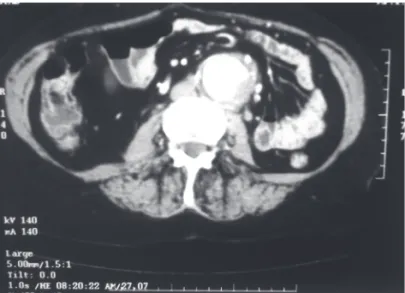
Conventional CT (Figure 4) was first used in 1980 in the diagnosis and preoperative and postoperative evalua-tions of abdominal aortic aneurysms.14,15 It provided clear images with information regarding the size and extent of
Table 4 - Comparison between nuclear magnetic resonance
(NMR) measurements and surgical observation (SO) (n = 20).
Region NMR SO Statistical
Average ± Average ± comparison
SDMedian SDMedian
PN 2.6 ± 0.5 2.5 ± 0.5 P = 0.209
2.6 2.6
MDA 5.9 ± 1.0 5.9 ± 1.0 P = 0.502
5.8 5.8
LA 9.6 ± 2.6 10.0 ± 2.6 P = 0.165
9.5 9.4
DRI 1.9 ± 0.7 1.8 ± 0.7 P = 0.624
1.8 1.8
DLI 1.6 ± 0.5 1.5 ± 0.5 P = 0.131
1.5 1.3
PN = proximal neck; MDA = maximum transverse diameter of the aorta; LA = straight-line length of the aneurysm; DRI = diameter of the right common iliac artery; DLI = diameter of the left common iliac artery; * = statistically significant; SD = standard deviation.
the aneurysm, presence of intraluminal thrombi, calcifica-tions and anatomic anomalies, ruptures, as well as the pres-ence of an inflammatory component. Conventional CT has the advantage over the angiography of revealing not only the vessel lumen but also the walls and adjacent structures. The CCT exam takes only a few minutes, and intravenous iodide contrast medium injections can be used but may cause problems with nephrotoxicity and eventual allergic complications. In our study, we did not have complications resulting from the use of iodide contrasts.
Our data analysis revealed that, on one hand, CCT has good accuracy regarding the proximal neck, transverse di-ameter of the aorta, and common iliac artery measurements, which did not differ statistically from those achieved in the intraoperative setting. On the other hand, this method showed some inconsistency with intraoperative measure-ments in the evaluation of the aneurysm length, probably due to artefacts caused by breathing movements. Thus, CCT can be considered a valuable method not only for di-agnosis but also for conventional surgery planning. At present, with the improvement of endoluminal correction techniques for aortic aneurysms, CCT has become the most utilized method in a number of medical centers. However, errors in the length measurements of the aneurysms may compromise the success of the procedure.
Other advantages of CCT compared with USG are the good visualization of the suprarenal aorta, iliac arteries, and thoracoabdominal transition, as well as the fact that image interpretation does not depend on the examiner. Available in most hospitals, CCT is a rapid and convenient method for the patient since only a peripheral venous puncture is necessary. However, CCT is more expensive than the USG and may be subject to interference from breathing move-ments.
Helical computerized tomography (Figure 5) is an evo-lution of CCT. The images are constructed faster and re-quire lower radiation exposures and volume of contrast for the patient. In addition, HCT allows 3D reconstruction and eliminates interference from breathing movements, which is a frequent cause of distortions in other imaging technolo-gies.16,17
In our study population, we did not observe any differ-ence between measurements from HCT images and the intraoperative measurements of the proximal neck of the aorta, the transverse diameter of the aorta and iliac arteries, and the longitudinal extent of the aneurysms. Consequently, with exact measurements of the arteries, we were able to plan for any type of abdominal aortic aneurysm treatment (conventional or endovascular).
Nuclear magnetic angioresonance (NMR) (Figure 6) is a recently developed technique that utilizes a powerful mag-netic field and the different tissue characteristics to gener-ate images in multiple planes.3,18,19 Developed at the end of the 1980s, it is relatively noninvasive and provides infor-Figure 4 - Conventional tomographic imaging of an abdominal aortic
aneurysm.
mation about the arterial wall morphology, the adjacent structures, and the physiology of arterial flow. It does not result in adverse effects from radiation, radio-opaque con-trasts, or arterial catheterization punctures that can be caused by other imaging techniques. It reveals the vessel structures, with not only the aneurysm lumen but also the walls and perivascular structures being well delineated. Patients with pacemakers or metallic devices cannot be submitted to NMR because the magnetic field may be harmful in such condi-tions.
Like the HCT technique, the NMR technique used in this study allowed precise measurements to be made that did not differ significantly from the measurements made during surgery
CONCLUSIONS
1. USG is a reliable technology for the diagnosis and follow-up of abdominal aortic aneurysm patients; however, it insufficient for endovascular surgery planning.
2. CCT is potentially faulty in measuring the length (extent) of the aortic dilatation and is therefore not suit-able for the endovascular surgery planning.
3. HCT and NMR are precise methods for evaluating all the studied parameters, including the proximal neck of
Figure 6 - Nuclear magnetic resonance imaging of an abdominal aortic aneurysm – longitudinal reconstruction.
the aorta and the maximum transverse diameters of the aorta and iliac arteries. Therefore, they are technologies of fun-damental value, and either of these methods can be used by themselves for endovascular surgery planning.
RESUMO
AZEVEDO F das C de e col. Comparação entre ultras-sonografia, tomografia computadorizada e ressonância nuclear magnética com medidas intra-operatórias na ava-liação dos aneurismas de aorta abdominal. CLINICS
60(1):21-28, 2005.
OBJETIVO: Estudar os métodos mais freqüentemente
empregados na avaliação dos aneurismas de aorta abdomi-nal – ultrassonografia, tomografia computadorizada conven-cional, tomografia computadorizada helicoidal e angio-res-sonância nuclear magnética – comparando as medidas fornecidas por estes exames radiológicos no pré-operatório com medidas realizadas durante a operação.
MÉTODO: Foram incluídos no estudo pacientes
por-tadores de aneurisma da aorta abdominal com indicação de
tratamento cirúrgico eletivo por via transperitoneal. O di-agnóstico inicial da dilatação aórtica foi feito com ultra-sonografia e, uma vez indicado o tratamento cirúrgico, era então solicitado um outro exame radiológico complemen-tar, já que não é nossa rotina operar esses pacientes com base apenas na ultra-sonografia. Sessenta pacientes foram divididos em 3 grupos de acordo com o exame complemen-tar realizado (tomografia computadorizada convencional, tomografia computadorizada helicoidal ou angio-ressonân-cia nuclear magnética). As ultra-sonografias dos 20 primei-ros pacientes foram incluídas em um 4° grupo.
radiológi-cos. As mesmas medidas eram realizadas por ocasião da ope-ração com o auxílio de um paquímetro e , então, compara-das aos valores indicados pelos exames de imagem.
RESULTADOS: As medidas do diâmetro transverso
má-ximo do aneurisma variaram de 4.5 a 13.6 cm no intra-ope-ratório, não apresentando diferença estatisticamente signi-ficativa em relação a nenhum dos exames radiológicos es-tudados. A ultra-sonografia, entretanto, superestimou as medidas do colo proximal da aorta e dos diâmetros trans-versos das artérias ilíacas, em comparação com os valores auferidos durante o tratamento cirúrgico. O comprimento dos aneurismas medidos pela tomografia computadorizada convencional era menor em relação às medições feitas com o paquímetro de maneira estatisticamente significativa. Tanto a tomografia computadorizada helicoidal quanto a angio-ressonância nuclear magnética proporcionaram me-didas sem diferença significante do ponto de vista
estatís-tico para todos os parâmetros estudados, quando confron-tados com os valores obtidos no intra-operatório.
CONCLUSÕES: A ultra-sonografia consiste em
méto-do valioso para o diagnóstico e seguimento clínico de pa-cientes com aneurisma de aorta abdominal, sendo, porém, insuficiente para o planejamento de tratamento por técnica endovascular. A tomografia computadorizada convencional pode induzir a erro na estimativa da extensão crânio-cau-dal do aneurisma. A tomografia computadorizada helicoi-dal e a angio-ressonância nuclear magnética geraram me-didas precisas de todos os parâmetros estudados, sendo am-bos de grande importância para a programação operatória.
UNITERMOS: Aneurisma de aorta. Ultra-sonografia.
Tomografia computadorizada. Ressonância nuclear mag-nética. Medidas intra-operatórias.
REFERENCES
1 . Eriksson I, Hemmingsson A, Lindgren PG. Diagnosis of abdominal aortic aneurysms by aortography, computed tomography and ultrasound. Acta Radiologica Diagnosis 1980;21:209-14. 2 . La Roy LL, Cormier PJ, Matalon TAS, Patel SK, Turner DA,
Silver B. Imaging of abdominal aortic aneurysms. AJR 1989;152:785–92.
3 . Amparo EG, Hoddick WK, Hricak H, Sollitto R, Justich E, Filly RA, et al. Comparison of magnetic resonance imaging and ultrasonography in the evaluation of abdominal aortic aneurysms. Radiology 1985;154:451-6.
4 . Pysklywec M, Evans MF. Diagnosing abdominal aortic aneurysm. How good is the physical examination? Canadian Family Physician 1999;45:452-8.
5 . Lederle FA, Walker JM, Reinke DB. Selective screening for abdominal aortic aneurysms with physical examination and ultrasound. Arch Intern Med 1988;148:1753-6.
6 . Parodi JC, Palmaz JC, Barone HD. Transfemoral intraluminal graft implantation for abdominal aortic aneurysms. Ann Vasc Surg 1991;5:491-9.
7 . Moritz JD, Rotermmund S, Keating DP, Oestmann JW. Infra-renal abdominal aortic aneurysms: implications of CT evaluation of size and configuration for placement of endovascular aortic graft. Radiology 1996;198(2):466.
8 . Gomes MN, Choyke PL. Pre-operative evaluation of abdominal aortic aneurysms: ultrasound or computed tomography? J Cardiovas Surg 1987;28:159-66.
9 . Pavone P, Di Cesare E, Di Renzi P, Marsili L, Ventura M, Spartera C, et al. Abdominal aortic aneurysm evaluation: comparison of US, CT, MRI, and angiography. Magn Reson Imaging. 1990;8(3):199-204.
10. Tennant WG, Hartnell GG, Baird RN, Horrocks M. Radiologic investigation of abdominal aortic aneurysm disease: comparison of three modalities in staging and the detection of inflammatory change. J Vasc Surg. 1993;17(4):703-9.
11. Fox AD, Whiteley MS, Murphy P, Budd JS, Horrocks M. Comparison of magnetic resonance imaging measurements of abdominal aortic aneurysms with measurements obtained by other imaging techniques and intra-operative measurements: possible implications for endovascular grafting. J Vasc Surg 1996;24(4):632-8.
12. Baud JM, Mas D, Pichot O, Laroche IP, Viarel A, Crebillier M, Ginaud M, Ledemeney M. Critères de quantification et de caractérisation des anéurysmes de láorte abdominale par l’echographie. J Mal Vasc 1997;22(5):313-20.
14. Axelbaum SP, Schellinger D, Gomes MN and others. Computed tomographic evaluation of aortic aneurysms. Am J Roentgenol 1976;127:75.
15. Todd GJ, Nowygrod R, Benvenisty A, Buda J, Reemtsma K. The accuracy of CT scanning in the diagnosis of abdominal and thoracoabdominal aortic aneurysms. J Vasc Surg. 1991;13(2):302-10.
16. Kalender, WA, Seissler W, Klotz E, Vock P. Spiral volumetric CT with single-breath-hold technique, continuous transport and continuous scanner rotation. Radiology 1990;176-83.
17. Rubin GD, Dake MD, Napel SA, McDonnell CH, Jeffrey Jr. RB. Three-dimensional spiral angiography of the abdomen: initial clinical experience. Radiology 1993;186:147-52.
18. Wolosker N, Nakano L, D’Hippolito G, Rosoky RA, Borri ML, Wolosker AM. Gadolinium magnetic angioresonance in the study of aortoiliac disease. Angiology. 2003;54(2):163-8. 19. Lee JKT, Ling D, Keiken JP, Glazer HS, Sicard GA, Totty WG, et