A tale of two hemispheres
Contrasting socioemotional dysfunction in
right- versus left-lateralised semantic dementia
Muireann Irish1, Fiona Kumfor2, John R. Hodges3, Olivier Piguet4
ABSTRACT. Objective: Semantic dementia, a subtype of frontotemporal lobar degeneration, is characterised by cross-modal loss of conceptual knowledge attributable to progressive degeneration of the left anterior temporal lobe. Much less is known regarding the clinical presentation of SD patients with predominantly right-lateralised atrophy. Recent reports emphasise marked socioemotional and behavioural disturbances in such cases. Given the importance of the right anterior temporal lobes in social cognition, we hypothesised that socioemotional functioning would be disproportionately affected in right versus left-lateralised SD cases. Methods: We assessed well-characterised cases of predominantly right (n=10) and left (n=12) SD and 20 matched healthy controls on tests of emotion processing and interpersonal functioning. Results: Right SD cases showed disproportionate difficulties in the recognition of positive and negative facial emotions, specifically happiness and anger, compared with left SD cases. Deficits in anger recognition persisted in right SD despite covarying for facial and semantic processing. On a contextually rich task of emotion recognition using multimodal videos, no subgroup differences were evident. Finally, empathic concern was rated as significantly lower by caregivers of right versus left SD cases. Overall, the extent of socioemotional disturbance was associated with the degree of behavioural changes in SD. Conclusion: Our results reveal considerable overlap in the extent to which socioemotional processes are disrupted in left and right-lateralised cases of SD. Notably, however, right SD cases show disproportionate deficits for recognition of facial emotions and the capacity for empathic concern, supporting a specialised role for the right anterior temporal lobes in mediating these cognitive functions.
Key words: semantic dementia, emotion processing, frontotemporal dementia, hemispheric lateralisation.
UM CONTO SOBRE DOIS HEMISFÉRIOS: CONTRASTANDO A DISFUNÇÃO SOCIOEMOCIONAL NA DEMÊNCIA SEMÂNTICA COM ATROFIA LATERALIZADA À DIREITA VERSUS ESQUERDA
RESUMO. Objetivo: A demência semântica (DS), um subtipo de degeneração lobar frontotemporal, é caracterizada por perda multimodal do conhecimento conceitual atribuída à degeneração progressiva do região anterior do lobo temporal esquerdo. Sabe-se menos sobre o quadro clínico de pacientes com DS em que a atrofia é localizada predominantemente à direita. Relatos recentes têm enfatizado marcantes distúrbios socioemocionais e comportamentais em tais casos. Dada a importância da região anterior do lobo temporal direito na cognição social, aventamos a hipótese de que o funcionamento socioemocional seria desproporcionalmente afetado nos casos de DS com atrofia lateralizada à direita. Métodos: Foram avaliados os desempenhos de casos bem caracterizados de DS com atrofia do lobo temporal predominantemente à direita (n=10) e à esquerda (n=12) e 20 controles saudáveis em testes de processamento de emoções e funcionamento interpessoal. Resultados: Casos de DS com atrofia predominante à direita apresentaram dificuldades desproporcionadas no reconhecimento de emoções faciais positivas e negativas, especificamente expressões de felicidade e raiva, em comparação com os casos de atrofia à esquerda. Os déficits no reconhecimento de raiva persistiram depois de excluídas as covariações com processamento facial e semântico. Em uma tarefa contextualmente rica de reconhecimento de emoções através de vídeos multimodais, não houve diferenças entre os subgrupos. Por fim, preocupação empática foi classificada por cuidadores como significativamente menor nos casos com atrofia à direita. Em geral, o grau de perturbação socioemocional foi associado com o grau de alterações comportamentais na DS. Conclusão: Nossos resultados revelam uma considerável
1School of Psychology, University of New South Wales, Sydney, Australia. Neuroscience Research Australia, Sydney, Australia, and School of Medical Sciences,
Uni-versity of New South Wales, Sydney, Australia. PhD, Research Fellow at the School of Psychology, UniUni-versity of New South Wales, Sydney, Australia. 2Masters, PhD,
Candidate at Neuroscience Research Australia, Randwick, Sydney, Australia. 3Professor and Senior Principal Research Fellow at Neuroscience Research Australia,
Randwick, Sydney, Australia. 4Associate Professor and Senior Research Fellow at Neuroscience Research Australia, Randwick, Sydney, Australia. Olivier Piguet. Neuroscience Research Australia, Barker Street, Randwick, Sydney, Australia, NSW 2031. E-mail: [email protected] Disclosure: The authors report no conflicts of interest.
sobreposição na medida em que os processos socioemocionais são rompidos tanto em casos com atrofia predominante à direita como à esquerda. Notavelmente, entretanto, os casos com DS com atrofia predominante à direita apresentam déficits desproporcionais no reconhecimento de emoções faciais e na capacidade de preocupação empática, dando suporte à hipótese de um papel especializado das regiões anteriores do lobo temporal direito na mediação dessas funções cognitivas. Palavras-chave: demência semântica, processamento de emoção, demência frontotemporal, lateralização hemisférica.
INTRODUCTION
S
emantic dementia (SD) is a clinical syndrome associ-ated with focal degeneration of the anterior temporal lobes of the brain, manifesting in the progressive cross modal deterioration of general conceptual knowledge.1,2Patients with SD present with severe semantic impair-ments due to asymmetrical, primarily left-sided, brain atrophy including the anterior and medial portions of the temporal lobe.3,4 Extensive clinical and anatomical
characterisations of predominantly left-sided SD cases concord with the lateralisation of verbal skills and pho-nological representations to the left hemisphere5 and
have proved particularly illuminating for our under-standing of the complex cognitive architecture of the semantic and episodic memory systems of the brain.6
In contrast, however, a dearth of information exists re-garding the less common presentation of SD with pre-dominant right-lateralised atrophy.
To date, clinical data on right SD have been largely gleaned from individual or case series reports, the ma-jority of which emphasise the presence of prosopagno-sia, loss of empathy, behavioural disinhibition, and dis-ruptions to interpersonal functioning.7-11 Recent group
studies have revealed episodic memory and spatial navi-gation deicits,12 and alterations in food preferences13 in
this group. Eccentric social behaviour, with alterations in dressing, personal hygiene, sociopathic behaviours, irritability, and impulsivity, appear more frequent in patients with predominantly right sided pathology in comparison with left-sided SD cases.7 Together with loss
of empathy and insight, disinhibition, and diiculties in afect regulation, such alterations in social comport-ment may bias the clinician to misdiagnose right SD as behavioural variant frontotemporal dementia, particu-larly when structural neuroimaging is not available.10
he emergence of lorid socioemotional and behav-ioural changes in the majority of right SD cases resonates well with the position that the right anterior temporal lobe plays a pivotal role in the abstraction of conceptual knowledge from the social domain.14,15 Despite recent
group studies involving large samples of right SD cas-es10,12 much remains to be elucidated regarding the
clini-cal features that may be speciic to this syndrome. While the evidence to date suggests that socioemotional
dei-cits may be particular to right SD,8,13 deicits in emotion
processing are widely reported in left SD cases across a range of modalities including facial stimuli,16,17 musical
excerpts,18 and emotional words.19 Importantly, while
left SD patients do show marked impairments in emo-tion processing, such deicits appear attributable, in part, to the verbal demands of the tasks used.17 Further,
changes in the capacity for empathic concern have been documented in left SD.20 hese changes, which
encom-pass both the cognitive and afective aspects of interper-sonal functioning,21,22 correlate with the extent of
atro-phy in right, rather than left, temporal lobe structures.20
he extent to which social functioning is diferential-ly compromised in right versus left lateralised cases of SD remains unknown. Current evidence suggests that emotion recognition and interpersonal functioning may be disproportionately afected in right SD compared to left SD. To our knowledge, however, group studies com-paring socioemotional functioning in right versus left SD cases have not been conducted. he objective of this study was to investigate proiles of socioemotional dys-function in a sample of predominantly right-lateralised cases of SD and to contrast their performance with a well-characterised cohort of age-, education- and dis-ease-matched left-sided SD cases. We predicted that, right SD cases would show disproportionate deicits in comparison to the left-sided cases for those functions largely ascribed to the right anterior temporal lobes, namely emotion recognition, and the capacity for em-pathic concern.
METHODS
Participants. Twenty-two patients with semantic
de-mentia (SD) and 20 older age- and education-matched healthy controls took part in this study. Diagnosis of SD cases was established in line with current clinical diagnostic criteria23 by consensus among a
contrast, patients diagnosed with right SD presented with loss of semantic knowledge, marked prosopagno-sia, and behavioural changes, with evidence of extensive neural atrophy predominantly in the right temporal lobe on structural MRI. Figure 1 displays representative scans for right and left SD cases.
Controls scored 0 on the sum of boxes score of the Clinical Dementia Rating scale (CDR)25 and 88 or above
on the Addenbrooke’s Cognitive Examination-Revised (ACE-R).25 Exclusion criteria included prior history of
mental illness, signiicant head injury, movement disor-ders, cerebrovascular disease, alcohol or substance abuse, and limited english proiciency. Ethical approval was ob-tained from the Southern Eastern Sydney and Illawarra Area Health Service and the University of New South Wales ethics committees. Informed consent was ob-tained from all participants, or their person responsible.
General cognitive screening. Participants completed a
neu-ropsychological battery of tests to assess general cogni-tive functioning (ACE-R),25 visual episodic memory (Rey
Complex Figure),26 semantic processing including an
index of Naming and Comprehension,27 speed of
pro-cessing (Trail Making Test Part A),28 verbal luency,29
(FAS),29 and measures of facial matching and facial
identiication.17
Emotion processing. Ekman 60 task30 – Recognition of
60 facial expressions across six basic emotions (anger, disgust, fear, happiness, sadness, surprise) was assessed using stimuli from the Pictures of Facial Afect series.31
Stimuli were presented pseudorandomly for 5 seconds on a computer screen, and participants were required to select the label that best described the emotional ex-pression. Emotion labels were present throughout test-ing and selection was untimed. he maximum score for this task is 60 points.
he Awareness of Social Inference Test (TASIT)32 – his
task assesses the perception of emotions within an eco-logically valid setting and consists of 24 short video clips in which an actor portrays one of six basic emotions (an-ger, disgust, fear, sadness, surprise, happy) as well as a Neutral, non-emotional, condition. Participants were required to view each video clip, following which a pause occurred in which participants completed the accompa-nying questions. A maximum of 5 points was awarded for each emotion category.
Interpersonal Reactivity Interview (IRI)33 – Spouses of
SD patients completed the IRI as an index of the
pa-tient’s present interpersonal functioning. he IRI is a 28-item questionnaire consisting of four 7-item sub-scales; Perspective Taking (PT; the capacity to imagine the cognitive perspective of another person), empathic concern (EC; the ability to perceive another person’s emotional state), fantasy (FS; the capacity to project oneself into experiences of imaginary characters), and personal distress (PD, the tendency towards feeling anx-iety in response to experiencing others in distress).
Statistical analyses. Data were analysed using IBM SPSS
Statistics version 20. For general cognitive screening and performance on the Ekman 60 task, multivariate analyses of variance (ANOVA) were run, with Sidak post hoc comparisons used to explore group diferences. Giv-en the smaller sample size tested on the TASIT and IRI, non-parametric Kruskal-Wallis tests were run for over-all group comparisons, and Mann-Whitney U tests were employed for post hoc comparisons. Finally, Spearman rank correlations, corrected for multiple comparisons (p<0.01) explored possible relationships between the experimental variables.
RESULTS
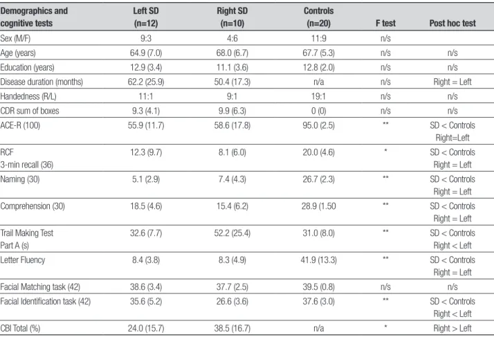
Demographics. Demographic and clinical data are
pre-sented in Table 1. he groups were matched for age (F(2, 39)=0.951, p=0.395) and years in education (F(2,39)= 1.376, p=0.265). Chi-squared tests revealed that sex (χ2(2)=2.880, p=0.247) and handedness (χ2(4)=3.635,
p=0.458) did not difer between the participant groups. Further, no signiicant diferences were evident between the left and right SD cases for disease duration (i.e. the time elapsed between symptom onset and testing, F(1, 20)=1.521, p=0.232) or disease severity (CDR Sum of Boxes, F(1, 16)=0.049, p=0.828; ACE-R total score, F(1, 20)=0.181, p=0.675).
General cognitive functioning. Neuropsychological
test-ing results are presented in Table 1. On the overall
Figure 1. Structural T1-weighted coronal images of the representative pat-terns of atrophy in semantic dementia patients with predominantly right (R) or left (L) temporal lobe atrophy.
screening ACE-R measure, group diferences were evi-dent (F(2, 39)=65.79, p<0.0001), with both SD groups scoring signiicantly lower than controls (all p values <0.0001). Striking semantic processing deicits were ev-ident on the Naming (F(2, 38)=240.86, p<0.0001) and Comprehension (F(2, 38)=47.31, p<0.0001) tasks with controls scoring signiicantly higher than both left and right SD cases (all p values <0.0001) and no diferences between the SD subgroups (all p values >0.2). On a non-verbal test of episodic memory recall, a group efect was again observed (F(2, 35)=9.23, p=0.001) with both left (p=0.011) and right (p=0.002) SD cases showing sig-niicant impairments relative to controls, but no sub-group diferences (p=0.531). Overall sub-group diferences were evident on the letter luency task (F(2, 35)=51.35, p<0.0001) with severe luency deicits in both SD groups relative to controls (all p values <0.0001) but no diference between the SD subgroups (p>0.9). Similar-ly, signiicant impairments were observed on the Trail Making test Part A (F(2, 35)=8.53, p=0.001), driven by diiculty exclusively in the right SD group relative to
controls (p=0.001). In contrast, left SD cases scored in line with controls for Trails Part A (p=0.983), perform-ing signiicantly better than their right-sided counter-parts (p=0.006). Finally, on a test of facial perception, no group diferences were evident (p=0.153), however, signiicant impairments were found on a facial identii-cation discrimination task (F(2, 36)=21.01, p<0.0001) driven exclusively by severe deicits in the SD right group (p<0.0001) with respect to controls (left SD, p=0.417), consistent with previous reports of prosopag-nosia in right SD.
Caregiver ratings of behavioural changes on the Cambridge Behavioural Inventory (CBI)34 revealed a
sig-niicant diference between the SD subgroups (U=32.0, p=0.032) with right SD cases showing greater behav-ioural changes in comparison to the left SD subgroup.
In summary, left and right SD cases displayed com-parable impairments in semantic processing, and epi-sodic memory, with disproportionate speed of process-ing, facial identiication, and behavioural disturbance evident in the right SD group.
Table 1. Demographic and clinical characteristics of study cohorts (standard deviations in brackets).a,b
Demographics and cognitive tests
Left SD (n=12)
Right SD (n=10)
Controls
(n=20) F test Post hoc test
Sex (M/F) 9:3 4:6 11:9 n/s
Age (years) 64.9 (7.0) 68.0 (6.7) 67.7 (5.3) n/s n/s
Education (years) 12.9 (3.4) 11.1 (3.6) 12.8 (2.0) n/s n/s
Disease duration (months) 62.2 (25.9) 50.4 (17.3) n/a n/s Right = Left
Handedness (R/L) 11:1 9:1 19:1 n/s n/s
CDR sum of boxes 9.3 (4.1) 9.9 (6.3) 0 (0) n/s n/s
ACE-R (100) 55.9 (11.7) 58.6 (17.8) 95.0 (2.5) ** SD < Controls
Right=Left RCF
3-min recall (36)
12.3 (9.7) 8.1 (6.0) 20.0 (4.6) * SD < Controls
Right = Left
Naming (30) 5.1 (2.9) 7.4 (4.3) 26.7 (2.3) ** SD < Controls
Right = Left
Comprehension (30) 18.5 (4.6) 15.4 (6.2) 28.9 (1.50 ** SD < Controls
Right = Left Trail Making Test
Part A (s)
32.6 (7.7) 52.2 (25.4) 31.0 (8.0) ** SD < Controls
Right < Left
Letter Fluency 8.4 (3.8) 8.3 (4.9) 41.9 (13.3) ** SD < Controls
Right = Left
Facial Matching task (42) 38.6 (3.4) 37.7 (2.5) 39.5 (0.8) n/s n/s
Facial Identification task (42) 35.6 (5.2) 26.6 (3.6) 37.6 (3.0) ** SD < Controls
Right < Left
CBI Total (%) 24.0 (15.7) 38.5 (16.7) n/a * Right > Left
aMaximum score for each test in brackets where applicable. bCDR data available for 11 left SD and 7 right SD; Trail Making test data available for 11 left SD and 8 right SD, Digits backwards available for 9
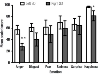
Ekman 60 performance. Figure 2 shows overall group per-formance on the Ekman 60 task. A repeated measures ANOVA revealed a signiicant main efect of group (F(2, 38)=28.46, p<0.0001) with both SD subgroups showing gross impairments on the Ekman 60 emotion recogni-tion task irrespective of valence ( all p values <0.0001). No signiicant overall diferences were evident between left and right SD subgroups (p=0.115). A main efect of valence was observed (F(5, 190)=26.54, p<0.0001), which relected the fact that recognition of the emo-tion happiness was signiicantly higher, irrespective of group, in comparison with all other emotion categories (all p values <0.0001).
A signiicant group by valence interaction was found (F(10, 190)=3.02, p=0.001), which was driven by dif-ferential patterns of performance in each SD subgroup. Post hoc Sidak tests conirmed that in left SD, the recog-nition of all negative emotions was markedly disrupted relative to controls (anger, p<0.0001; disgust, p<0.0001; fear, p=0.028; sadness, p=0.020). Recognition of surprise was also signiicantly impaired in this group (p=0.025). In contrast, recognition of happiness was intact in the left SD cohort with respect to controls (p=0.900).
For right SD cases, striking impairments for all basic emotions were observed (anger, p<0.0001; dis-gust, p<0.0001; fear, p=0.006; sadness, p=0.004; sur-prise, p=0.025) including the recognition of happiness (p=0.002) relative to controls. Further post hoc analyses conirmed that right SD cases performed signiicantly poorer than left SD cases for the recognition of anger (p=0.005) and happiness (p=0.021), with no other sig-niicant diferences detected between the subgroups (all p values >0.1).
To investigate the contribution of perceptual pro-cesses on facial emotion recognition, an analysis of covariance (ANCOVA) was run with performance on the facial matching task included as a covariate. he main efect of diagnosis persisted (F(2, 35)=19.63, p<0.0001), with both left and right SD cases displaying signiicant overall impairments with respect to controls (all p values < .0001). A signiicant valence by group in-teraction was also evident (F(10, 175)=3.22, p=0.001) which was driven by severe impairments in the recogni-tion of speciic emorecogni-tions in each SD subgroup. Left SD cases continued to show marked deicits relative to con-trols for the recognition of anger (p<0.0001), disgust (p<0.0001), sadness (p=0.023) and surprise (p=0.016) with intact recognition of fear (p=0.067) and happiness (p=0.985). In contrast, right SD cases’ deicits remained present for the recognition of anger (p<0.0001), disgust (p<0.0001), fear (p=0.044), sadness (p=0.022), and
hap-piness (p=0.027), but recognition of surprise was intact (p=0.594). Looking between the SD subgroups, right SD cases scored signiicantly lower than left SD cases for the recognition of anger (p=0.034) with no other dif-ferences between subgroups found (all p values >0.05). A separate ANCOVA was also carried out using nam-ing performance as a covariate, to control for the pos-sible inluence of semantic processing on the labelling of emotions. he main efect of diagnosis persisted (F(2, 37)=5.610, p=0.007); however, post hoc tests revealed that SD groups did not difer signiicantly from controls for overall emotion recognition performance (Right SD, p=0.957; Left SD, p=0.279). Interestingly, right SD cases continued to score signiicantly lower than left SD cases (p=0.006), for the recognition of anger (p=0.001), dis-gust (p=0.012), and happiness (p=0.008).
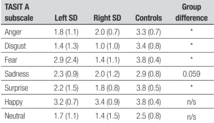
The Awareness of Social Inference Test (TASIT). A Kruskal
Wallis test revealed signiicant group diferences for the recognition of positive (χ2(2)=16.26, p<0.0001),
and negative (χ2(2)=26.73, p<0.0001) emotions on the
TASIT. Mann Whitney U tests demonstrated that left SD cases were signiicantly impaired for positive (U=37.5, p=0.001) and negative (U=3.5, p<0.0001) emotions rel-ative to controls. Likewise, right SD cases showed signif-icant impairments across positive (U=5.0, p=0.001) and negative (U=0.000, p<0.0001) emotions on the TASIT with respect to controls. No signiicant diferences were evident between the SD subgroups (positive, U=22.0, p=0.221; negative, U=16.5, p=0.080).
Looking at the subscales of the TASIT, a Kruskal Wallis test revealed overall group diferences for
recog-100
80
60
40
20
0
Mean scaled score
Left SD Right SD
Anger Disgust Fear Sadness Surprise Happiness Emotion
nition of the following emotions; anger (χ2(2)=15.61,
p<0.0001), fear (χ2(2)=15.94, p<0.0001), disgust
(χ2(2)=19.19, p<0.0001), and surprise (χ2(2)=19.82,
p<0.0001) with the suggestion of a group diference for sadness (χ2(2)=5.66, p=0.059) (Table 2). Mann Whitney
U tests conirmed that left SD cases were signiicantly impaired with respect to controls for recognition of anger (U=34.0, p<0.0001), fear (U=59.0, p=0.017), dis-gust (U=27.5, p<0.0001), surprise (U=38.5, p=0.001), and mild deicits observed for recognition of happiness (U=70.0, p=0.053). Similarly, right SD cases showed signiicant impairments relative to controls for the rec-ognition of surprise (U=2.5, p<0.0001), anger (U=11.5, p=0.006), fear (U=1.5, p<0.0001) and disgust (U=3.5, p<0.0001), but preserved recognition of happiness (U=38.0, p=0.447). No signiicant subgroup diferences were evident between the SD subgroups for any of the TASIT emotion categories (anger, U=29.0, p=0.479; dis-gust, U=26.0, p=0.360; fear, U=16.5, p=0.080; sadness, U=22.5, p=0.221; happiness, U=26.0, p=0.360; surprise, U=23.5, p=0.253).
Caregiver ratings of interpersonal changes. Investigating
the SD subgroups on carer rated measures of interper-sonal reactivity, Mann-Whitney U tests failed to reveal signiicant group diferences for caregiver ratings of perspective taking (U=36.5, p=0.165), fantasy (U=33.0, p=0.178), and personal distress (U=42.0, p=0.302) on the IRI. Right SD cases, however, were rated as demon-strating less empathic concern relative to left SD cases (U=27.0, p=0.047) (Figure 3).
Correlations between experimental measures. Spearman
rank correlations for the overall SD cohort (n=22), ad-justed for multiple comparisons at p<0.01 level, are pre-sented in Table 3. Performance on the Ekman 60 task was signiicantly related to global cognitive functioning, and total TASIT performance, with higher performance on the Ekman 60 associated with lower incidences of behavioural change on the CBI. Similarly, total TASIT performance was signiicantly inversely related to de-gree of behavioural change on the CBI. Further, ratings of empathic concern on the IRI inversely correlated with behavioural changes on the CBI.
DISCUSSION
he role of the anterior temporal lobes in mediating successful social interactions remains a source of debate within neuropsychology. Using two well-characterised groups of patients with SD, in which the predominant burden of brain atrophy was lateralised to either the left
Table 2. Performance of left and right SD cases and controls on subscales of the TASIT emotion recognition task.
TASIT A
subscale Left SD Right SD Controls
Group difference
Anger 1.8 (1.1) 2.0 (0.7) 3.3 (0.7) *
Disgust 1.4 (1.3) 1.0 (1.0) 3.4 (0.8) *
Fear 2.9 (2.4) 1.4 (1.1) 3.8 (0.4) *
Sadness 2.3 (0.9) 2.0 (1.2) 2.9 (0.8) 0.059
Surprise 2.2 (1.5) 1.8 (0.8) 3.8 (0.5) *
Happy 3.2 (0.7) 3.4 (0.9) 3.8 (0.4) n/s
Neutral 1.7 (1.1) 1.4 (1.5) 2.5 (0.8) n/s
Standard deviations are shown in brackets. TASIT data available for 5 right SD and 12 left SD patients. *p<0.0001 based on non-parametric Kruskal Wallis test.
20
15
10
5
0
Total score
Left SD Right SD
Empathic
concern PerspectiveIRI subscaleFantasy Distress
Figure 3. Caregiver ratings of interpersonal changes on the Interpersonal Reactivity Interview (IRI) in left and right SD cases. IRI data available for 11 left SD and 9 right SD cases. Error bars depict standard error of the mean. *p<0.05.
Table 3. Spearman rank correlations showing relationships between experi-mental measures in the combined SD groups (n=22).
ACE-R Total TASIT Total CBI Total
Ekman 60 0.546* 0.874** –0.651**
TASIT 0.328 1.000 –0.606*
IRI Empathy 0.100 0.560 –0.596*
Correlations were adjusted for multiple comparisons using a corrected alpha level of 0.01; *p<0.005; ** p<0.001
he inding of marked alterations in the recognition of basic facial emotions in SD resonates with previous re-ports in the literature, in particular for the recognition of negative emotions.16,17 Left SD cases displayed marked
diiculties in the recognition of all negative emotions on the Ekman 60, as well as surprise, deicits which were not related to naming or general semantic processing capac-ity. In contrast, right SD cases displayed profound dei-cits in the recognition of all basic facial emotions, scoring signiicantly poorer than the left SD group for the rec-ognition of anger and happiness. Importantly, while Ek-man 60 perforEk-mance correlated signiicantly with facial identity discrimination in right SD, our covariate analy-sis suggests that general face processing disturbances, and semantic naming impairments, do not fully account for the marked emotion recognition deicits found in this group. Degeneration of the right anterior temporal lobe appears critical in the genesis of global emotion pro-cessing diiculties in right SD. he right amygdala is the most likely candidate driving such disruption, a struc-ture which has previously been implicated in disrup-tion of negative emodisrup-tion recognidisrup-tion,35 and behavioural
changes including disinhibition and depression in SD.36
Findings from the Ekman 60 task were largely replicated on the TASIT, with a number of important exceptions. Both SD subgroups displayed signiicant impairments for the recognition of negative emotions, as well as sur-prise. Subgroup analyses, however, uncovered a rela-tively spared capacity to recognise happiness in the right SD group. Unlike on the Ekman 60, diferences between SD groups were not evident on the TASIT, a inding that likely relates to the provision of rich contextual informa-tion on this task. Right SD patients may beneit from the additional multimodal information provided on the TASIT, such as tone, prosody, and gesture, thus reducing diferences between SD subgroups. hese indings lend support to the proposal that the right temporal lobe is specialised for the processing of facial stimuli.37
Given the evidence pointing to the importance of right temporal structures in facilitating interpersonal behaviours including empathy,8,20,38-39 the
dispropor-tionate deicits found on the empathic concern subscale of the IRI in the right SD cases are not surprising. Pa-tients with right predominant SD are typically held to show marked reduction of interpersonal functioning with reports of “cold-heartedness” and loss of warmth. he inability to share emotional experiences in this manner, in turn, likely impacts on the capacity for per-spective taking, and the suppression of one’s own view-point, particularly as the pathological process begins to encroach into adjacent frontal regions.20 he status
of complex self-projective social cognitive functions in SD remains poorly understood.40 Recent evidence,
how-ever, points towards striking deicits in theory of mind in left SD cases.41 Whether SD patients with
predomi-nantly right-sided pathology show theory of mind dei-cits of a greater magnitude than left SD cases remains to be established, but this seems a plausible assumption.
To our knowledge, this study represents the irst concerted efort to investigate diferences in interper-sonal functioning in left versus right SD. Given the size of our sample, further investigations in larger indepen-dent samples will be important to conirm our indings. Another important consideration relates to the disease staging of our SD participants. Over time, the patho-logical process in SD spreads from one anterior lobe to the other, resulting in bilateral insult to the amygdalae, as well as encroachment of atrophy into ventromedial prefrontal areas.42,43 With disease progression,
symp-toms undetected at baseline become evident, resulting in a mixed clinical presentation.13,42 Finally, the binary
classiication into left or right SD obscures the fact that a degree of bilateral atrophy is often present in these pa-tients.2,4 Future studies incorporating automated
neu-roimaging analyses, such as voxel-based morphometry, to quantify the extent of left and right anterior lobe at-rophy in each subgroup are thus warranted.
In summary, we have demonstrated a considerable overlap in the extent to which socioemotional processes are disrupted in SD cases with predominant left or right temporal lobe atrophy. Despite these common features, however, right SD cases show disproportionate deicits in the recognition of basic emotions, and in their capac-ity for empathic concern. Future studies investigating associations between regional brain integrity and per-formance on emotion processing tasks will provide valu-able information regarding the relative contribution of left versus right anterior temporal structures to socio-emotional functioning in SD.
Grant support. MI is supported by an Australian Research
Council (ARC) Discovery Early Career Researcher Award (DE130100463). FK is supported by an Australian Post-graduate Award. OP is supported by a National Health and Medical Research Council of Australia Career Devel-opment Fellowship (APP1022684).
Acknowledgements. he authors are grateful to the
REFERENCES
1. Hodges JR, Patterson K. Semantic dementia: a unique clinicopatho-logical syndrome. Lancet Neurol 2007;6:1004-1014.
2. Mion M, Patterson K, Acosta-Cabronero J, et al. What the left and right anterior fusiform gyri tell us about semantic memory. Brain 2010; 133:3256-3268.
3. Chan D, Fox N, Scahill R, et al. Patterns of temporal lobe atrophy in semantic dementia and Alzheimer’s disease. Ann Neurol 2001;49: 433-442.
4. Galton C, Patterson K, Graham K, et al. Differing patterns of tempo-ral atrophy in Alzheimer’s disease and semantic dementia. Neurology 2001;57:216-225.
5. Lambon Ralph MA, McClelland JL, Patterson K, Galton CJ, Hodges JR. No right to speak? The relationship between object naming and se-mantic impairment: neuropsychological evidence and a computational model. J Cogn Neurosci 2001;13:341-356.
6. Irish M, Addis DR, Hodges JR, Piguet O. Considering the role of seman-tic memory in episodic future thinking: evidence from semanseman-tic demen-tia. Brain 2012;135:2178-2191.
7. Edwards-Lee T, Miller BL, Benson DF, et al. The temporal variant of frontotemporal dementia. Brain 1997;120:1027-1040.
8. Perry RJ, Rosen HR, Kramer JH, Beer JS, Levenson RL, Miller BL. Hemispheric dominance for emotions, empathy and social behaviour: evidence from right and left handers with frontotemporal dementia. Neurocase 2001;7:145-160.
9. Gainotti G, Barbier A, Marra C. Slowly progressive defect in recognition of familiar people in a patient with right anterior temporal atrophy. Brain 2003;126:792-803.
10. Thompson SA, Patterson K, Hodges JR. Left/right asymmetry of atro-phy in semantic dementia: behavioral-cognitive implications. Neurology 2003;61:1196-1203.
11. Gorno-Tempini ML, Rankin KP, Woolley JD, Rosen HJ, Phengrasamy L, Miller BL. Cognitive and behavioral profile in a case of right anterior temporal lobe neurodegeneration. Cortex 2004;40:631-644.
12. Chan D, Anderson V, Pijnenburg Y, et al. The clinical profile of right tem-poral lobe atrophy. Brain 2009;132:1287-1298.
13. Henry ML, Wilson SM, Ogar JM, et al. Neuropsychological, behavioral, and anatomical evolution in right temporal variant frontotemporal de-mentia: A longitudinal and post-mortem single case analysis. Neuro-case. In press.
14. Ross LA, Olson IR. Social cognition and the anterior temporal lobes. Neuroimage 2010;49:3452-3462.
15. Zahn R, Moll J, Krueger F, Huey ED, Garrido G, Grafman J. Social con-cepts are represented in the superior anterior temporal cortex. Proc Natl Acad Sci USA 2007;104:6430-6435.
16. Kumfor F, Miller L, Lah S, et al. Are you really angry? The effect of in-tensity on facial emotion recognition in frontotemporal dementia. Soc Neurosci 2011;6:502-514.
17. Miller LA, Hsieh S, Lah S, Savage S, Hodges JR, Piguet O. One size does not fit all: face emotion processing impairments in semantic de-mentia, behavioural-variant frontotemporal dementia and Alzheimer’s disease are mediated by distinct cognitive deficits. Behav Neurol 2012;25:53-60.
18. Hsieh S, Hornberger M, Piguet O, Hodges JR. Brain correlates of musi-cal and facial emotion recognition: evidence from the dementias. Neu-ropsychologia 2012;50:1814-1822.
19. Hsieh S, Foxe D, Leslie F, Savage S, Piguet O, Hodges JR. Grief and joy: emotion word comprehension in the dementias. Neuropsychology 2012;26:624-630.
20. Rankin KP, Gorno-Tempini ML, Allison SC, et al. Structural anatomy of empathy in neurodegenerative disease. Brain 2006;129:2945-2956. 21. Rankin KP, Kramer JH, Miller BL. Patterns of cognitive and emotional
empathy in frontotemporal lobar degeneration. Cogn Behav Neurol 2005;18:28-36.
22. Eslinger PJ, Moore P, Anderson C, Grossman M. Social cognition, execu-tive functioning, and neuroimaging correlates of empathic deficits in fron-totemporal dementia. J Neuropsychiatry Clin Neurosci 2011;23:74-82. 23. Gorno-Tempini ML, Hillis AE, Weintraub S, et al. Classification of prima-ry progressive aphasia and its variants. Neurology 2011;76:1006-1014. 24. Morris J. Clinical dementia rating: a reliable and valid diagnostic and
staging measure for dementia of the Alzheimer type. Int Psychogeriatr 1997;9:173-176.
25. Mioshi E, Dawson K, Mitchell J, Arnold R, Hodges J. The Addenbrooke’s Cognitive Examination Revised (ACE-R): a brief cognitive test battery for dementia screening. Int J Geriatr Psychiatry 2006;21:1078-1085. 26. Meyers J, Meyers K. The Meyers Scoring System for the Rey Complex
Figure and the Recognition Trial: Professional Manual. Odessa, FL: Psy-chological Assessment Resources; 1995.
27. Savage, S., Hsieh, S., Leslie, F., Foxe, D., Piguet, O., Hodges, J.R. Dis-tinguishing subtypes in primary progressive aphasia: Application of the Sydney Language Battery. Dem Ger Cogn Disord. In press, accepted 3/12/2012.
28. Reitan R. Validity of the Trail Making Test as an indicator of organic brain damage. Percept Mot Skills 1958;8:271-276.
29. Strauss E, Sherman EMS, Spreen O. A compendium of neuropsycho-logical tests: Administration, norms, and commentary: Oxford University Press, USA; 2006.
30. Young AW, Perrett D, Calder AJ, Sprengelmeyer R, Ekman P. Facial Expressions of Emotion - Stimuli and Tests (FEEST): Thames Valley Test Company; 2002.
31. Ekman P, Friesen WV. Pictures of Facial Affect. San Francisco: CA: Con-sulting Psychologists Press; 1976.
32. McDonald S, Flanagan S, Rollins J, Kinch J. TASIT: A new clinical tool for assessing social perception after traumatic brain injury. J Head Trau-ma Rehabil 2003;18:219-238.
33. Davis MH. Measuring individual differences in empathy: Evidence for a multidimensional approach. J Pers Soc Psychol 1983;44:113-126. 34. Wedderburn C, Wear H, Brown J, et al. The utility of the Cambridge
Be-havioural Inventory in neurodegenerative disease. J Neurol Neurosurg Psychiatry 2008;79:500-503.
35. Rosen HJ, Perry RJ, Murphy J, et al. Emotion comprehension in the temporal variant of frontotemporal dementia. Brain 2002;125:2286-2295.
36. Liu W, Miller BL, Kramer JH, et al. Behavioral disorders in the frontal and temporal variants of frontotemporal dementia. Neurology 2004;62: 742-748.
37. Adolphs R, Tranel D, Damasio H. Emotion recognition from faces and prosody following temporal lobectomy. Neuropsychology 2001;15: 396-404.
38. Mendez MF, Perryman KM. Disrupted facial empathy in drawings from artists with frontotemporal dementia. Neurocase 2003;9:44-50. 39. Sollberger M, Stanley CM, Wilson SM, et al. Neural basis of
interper-sonal traits in neurodegenerative diseases. Neuropsychologia 2009;47: 2812-2827.
40. Irish M, Piguet O, Hodges JR. Self-projection and the default network in frontotemporal dementia. Nat Rev Neurol 2012;8:152-161.
41. Duval C, Bejanin A, Piolino P, et al. Theory of mind impairments in pa-tients with semantic dementia. Brain 2012;135:228-241.
42. Seeley WW, Bauer AM, Miller BL, et al. The natural history of temporal variant frontotemporal dementia. Neurology 2005;64:1384-1390. 43. Brambati SM, Rankin KP, Narvid J, et al. Atrophy progression in