Rev Odontol UNESP. 2014 Sep.-Oct.; 43(5): 343-350 © 2014 - ISSN 1807-2577 Doi: http://dx.doi.org/10.1590/rou.2014.055
Ef ect of laser therapy on the inl ammatory response
induced by endodontic medications implanted
into the subcutaneous tissue of rats
Efeito da laserterapia na resposta inl amatória induzida por medicamentos
endodônticos implantados em tecido subcutâneo de ratos
Felipe de Souza MATOS
a*, Andréa Ferreira SOARES
a, Ricardo Luiz Cavalcanti de ALBUQUERQUE
JÚNIOR
b, Samuel de Oliveira RIBEIRO
a, Gustavo Danilo Nascimento LIMA
a,
Sônia Maria Alves NOVAIS
a, Maria Amália Gonzaga RIBEIRO
aaUFS – Universidade Federal de Sergipe, Aracaju, SE, Brasil
bUNIT – Universidade Tiradentes, Aracaju, SE, Brasil
Resumo
Introdução: Medicamentos endodônticos apresentam componentes tóxicos que provocam algum grau de reação inflamatória. Objetivo: Este estudo avaliou o efeito da laserterapia na resposta inflamatória causada por medicações intracanais, em tecido subcutâneo de ratos, por meio da análise quantitativa de mastócitos. Material e método: Tubos de polietileno contendo as medicações foram implantados no dorso de 60 ratos, distribuídos em seis grupos: HS (pasta de hidróxido de cálcio P.A.); HL (pasta de hidróxido de cálcio P.A. e laserterapia); HPS (pasta de hidróxido de cálcio P.A. com paramonoclorofenol canforado); HPL (pasta de hidróxido de cálcio P.A. com paramonoclorofenol canforado e laserterapia); IS (iodofórmio e soro fisiológico) e IL (iodofórmio, soro fisiológico e laserterapia). Os animais foram eutanasiados oito e quinze dias após a cirurgia, as peças cirúrgicas foram removidas, processadas para inclusão em parafina e os cortes histológicos corados em Azul de Toluidina 0.2%, para quantificação dos mastócitos. A análise de variância (ANOVA) e o teste de Tukey post hoc foram aplicados para determinar diferenças significativas entre os grupos quanto ao número de mastócitos (p<0.05). Resultado: Nos grupos HL, HPL e IL houve uma diminuição de mastócitos em ambos os períodos experimentais em relação aos grupos HS, HPS e IS, porém não se observou diferença estatística significativa entre o grupo HPS e o HPL aos oito dias. Conclusão: A laserterapia foi eficaz em modular a intensidade da resposta inflamatória induzida pelos medicamentos endodônticos a partir da redução significativa na quantidade de mastócitos.
Descritores: Endodontia; laserterapia; mastócitos; inflamação; subcutâneo.
Abstract
Introduction: Endodontic medications contain toxic components that cause varying degrees of inflammation.
Objective: This study evaluated the effect of laser therapy on the inflammatory response induced by intracanal medications implanted into the subcutaneous tissue of rats using a quantitative analysis of mast cells. Material and method: Polyethylene tubes containing the medications were implanted in the dorsum of 60 rats divided into six groups, including HS (P.A. calcium hydroxide paste), HL (P.A. calcium hydroxide paste and laser therapy), HPS (P.A. calcium hydroxide paste with camphorated paramonochlorophenol), HPL (P.A. calcium hydroxide paste with camphorated paramonochlorophenol and laser therapy), IS (iodoform with saline) and IL (iodoform with saline and laser therapy). The animals were euthanized eight or fifteen days after surgery, and samples were removed and embedded in paraffin. Histological sections were stained with 0.2% toluidine blue for the quantification of mast cells. Analysis of variance (ANOVA) and Tukey’s post-hoc test were applied to determine significant differences in the number of mast cells between groups (p<0.05). Result: There was a decrease in mast cells for the HL, HPL and IL groups when compared with the HS, HPS and IS groups at both time points. There was no statistically significant difference between the HPS and HPL groups at the eight-day time point. Conclusion: Laser therapy was effective at modulating the inflammatory response induced by endodontic medications by significantly reducing the number of mast cells.
INTRODUCTION
he presence of microorganisms is considered the principal factor in the development and progression of pulp and periapical pathologies. hus, the success of endodontic therapy depends on the complete elimination of these pathogens from the interior root canal system1.
Biomechanical preparation combined with the action of auxiliary chemical substances with antimicrobial properties has been efective in reducing bacterial levels by providing a deep cleaning of the root canals. However, due to the presence of numerous dentinal tubules that are inaccessible to chemical-mechanical preparations, it is oten necessary to use endodontic medications between sessions to eliminate or reduce the number of persistent microorganisms in the root dentin2.
Among the endodontic medications commercially available, calcium hydroxide (Ca(OH)2) is considered the intracanal medication of choice because it has multiple beneicial properties, such as satisfactory bactericidal and bacteriostatic actions, antiexudative actions and induction of mineralization3.
To improve these bactericidal properties, calcium hydroxide has also been used in combination with camphorated paramonochlorophenol (PMCC) to form a paste with a greater spectrum of antimicrobial action4.
Another medication oten used as an intracanal dressing for endodontic treatment is iodoform, a chemical with a variety of advantageous properties for bone repair, such as antimicrobial, anti-inlammatory, analgesic, deodorant and detergent activities5.
Despite their beneicial properties and biocompatibility with periradicular tissues, most medications used in endodontic treatments may present potentially toxic or irritating components that cause inlammation, particularly when extravasated to the periapical space6.
he inlammatory process that occurs in the presence of toxic components is a biological response of the organism that is characterized by the movement of specialized cells to the injury site, triggering a series of events in an attempt to restore the integrity and function of damaged tissues7. Among these
cells, mast cells play a fundamental role in the immune response and tissue repair. he cytoplasm of mast cells contains a broad spectrum of pro-inlammatory mediators, and immunoregulatory and angiogenic molecules that participate in inlammatory reactions as well as tissue protection and repair8,9.
Low-level laser therapy has been shown to be an important therapeutic tool because it can modulate the inlammation response through edema reduction, minimization of painful symptoms and cellular biostimulation to ensure rapid and eicient tissue regeneration10,11. herefore, the aim of this study
was to evaluate the efect of laser therapy on the inlammatory response caused by the subcutaneous administration of intracanal medications in rats using a quantitative analysis of mast cells.
MATERIAL AND METHOD
1. Ethical Aspects and Experimental Design
Based on the ethical principles of animal experimentation established by the Brazilian College of Animal Experimentation (COBEA) and the standards of the Brazilian Society of Laboratory Animal Science (SBCAL), this study was completed ater approval from the University Research Ethics Committee (protocol 180211 - CEP).
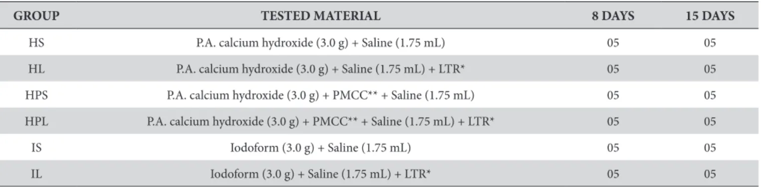
In this study, sixty male rats (Rattus norvegicus, Wistar strain) with an average weight between 250 and 300 g were used. he animals were randomly divided into six groups (n = 10) according to the medication used and the application of laser therapy (Table 1). he animals were euthanized eight or iteen days ater the surgical procedure.
2. Preparation of Medications and Tubes
he endodontic medications were handled in sterile glass plates following the manufacturer’s recommendations. With the aid of a sterile lentulo (#25 Dentsply-Maillefer, RJ, Brazil) they were inserted in polyethylene tubes with a 4.0 mm internal diameter and 8.0 mm length. Prior to this procedure, each tube was sealed at one end with Cyanoacrylate Ester gel (Super Bonder, Aachen, Germany)12,13 to avoid overlow of the materials
to be tested and then autoclaved in metal boxes at a temperature of 120°C for 20 minutes14.
3. Surgical Procedure
he animals were anesthetized with an intraperitoneal injection of ketamine hydrochloride (Ketamine 10%; Agener National Pharmaceutical Chemical Union, Embu, SP, Brazil) at
Table 1. he distribution of groups based on the materials tested, experimental periods and number of animals
GROUP TESTED MATERIAL 8 DAYS 15 DAYS
HS P.A. calcium hydroxide (3.0 g) + Saline (1.75 mL) 05 05
HL P.A. calcium hydroxide (3.0 g) + Saline (1.75 mL) + LTR* 05 05
HPS P.A. calcium hydroxide (3.0 g) + PMCC** + Saline (1.75 mL) 05 05
HPL P.A. calcium hydroxide (3.0 g) + PMCC** + Saline (1.75 mL) + LTR* 05 05
IS Iodoform (3.0 g) + Saline (1.75 mL) 05 05
IL Iodoform (3.0 g) + Saline (1.75 mL) + LTR* 05 05
a dose of 0.1 mL/100 g and with the muscle relaxant thiazine hydrochloride (Rompun 2%; Bayer S.A. - Animal Health, SP, Brazil) at a dose 0.01 mL/100 g that was diluted in saline at a ratio of 1:9 and administered at a dosage of 0.25 mL/100 mg. A trichotomy and antisepsis were performed in the dorsal region of the animals with a 10% povidone-iodine topical solution (LM PHARMA).
For the insertion of polyethylene tubes containing medication, two incisions 5 mm in width were made on the back of animals, one next to the pelvic region and another next to the scapular region. he tubes were longitudinally implanted with the open end of the tube facing the head of the animal and the sealed end of the tube facing the caudal region14. he wounds were sutured
with 4-0 polyamide monoilament thread, and another antisepsis was performed with iodine alcohol15.
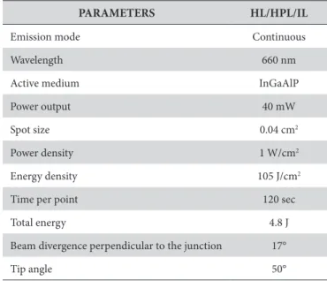
4. Laser herapy Protocol
he experimental groups HL, HPL and IL underwent laser therapy and received a total of four irradiations with a 48-hour interval between sessions. he irst session was immediately ater surgery following the protocol described by Ribeiro et al.16,17. As
described in Table 2, the laser device was used according to the parameters of the irradiation Laser Diode (Twin Laser-InGaAlP/ MMOPTICS, São Carlos, SP, Brazil) with a wavelength in the red spectrum (ƛ660 nm). he irradiation was applied by contact at two points, one caudal and the other radial, of the surgical wound perpendicular to the long axis of the back of the animal for 120 sec per point.
5. Animal Euthanasia and Sample Preparation
Eight and iteen days ater subcutaneous implantation of the polyethylene tubes, the animals were euthanized with an overdose of hio-Pental (Barbiturate-CRYSTALIA) at a dose
of 0.43 mL/kg. he specimens were removed from each animal
via a lozenge incision around the wound with a margin of 1 cm. hen, the samples were ixed in 10% formalin for 24 hours and processed for parain embedding. Histological sections with a thickness of 5 μm were cut and then stained with 0.2% toluidine blue for the quantitative analysis of mast cells18.
6. Quantitative Analysis of Mast Cells
Mast cell counts were performed using 10 histologic ields in the area adjacent to the open end of the tube at an original magniication of 400X. A total of 10 histological sections per group were counted. he sections were examined under a standard light microscope (LEICA DM500) coupled to a computer (Pentium 133 MHz) with an image capture system. LAS EZ 2.1.0 (Leica Microsystems) sotware was used to obtain digital images from the histological ields analyzed.
7. Statistical Analysis
To assess the normality of the data, the Kolmogorov-Smirnov test in Minitab 15.0 sotware was used (Minitab Inc., PA). Statistical analysis of the mean number of mast cells obtained for each group in both experimental periods was performed using analysis of variance (ANOVA) and the Tukey test (p<0.05) with the SPSS program (Version 16; SPSS, Chicago, IL).
RESULT
Histology using toluidine blue revealed globular and large mast cells that were highly granulated and stained violet due to the metachromatic property of their cytoplasmic granules. here was a wide distribution of cells in the area adjacent to the open end of the tube that contained the medications (Figure 1).
he Kolmogorov-Smirnov test was used to assess the normality of the data. he results showed the data had a normal parametric distribution (p=0.110). Subsequently, an analysis of
Table 2. he irradiation parameters for a low laser power intensity
PARAMETERS HL/HPL/IL
Emission mode Continuous
Wavelength 660 nm
Active medium InGaAlP
Power output 40 mW
Spot size 0.04 cm2
Power density 1 W/cm2
Energy density 105 J/cm2
Time per point 120 sec
Total energy 4.8 J
Beam divergence perpendicular to the junction 17°
Tip angle 50°
Source: Tomaz et al. 13 (2013).
variance (ANOVA) and Tukey test with a 5% signiicance level was used.
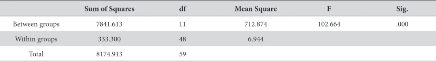
Table 3 shows that analysis of variance (ANOVA) revealed a signiicant diference between the groups (p<0.001). hus, the Tukey’s post-hoc test with a signiicance level of 5% was applied.
Table 4 shows the descriptive statistics, including the mean, standard deviation and standard error, for the mast cells counted in each group from both experimental periods and the results of the Tukey’s post-hoc test (p<0.05).
he HL and IL groups showed a signiicant reduction in the number of mast cells ater eight days of treatment when compared with the HS and IS groups (Figure 2). Despite a decrease in the number of mast cells in the HPL group, there was no signiicant diference when compared with the HPS group. he laser therapy signiicantly reduced the number of mast cells in the HL, HPL and IL groups ater 15 days of treatment when compared with their respective groups that did not receive laser therapy (HS, HPS and IS).
he HPS group showed the greatest number of mast cells ater the eight-day experimental period, followed by the IS and HS groups. here were signiicant diferences between the three groups. he HPS group maintained the highest number of mast cells ater the 15-day experimental period, followed by the HS and IS groups. he signiicant diferences between the groups remained (Figure 2).
Among the groups treated with laser therapy, the HPL group had the highest number mast cells when compared to the HL and IL groups in both experimental periods. he number of mast cells in the HL and IL groups was similar in both experimental periods.
DISCUSSION
he biological and physicochemical properties of the intracanal medications used between endodontic treatments are important for the maintenance and restoration of apical and periapical tissue integrity4. he presence of toxic components
in the majority of intracanal medications produces a varying degree of inlammation and tissue damage in the periradicular area depending on the biocompatibility of these individual substances5.
In this study, laser therapy was used to minimize the irritation caused by medications that contained P.A. calcium hydroxide, P.A. calcium hydroxide with camphorated paramonochlorophenol (PMCC) or iodoform by modulating the inlammatory response. A quantitative analysis of mast cells was used to evaluate medication-induced inlammation and the eicacy of laser therapy for mitigating this response. Analysis of mast cells is a valid and simple method to determine the extent of inlammation caused by biomaterials because these cells are directly involved
Table 3. Analysis of variance (ANOVA)
Sum of Squares df Mean Square F Sig.
Between groups 7841.613 11 712.874 102.664 .000
Within groups 333.300 48 6.944
Total 8174.913 59
Table 4. Descriptive statistics and Tukey test
Groups N Mean Std. deviation Std. error Homogeneous groups*
HS 8 days 5 28.70 2.04939 .91652 c
HL 8 days 5 20.00 2.44949 1.09545 ab
HPS 8 days 5 38.70 1.98746 .88882 d
HPL 8 days 5 35.80 2.56418 1.14673 d
IS 8 days 5 34.60 3.11047 1.39104 d
IL 8 days 5 23.30 2.86356 1.28062 bc
HS 15 days 5 36.70 2.56418 1.14673 d
HL 15 days 5 17.00 2.47487 1.10680 a
HPS 15 days 5 58.00 3.04138 1.36015 e
HPL 15 days 5 23.00 3.06186 1.36931 bc
IS 15 days 5 23.30 2.51496 1.12472 bc
IL 15 days 5 15.80 2.65989 1.18954 a
in the release of important chemical mediators that stimulate the inlammatory response19.
he tested medications were placed in polyethylene tubes to simulate a clinical condition of extravasation of endodontic material into the apical periodontium. hus, the material comes in contact with the subcutaneous tissue only at the open end of the tube, which corresponds to the root apex13,20. his methodology
was introduced by Torneck21, who observed minimal or no tissue
reaction caused by the tube, demonstrating its acceptability for the evaluation of endodontic materials.
Each tube was sealed at one end with a cyanoacrylate ester gel (Super Bonder) to avoid the extravasation of the material and establish an interface between the subcutaneous tissue and the material being investigated13,14. he cyanoacrylate ester is
a synthetic biological adhesive that has proven to be secure and eicacious based on its biological compatibility and high adhesion capacity in wet environments12,22.
he choice of experimental periods (eight and iteen days) was based on criteria established by the ISO 10993-623
and ADA24,25. hese criteria indicate that the initial period for
histological analysis should be at least seven days post-surgery to eliminate any potential confounds from operative trauma. Based on the objectives of this study, these time periods were also chosen because they are common in the clinical use of intracanal medications, and these periods allow for observation of inlammatory and repair processes16.
A histological analysis showed that mast cells were concentrated at the material/tissue interface, suggesting that the tested materials cause tissue damage and that mast cells play a fundamental role in the organism’s defense by producing a local inlammatory reaction.
his cell distribution pattern was also observed by Rezzani et al.19 and Berbert et al.20. hese studies showed that the
frequency of mast cells was higher in areas near the implanted biomaterial, suggesting that the cells produced the inlammatory response induced by the studied materials. According to de Noronha Santos Netto et al.26, a signiicantly greater number
of mast cells was observed in lesions with inlammation based on an analysis of the frequency and distribution of mast cells in inlamed and non-inlamed cysts. hese data indicate that mast cells participate in various acute and chronic inlammatory responses.
he distribution of mast cells in tissue compartments is important because they can release chemical mediators that inluence the development, extension and duration of inlammatory reactions and, consequently, tissue repair27.
Speciically, histamine causes vasodilation, increases vascular permeability and, together with leukotrienes and prostaglandins, favors the recruitment of defense cells, such as neutrophils, eosinophils and macrophages28,29.
In the present study, laser therapy efectively reduced the number of mast cells at the endodontic medication/tissue interface, thus modulating the intensity of the local inlammatory response and likely accelerating tissue repair19. he absence of a
statistical signiicance between the HPS and HPL groups ater the eight-day period may be due to a cytotoxic efect caused by the association of the medications and the low permeability of calcium hydroxide in the tissue. hese efects could produce an intense inlammatory action during the irst eight days that counteracts the anti-inlammatory potential of the laser. By iteen days of treatment, a greater difusion of these substances in
the interstitium would result in more evident efects of the laser radiation16.
Similar results were found in the work of Berbert et al.20, who
observed a signiicant decrease in the density of mast cells ater treatment with a red laser (ƛ685 nm) and infrared light (ƛ830 nm) in rats that were subcutaneously exposed to endodontic sealer. Meireles et al.30,31 and Ribeiro et al.16,17 also evaluated the efect of
a red laser (ƛ660 nm) in the repair process. hese authors showed that the use of the laser therapy attenuated the inlammatory reaction found in the subcutaneous tissue wounds of rats.
Ribeiro32 quantitatively evaluated mast cells during the repair
process of subcutaneous wounds in the backs of rats ater laser photobiomodulation at ƛ660 nm and found that the overall number of mast cells decreased ater laser therapy. However, no signiicant diference was observed in the number of mast cells among the groups at the diferent times analyzed.
In this study, the signiicant decrease in the number of mast cells in groups treated with laser therapy is likely due to the photobiomodulator properties of the laser that promotes analgesic and anti-inlammatory efects and accelerates tissue repair17.
here are many diferent therapeutic actions of lasers on tissues, including increased local microcirculation33, reduction of the
number of inlammatory cells34, inhibition of cyclooxygenase-2
(COX-2) and proinlammatory cytokine synthesis35,36, increase
in collagen synthesis and stimulation of the proliferation of epithelial cells and ibroblasts37,38.
P.A. calcium hydroxide with camphorated paramonochlorophenol (PMCC) produced a more aggressive tissue reaction when compared with the other medications studied in both experimental periods. his result is likely due to the presence of paramonochlorophenol because it is considered a potent cytotoxic agent despite its bactericidal properties. Moreover, PMCC is a phenolic compound that releases free radicals, and the low surface tension and lipid solubility of PMCC leads to high difusion rates in the tissue39. he cytotoxic potential
of calcium hydroxide is based on its alkalizing action resulting from its ionization in hydroxyl ions that cause a zone of surface protein denaturation in the surrounding tissue4.
he smallest number of mast cells was found in the group treated with P.A. calcium hydroxide for eight days. his efect was likely due to the excellent biological properties of P.A. calcium hydroxide, including its biocompatibility, capacity to aid in the repair of periapical lesions, antiexudative action and induction of mineralization3. Additionally, calcium hydroxide is well-tolerated
by tissues because of its low solubility, which limits its cytotoxicity in the area that is in direct contact with the substance4. On the
other hand, the lowest frequency of mast cells was found in the group treated with iodoform at 15 days. his result can be explained by the rapid elimination of iodoform by the organism, gradually minimizing any toxic efects in the adjacent tissue5,6.
his work demonstrates that the endodontic medications studied present diferent cytotoxic potentials and that laser therapy is efective at reducing the number of mast cells at the medication/ tissue interface. hus, these data suggest that laser therapy has the capacity to modulate the intensity of the inlammatory reaction. However, new studies are needed to precisely demonstrate the relationship between the number of mast cells, the biological response to toxic components of medications and the efect of laser therapy. hese future studies may provide data that can support the justiication of laser therapy in clinical use.
CONCLUSION
he use of laser therapy with endodontic medications, including P.A. calcium hydroxide, P.A. calcium hydroxide with camphorated paramonochlorophenol (PMCC) and iodoform, that were implanted subcutaneously in rats signiicantly reduced the number of mast cells, thus modulating the local inlammatory response.
Based on the signiicant increase in the number of mast cells, P.A. calcium hydroxide with camphorated paramonochlorophenol (PMCC) was more irritating to the subcutaneous tissue of rats during both experimental periods when compared with the other medications. Again based on the number of mast cells, the P.A. calcium hydroxide paste and the iodoform demonstrated the highest level of biocompatibility ater eight days and iteen days of treatment, respectively.
REFERENCES
1. El KarimI, KennedyJ, HusseyD. The antimicrobial effects of root canal irrigation and medication.Oral Surg Oral Med Oral Pathol Oral Radiol Endod. 2007April; 103(4): 560-9. http://dx.doi.org/10.1016/j.tripleo.2006.10.004. PMid:17223590.
2. RôçasIN, SiqueiraJFJr. Identification of bacteria enduring endodontic treatment procedures by a combined reverse transcriptase-polymerase chain reaction and reverse-capture checkerboard approach.J Endod. 2010January; 36(1): 45-52. http://dx.doi.org/10.1016/j.joen.2009.10.022. PMid:20003934.
3. FerracaneJL, CooperPR, SmithAJ. Can interaction of materials with the dentin-pulp complex contribute to dentin regeneration?Odontology. 2010 February; 98(1): 2-14. http://dx.doi.org/10.1007/s10266-009-0116-5. PMid:20155502.
4. LopesHP, SiqueiraJFJr. Endodontia: biologia e técnica. Rio de Janeiro: Guanabara Koogan; 2010.
5. FernandesKPS, PuertasKV, BussadoriSK, PavesiVCS, MartinsMD. Análise comparativa in vivo da biocompatibilidade de pastas de iodofórmio.Rev ABO Nac.2008; 15(6): 342-6.
6. CerqueiraDF, Mello-MouraAC, SantosEM, Guedes-PintoAC. Cytotoxicity, histopathological, microbiological and clinical aspects of an endodontic iodoform-based paste used in pediatric dentistry: a review.J Clin Pediatr Dent. 2008; 32(2): 105-10. PMid:18389674.
8. MichailidouEZ, MarkopoulosAK, AntoniadesDZ. Mast cells and angiogenesis in oral malignant and premalignant lesions.Open Dent J. 2008; 2(1): 126-32. http://dx.doi.org/10.2174/1874210600802010126. PMid:19444318.
9. MoonTC, St LaurentCD, MorrisKE, MarcetC, YoshimuraT, SekarY, et al. Advances in mast cell biology: new understanding of heterogeneity and function.Mucosal Immunol. 2010March; 3(2): 111-28. http://dx.doi.org/10.1038/mi.2009.136. PMid:20043008.
10. MarinhoRR, MatosRM, SantosJS, RibeiroMAG, SmaniottoS, BarretoEO, et al. Potentiated anti-inflammatory effect of combined 780 nm and 660 nm low level laser therapy on the experimental laryngitis.J Photochem Photobiol B. 2013April; 121: 86-93. http://dx.doi.org/10.1016/j.jphotobiol.2013.02.012. PMid:23524249.
11. MaiaMLM, BonjardimLR, QuintansJS, RibeiroMAG, MaiaLGM, ContiPCR. Effect of low-level laser therapy on pain levels in patients with temporomandibular disorders: a systematic review. J Appl Oral Sci. 2012 November-December; 20(6): 594-602. http://dx.doi.org/10.1590/S1678-77572012000600002. PMid:23329239.
12. SouzaSC, BrigliaC, CostaSRMR. Reparo de feridas cutâneas usando cola cirúrgica de baixo custo.An Bras Dermatol. 2012; 87(2): 241-9. PMid:22570028.
13. TomazPJS, FariasMP, PivaMR, Albuquerque JúniorRLC, RibeiroMAG. Effect of laser therapy in inflamed tissue by medications based on iodoform laser therapy in inflamed tissue.Am J Appl Sci. 2013; 10(1): 81-8. http://dx.doi.org/10.3844/ajassp.2013.81.88.
14. GarciaLFR, LiaRCC, LopesRA, OliveiraDA, Pires-de-SouzaFCP, SantosHSL. Análise morfológica e morfométrica do tecido subcutâneo de ratos submetidos à ação de pasta de hidróxido de cálcio e óleo de Ricinus communis.Ciênc Odontol Bras.2008; 11(3): 47-54.
15. SassiotoMCP, InouyeCM, AydosRD, FigueiredoAS, PontesERJC, TakitaLC. Estudo do reparo ósseo com matriz óssea bovina desvitalizada e calcitonina em ratos.Acta Cir Bras. 2004; 19(5): 495-503. http://dx.doi.org/10.1590/S0102-86502004000500007.
16. RibeiroMAG, AlbuquerqueRLJr, RamalhoLM, PinheiroAL, BonjardimLR, Da CunhaSS. Immunohistochemical assessment of myofibroblasts and lymphoid cells during wound healing in rats subjected to laser photobiomodulation at 660 nm.Photomed Laser Surg. 2009February; 27(1): 49-55. http:// dx.doi.org/10.1089/pho.2007.2215. PMid:19250051.
17. RibeiroMAG, Albuquerque-JúniorRLC, BarretoALS, OliveiraVGAM, SantosTB, DantasCDF. Morphological analysis of second-intention wound
healing in rats submitted to 16 J/cm2 λ 660-nm laser irradiation.Indian J Dent Res. 2009; 20(3): 390. http://dx.doi.org/10.4103/0970-9290.57360.
18. MichalanyJ. Técnica histológica em anatomia patológica. São Paulo: Pedagógica e Universitária; 1980.
19. RezzaniR, RodellaL, TartagliaGM, PaganelliC, SapelliP, BianchiR. Mast cells and the inflammatory response to different implanted biomaterials.Arch Histol Cytol. 2004September; 67(3): 211-7. http://dx.doi.org/10.1679/aohc.67.211. PMid:15570886.
20. BerbertFLCV, Sivieri-AraújoG, RamalhoLTO, PereiraSAL, RodriguesDBR, de AraújoMS. Quantification of fibrosis and mast cells in the tissue response of endodontic sealer irradiated by low-level laser therapy.Lasers Med Sci. 2011November; 26(6): 741-7. http://dx.doi.org/10.1007/s10103-010-0797-6. PMid:20549281.
21. TorneckCD. Reaction of rat connective tissue to polyethylene tube implants. I.Oral Surg Oral Med Oral Pathol. 1966March; 21(3): 379-87. http://dx.doi. org/10.1016/0030-4220(66)90077-6. PMid:5216747.
22. BozkurtMK, SaydamL. The use of cyanoacrylates for wound closure in head and neck surgery.Eur Arch Otorhinolaryngol. 2008March; 265(3): 331-5. http://dx.doi.org/10.1007/s00405-007-0454-2. PMid:17899144.
23. ISO 10993-6. Biological evaluation of medical devices – part 6: tests for local effects after implantation. 2007. Available from: http://www.iso.org.
24. American Dental Association. Standardization programs for dental materials and devices: Council on Dental Materials and Devices.J Am Dent Assoc. 1972 February; 84(2): 375-81. PMid:4500355.
25. American National Standards. American Dental Association. Document nº 41 for recommended standard practices for biological evaluation of dental materials. New York: ANSI/ADA; 1982.
26. de Noronha Santos NettoJ, PiresFR, da FonsecaEC, SilvaLE, de Queiroz Chaves LourençoS. Evaluation of mast cells in periapical cysts, dentigerous cysts, and keratocystic odontogenic tumors.J Oral Pathol Med. 2012 September; 41(8): 630-6. http://dx.doi.org/10.1111/j.1600-0714.2012.01126.x. PMid:22280463.
27. GalliSJ, TsaiM. Mast cells: versatile regulators of inflammation, tissue remodeling, host defense and homeostasis.J Dermatol Sci. 2008January; 49(1): 7-19. http://dx.doi.org/10.1016/j.jdermsci.2007.09.009. PMid:18024086.
28. RodewaldHR, FeyerabendTB. Widespread immunological functions of mast cells: fact or fiction?Immunity. 2012July; 37(1): 13-24. http://dx.doi. org/10.1016/j.immuni.2012.07.007. PMid:22840840.
29. PejlerG, KnightSD, HenningssonF, WernerssonS. Novel insights into the biological function of mast cell carboxypeptidase A.Trends Immunol. 2009 August; 30(8): 401-8. http://dx.doi.org/10.1016/j.it.2009.04.008. PMid:19643669.
30. MeirelesGC, SantosJN, ChagasPO, MouraAP, PinheiroAL. Effectiveness of laser photobiomodulation at 660 or 780 nanometers on the repair of third-degree burns in diabetic rats.Photomed Laser Surg. 2008February; 26(1): 47-54. http://dx.doi.org/10.1089/pho.2007.2051. PMid:18248161.
31. MeirellesGC, SantosJN, ChagasPO, MouraAP, PinheiroAL. A comparative study of the effects of laser photobiomodulation on the healing of third-degree burns: a histological study in rats.Photomed Laser Surg. 2008April; 26(2): 159-66. http://dx.doi.org/10.1089/pho.2007.2052. PMid:18338966.
32. RibeiroMAG. Estudo morfológico, histoquímico e imuno-histoquímico do processo de reparo subcutâneo em ratos submetidos a fotobiomodulação à laser
ƛ660nm[tese doutorado]. Salvador: Universidade Federal da Bahia; 2006.
33. LinsRDAU, DantasEM, LucenaKCR, CatãoMHCV, Granville-GarciaAF, Carvalho NetoLG. Efeitos bioestimulantes do laser de baixa potência no processo de reparo.An Bras Dermatol. 2010; 85(6): 849-55. http://dx.doi.org/10.1590/S0365-05962010000600011. PMid:21308309.
35. SakuraiY, YamaguchiM, AbikoY. Inhibitory effect of low-level laser irradiation on LPS-stimulated prostaglandin E2 production and cyclooxygenase-2 in human gingival fibroblasts.Eur J Oral Sci. 2000February; 108(1): 29-34. http://dx.doi.org/10.1034/j.1600-0722.2000.00783.x. PMid:10706474.
36. Esteves JuniorI, MassonIB, OshimaCTF, PaiottiAPR, LiebanoRE, PlaplerH. Low-level laser irradiation, cyclooxygenase-2 (COX-2) expression and necrosis of random skin flaps in rats.Lasers Med Sci. 2012May; 27(3): 655-60. http://dx.doi.org/10.1007/s10103-011-1011-1. PMid:22016040.
37. Dantas MDM, Cavalcante DRR, Araújo FEN, Barretto SR, Aciole GT, PinheiroAL, et al. Improvement of dermal burn healing by combining sodium alginate/chitosan-based films and low level laser therapy.J Photochem Photobiol B. 2011October; 105(1): 51-9. http://dx.doi.org/10.1016/j. jphotobiol.2011.06.009. PMid:21803596.
38. RodriguesSSMRG, MaiorBSS, AquinoDR, AnbinderAL. Effects of low power laser under diferente protocols, in the repair of skin wounds in rats.Clin Pesquisa Odontol UNITAU.2009; 1: 31-7.
39. LeonardoMR. Endodontia: tratamento de canais radiculares: princípios técnicos e biológicos. São Paulo: Artes Médicas; 2008.
CONFLICTS OF INTERESTS
he authors declare no conlicts of interest.
*CORRESPONDING AUTHOR
Felipe de Souza Matos
Rua Fátima Maria Chagas, 480, Bellagio Residence, Edifício San Lourenzo, Apto. 106, Bairro Jabotiana, 49095-793 Aracaju - SE, Brasil e-mail: [email protected]