Arq Neuropsiquiatr 2005;63(3-B):870-873
S e rviço de Neuro c i ru rgia do Hospital Santa Isabel, João Pessoa PB, Brasil:1N e u ro c i ru rgião;2N e u roanestesiologista;3C o o rd e n a d o r a
da Unidade de Ve rtigem do Departamento de Neurologia do Hospital das Clínicas-Faculdade de Medicina da Universidade de São Paulo, São Paulo SP, Brasil;4C o o rdenadora do Ambulatório de Distúrbios do Movimento do Hospital Universitário - da Universidade
Federal da Paraiba, João Pessoa PB, Brasil.
Received 20 January 2005, received in final form 31 March 2005. Accepted 17 May 2005.
Dr. José Alberto Gonçalves da Silva - Avenida Minas Gerais 1150 - 58030-092 João Pessoa PB - Brasil. The neural dysgenesis, named afterw a rds as
Chiari malformation (CM), was initially described by Cleland1(1883) and later by Chiari (1891)2, that noticed in many hydrocephalic patients that the c e rebellar tonsils migrated into the spinal canal. C h i a r i2also found at the necropsy of a 6 month-old infant an anatomic change in which the pons, medulla oblonga and the fourth ventricle were displaced down, to the level of the fifth cerv i c a l v e rtebra, in the spinal canal. Arn o l d3(1894) des-cribed a case of a lumbo-sacral myelomeningocele in which the cerebellar tonsils were found disloca-ted caudally to the mid cervical canal. This descrip-tion is identical to the malformadescrip-tion described by C h i a r i2. Chiari4(1894) re p o rted the anomalies of the hindbrain found in 63 cases of hydro c e p h a l u s and defined the spectrum of anomalies which is
now recognized as Chiari malformation types I, II, III and IV.
In the original description, type I was charac-terized by downward displacement of the cere-bellar tonsils and the medial portions of the inferi-or cerebellar lobes which accompanied the medul-la oblonga into the cervical spinal canal. The type I I showed downward displacement of the cere b e l-lar tonsils, vermis and, at least, a part of lengthe-ned fourth ventricle into the cervical spinal canal. In the type III, the hydrocephalic cerebellum, pons and medulla oblonga were inside a cervical menin-gocele (hydroencephaloceles cerebellaris cervica-lis), through a spina bifida of the first three cerv i-cal vertebrae. Finally, in the type IV, there was hy-poplasia of the cerebellum without herniation of cerebellar structures into the spinal canal.
RETROPULSION AND VERTIGO
IN THE CHIARI MALFORMATION
Case report
José Alberto Gonçalves da Silva
1, Maurus Marques de Almeida Holanda
1,
Cristiana Borges Pereira
3, Maria do Desterro Leiros
4,
Antônio Fernandes de Araújo
1, Everardo Bandeira
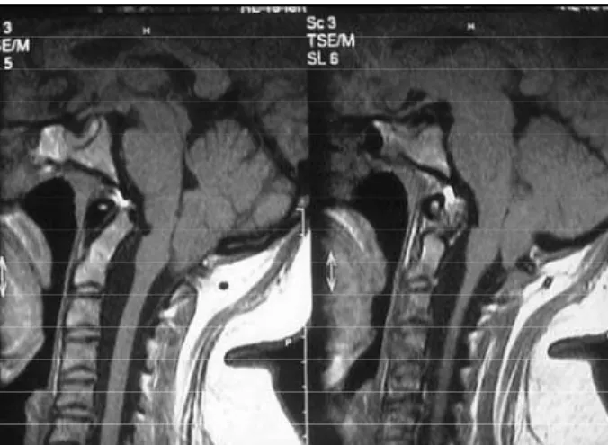
2ABSTRACT - We describe a rare case of a 30 year-old woman with intense vertiginous sensation, lack of body balance and a tendency to fall backwards, making it necessary for two people to sustain her. The magnetic resonance imaging of the craniocervical junction evidenced tonsilar herniation at the inferior lev-el of C1, and during the operation perf o rmed in sitting position, we observed crowding of the cereblev-ellar t o n-sils at the level of C3. After the osteo-dural-neural decompression, the symptomatology remitted on the s a m e day of the operation.
KEY WORDS: vertigo, retropulsion, Chiari malformation, cerebellar tonsils, posterior cranial fossa.
Retropulsão e vertigem na malformação de Chiari: relato de caso
RESUMO - Descrevemos um caso raro de mulher de 30 anos com intensa sensação vertiginosa, desequilí-brio do corpo e tendência à queda para trás, sendo necessário o auxílio de duas pessoas para ampará-la. A ressonância nuclear magnética da junção craniovertebral evidenciou herniação tonsilar ao nível da borda infe-rior de C1 e, durante a operação, em posição sentada, foi observado o deslocamento craniocaudal das tonsi-las cere b e l a res ao nível de C3. Após a descompressão ósteo-duro-neural, houve re g ressão da sintomatologia, no dia da operação.
Arq Neuropsiquiatr 2005;63(3-B) 871
O t h e rwise, Schwalbe and Gre d i g5, pupils of Ar-nold, considered that the anomalies pre v i o u s l y described by Cleland, Chiari and Arnold consisted of a specific anatomical syndrome they designat-ed “Arnold-Chiari” malformation. However, C a rm e l et al.6p roposed the denomination Cleland-Chiari m a l f o rmation, considering that Arnolds descrip-tion was identical to the anomalies previously des-cribed by Chiari. Unfortunately the description of Cleland was forgotten in the world literature, whi-le the concept “Chiari Malformation” was accept-ed in the same literature. It is a malformation of f re-quent occurrence, especially associated to the basi-lar impression (BI) and syringomyelia (SM). In nor-theast Brazil, the association of BI and CM pre s-ents high incidence, as observed in the studies of C a n e l a s7and Canelas et al.8, Caetano de Barro s9, Gonçalves da Silva1 0, Ta r i c c o1 1, Arru d a1 2 , 1 3, Carn e i ro F i l h o1 4, Gonçalves da Silva et al.1 5 , 1 6. The symptoma-tology is characterized by a cerebellar syndrome, nuchal pain, vertigo and horizontal, downbeat or rotatory nystagmus, among others.
The publication of this case is based on the ra-reness of the clinical picture characterized by re-tropulsion and vertigo, which is not found in the consulted literature.
CASE
A 30 year-old woman was assaulted by a vert i g i n o u s crisis of great intensity, lack of body balance and ten-dency to fall backwards, needing double support in or-der not to fall. She mentioned a shock sensation when attempting to move her head forw a rd or backward s , which began in the cervical region, spread down to her feet and, right after that, up to the head. The neuro l o-gical exam evidenced a marked postural instability - the patient could not remain in orthostatic position with-out the help of two people - suppression of the
nau-seous reflex and discrete hypopallesthesia in the inferi-or members. Nystagmus was not verified. The magneti c resonance image (MRI) evidenced the herniation of the cerebellar tonsils (Fig 1). She was submitted to posteri-or fossa surg e ry, through the osteo-dural-neural decom-p ression, technique used by Gonçalves da Silva1 2and G o
n-çalves da Silva et Holanda1 3, characterized by an larg e
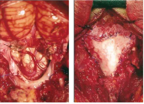
craniectomy, dissection of the cerebellar tonsils, which reached C3 level (the caudal limit of the laminectomy) (Fig 2) and the regional arteries, large opening of the f o u rth ventricle, intrapial aspiration of the cerebellar t o n-sils, suture of the residual pial sacs to the lateral dura-mater in ascending position (Fig 3) and, finally, a dural grafting was perf o rmed with the use of bovine pericar-dium (Fig 4). In the present study, the cerebellar tonsils w e re loose and did not show any adhesions to the circ u-mscribed tissues. The post-operative MRI revealed ab-sence of the herniated cerebellar tonsils and form a t i o n of great cisterna magna (Fig 5).
DISCUSSION
Among all symptoms and clinical signs observ e d in the CM, nuchal pain and the vestibular syndro-mes seem to be the dominant.
B r a n d t1 7described the several directions of falls in vestibular diseases: as lateral, in the vestibular neuritis and the Wa l l e n b e rg syndrome; forw a rd , in the benign paroxysmal positioning vertigo, and f o re-and-aft in the bilateral vestibulopathy. The a u t h o r, however, did not mention the vestibular s y n d rome in the CM. Paul et al.1 8, in a 71-case CM c a s u i s t ry, verified neck pain in 69% and lack of bal-ance in 40% of his patients. Williams19related, in a 54-case casuistry, 64.8% of migraine and 91.1% of vertigo. Caetano de Barro s9, among 21 patients, mentioned neck pain in 57.1% and vertigo in 2 3 . 8 % . Gonçalves da Silva1 5, in a casuistry of 245 BI and/or CM and SM cases, observed neck pain in 53% and vertigo in 54.6%.
872 Arq Neuropsiquiatr 2005;63(3-B)
geal irritation. In both of them the choleric attacks regressed after the operation.
In the presently related case, the acute sympto-matology of re t ropulsion and vertigo presents it-self as a rarity in the clinical aspects of CM. Possibly, the thick cerebellar tonsils, functioning as an ex-pansive process, could compress the vermis, which could likely be the cause of the re t ropulsion. De-J o n g2 4mentioned that in the cerebellar vermis and
medial line lesions, the patient may not be able to remain erect, falling both forw a rd and backward s .
On the other hand, the descending and ascend-ing shock sensation could originate from the pro-cess of friction of the cerebellar tonsils on the cere-bellum, medulla and especially the spinal cord, p ro-voking irritation of the dorsal funiculi on the occa-sion of front or back flexion of the head.
As for the surgical treatment, we opted for lar-ge craniectomy of the posterior fossa, taking under consideration that the posterior fossa, in BI and/or CM and SM cases, is notoriously reduced in its vol-ume, as described by Ackerm a n n2 5, Nyland and K ro g-n e s s2 6, Marin-Padilla2 7, Padilla and Marin-P a d i l l a2 8, Schady et al.2 9and Vega et al.3 0, among others. The large craniectomy involves the form a-tion of great cisterna magna, a fundamental condi-tion for the brainstem and cerebellum to migrate c r a n i a l l y. Let us cite as an illustration the studies of Badie et al.31, which demonstrated the smaller size of the posterior fossa in the CM and that it in-c reased in volume after the dein-compression. Milho-rat et al.3 2verified a decrease of 13.4 ml in the vol-ume of the posterior fossa and 40% (10.8 ml) in the cere b rospinal fluid volume of this region. Sa-huquillo et al.3 3c o m p a red the results obtained in 10 cases in which a reduced craniectomy was per-f o rmed with other 10 that were subjected to ex-tensive craniectomy. Cranial migration of the cere-Fig 2. Dissection of the cerebellar tonsils
with herniation up to C3, during the oper -ation in sitting position.
Fig 3.Tonsilectomy and large opening of the fourth ventricle and fixation of the re -sidual pial sac to the lateral dura-mater in cranial position.
Fig 4. Dura- mater graft.
Arq Neuropsiquiatr 2005;63(3-B) 873
bellum and brainstem was observed in all the lat-er patients, while in the cases whlat-ere reduced cra-niectomy was perf o rmed there was caudal migra-tion in 7 patients.
The dissection of the cerebellar tonsils and of the regional arteries, especially the posterior infe-rior cerebellar arteries, as well as the large open-ing of the fourth ventricle and intrapial aspiration of the cerebellar tonsils, as demonstrated by Bat-z d o rf3 4, take part in the decompression process of the posterior fossa, there f o re facilitating the circ u-lation of cere b rospinal fluid from the fourth ven-tricle to the recently created cisterna magna, and so, preventing the reappearance of the craniospi-nal pressure dissociation.
Of further importance is the observation that even though the tonsils were located at the level of C1 on the preoperative MRI, intraoperative ex-ploration, with the patient in sitting position re v e-led crowding of the tonsils at the level of C3. Pro-bably the cerebellar tonsils have the tendency to migrate downwards on the orthostatic position. In the future, the introduction of MRI carried out in the upright position, will enable us to detect a d i ff e rence on the topography of the cerebellar to-nsils, between the dorsal position placement and the ortostatic position.
REFERENCES
1. Cleland J. Contribution to the study of spina bifida, encefalocele, and anencephalus. Anat Physiol 1883;17:257-292.
2. Chiari H. Über Ve r ä n d e rungen des Kleinhirns infolge von Hydro c e-phalie des Grosshirns. Dtsch med Wschr 1891;17:1172-1175. 3. Arnold J. Myelocyste, Transposition von Gewebskeimen und Sympodie.
Beitr Path Anat allgem Path 1894;16:1-28.
4. Chiari H. Über Ve r ä n d e rungen des Kleinhirns, des Pons und der Medul-la Oblongata in Folge von congenitaler Hydrocephalie des Gro s s h i r n s . Dtsch Akd Wissenschaft 1895;63:71-85.
5. Schwalbe E, Gredig M. Über Entwicklungsstörungen des Kleinhirns, Hirnstamms und Halsmarks bei Spina bifida (Arnold`sche und Chiari`sche Missbildung). Beitr Path Anat Path 1907;4:132-194. 6. Carmel PW, Markesbery WR. Early descriptions of the Arnold-Chiari
Malformation: the contribution of John Cleland. J Neuro s u rg 1972; 93:543-547.
7. Canelas HM, Zaclis J, Tenuto RA, Cruz OR. Contribuição ao estudo das malformações occipitocervicais, particularmente da impressão basi-lar. Arq Neuropsiquiatr 1952;10:407-476.
8. Canelas HM, Zaclis J, Tenuto RA, Cruz OR. Malformações occipito-cervicais. A p ropósito de vinte novos casos. A rq Neuropsiquiatr 1956; 14:1-26.
9. Caetano de Barros M. Contribuição ao estudo da impressão basilar associada à malformação de Arnold-Chiari. Tese. Recife, 1959. 10. Gonçalves da Silva JA. Resultados do tratamento cirúrgico da impre s s ã o
basilar e malformação de Arnold-Chiari: estudo de 72 casos. Tese. João Pessoa, 1977.
11. Taricco MA. Tratamento cirúrgico da siringomielia associada à malfor-mação de Chiari do tipo I. Tese. São Paulo, 1994.
12. A r ruda JAM. Tratamento da siringomielia associada à malformação de Chiari: análise de 30 casos. Tese. São Paulo, 1996.
13. A r ruda JAM. Tratamento da siringomielia associada à malformação de Chiari: análise de 60 casos. Tese. São Paulo, 2001.
14. C a r n e i ro GS Filho. Tratamento circ u n f e rencial da invaginação basilar. Tese. Recife, 2001.
15. Gonçalves da Silva JA, et al. Malformações occipitocervicais. impre s s ã o b a s i l a r, ,malformação de Chiari, siringomielia, platibasia. Recife: Editora Universitária / UFPE, 2003:169-300.
16. Gonçalves da Silva JA, Holanda MMA. Basilar impression, Chiari mal-formation and syringomyelia: a re t rospective study of 53 surg i c a l l y treated patients. Arq Neuropsiquiatr 2003;61:368-375.
17. Brandt T. Vertigo: its multisensory syndromes. 2.Ed. London: Springer Verlag, 1999:13-21.
18. Paul KS, Lye RH, Strang FA, Dutton J. Arnold-Chiari malformation: review of 71 cases. J Neurosurg 1983;58:183-187.
1 9 . Williams B. Surgery for hindbrain related syringomyelia. Advances and technical standards in neuro s u rg e r y. Wien: Springer, 1993;20:107-164. 20. Rullan A. Associated laryngeal paralysis: presentation of a case of
bilate-ral abductor pabilate-ralysis in a patient with the Arnold-Chiari deformity. Arch Otolaryngol 1956;64: 207-212.
21. Gol A, Hellbusch LC. Surgical relief of pro g ressive upper limb paral-ysis in Arnold- Chiari malformation. J Neurol Neuro s u rg Psychiatry 1978;41:433-437.
22. Thomas M, Boyle R. A possible connection between basilar migraine and the Arnold- Chiari malformation. Neurology 1979;29:527-528. 23. Hudgins RJ. Paroxysmal rage as a presenting symptom of the Chiari I
malformation: report of two cases. J. Neurosurg 1999;91:328-329. 24. DeJong RN. Medullary and related syndromes. In The neurologic
exa-mination,.3.Ed. New York: Harper& Row, 1967:347-348.
25. Ackermann JF. Über die Kretinen, eine besondere Menschenabart in de Alpen. Gotha, in der Ettingerschen Buchhandlung, 1790. 26. Nyland H, Krogness KG. Size of posterior fossa in Chiari type I
malfor-mation in adults. Acta Neurochir 1978;40:233-242.
27. Marin-Padilla M. Cephalic axial skeletal-neural dysraphic disord e r s : embryology and pathology. Can J Neurol Sci 1991;18:153-169. 28. Marin-Padilla M, Marin-Padilla TM. Morphogenesis of
experimental-ly induced Arnold-Chiari malformation. J Neurol Sci 1981;50:29-55. 2 9 . Schady W, Metcalfe RA, Butler P. The incidence of craniocervical bony
anomalies in the adult Chiari malformation. J Neurol Sci 1987;82:193-203. 30. Vega A, Quintana F, Berciano J. Basichondrocranium anomalies in adult Chiari type I malformation: a morphometric study. J Neurol Sci 1990; 99:137-145.
3 1 . Badie B, Mendoza D, Batzdorf U. Posterior fossa volume and response to suboccipital decompression in patients with Chiari I malformation. Neurosurgery 1995;37:214-218.
32. Milhorat TH, Chou MW, Trinidad EM, et al. Chiari malformation re d e-fined:clinical and radiographic findings for 364 symptomatic patients. Neurosurgery 1999;44:1005-1017.
33. Sahuquillo J. Rubio E, Poca MA, Rovira A, Rodriguez-Baeza A, Cervera C. Posterior fossa re c o n s t ruction: a surgical technique for the tre a t m e n t of Chiari I malformation and Chiari I/syringomyelia: complex-pre l i m i-nary results and magnetic resonance imaging. Quantitative assessment of hindbrain migration. Neurosurgery 1994;35:874-884.