ABSTRACT
Analysis of the dentin-pulp complex in teeth
submitted to orthodontic movement in rats
Camila da Siveira MASSARO1, Renata Bianco CONSOLARO2, Milton SANTAMARIA JUNIOR3, Maria Fernanda Martins-Ortiz CONSOLARO4, Alberto CONSOLARO5
1- Undergraduate student, Bauru School of Dentistry, University of São Paulo, SP, Brazil.
2- DDS, MSc, PhD student of Oral Pathology, Bauru School of Dentistry, University of São Paulo, SP, Brazil. 3- DDS, MSc, PhD, Orthodontics Practice in Ribeirão Preto, SP, Brazil.
4- DDS, MSc, PhD, Professor of Orthodontics and Oral Biology, University of Sacred Heart, Bauru, SP, Brazil.
5- Professor of Oral Pathology, Department of Stomatology, Bauru School of Dentistry, University of São Paulo, SP, Brazil.
Corresponding address: Camila da Silveira Massaro/Prof. Dr. Alberto Consolaro - Faculdade de Odontologia de Bauru - USP - Disciplina de Patologia - Alameda Octávio Pinheiro Brisolla, 9-75 - Vila Universitária - 17012-901 - Bauru, SP - Brasil - Phone: +55 (14) 3235-8248 - Fax: +55 (14) 3235-8251 - e-mail: [email protected]/[email protected]
5HFHLYHG$XJXVW0RGL¿FDWLRQ)HEUXDU\$FFHSWHG0DUFK
I
n order to microscopically analyze the pulpal effects of orthodontic movement, 49 closed coil spring anchored to the maxillary incisors, placed for the achievement of mesial movement. Material and Methods: Ten animals were used as the control group and were not submitted to orthodontic force; the other animals were divided into groups according to the study period of tooth movement, namely 1, 2, 3, 4, 5, 6 and 7 days. The investigation of congestion. Statistical evaluation was performed between control and experimental groups and between periods of observation using non-parametric chi-square, Kruskal-Wallis and ! changes between control and experimental groups nor between periods of observation. The control group, at 3 and 5 days, revealed greater hyalinization of the periodontal ligament "#$$%& % ' "#$$%& Conclusion: No morphological change from the effect of induced tooth movement could ( ) * detectable in optical microscopy, was found in experimental groups.Key words: Dental pulp. Rat. Tooth movement. Root resorption.
INTRODUCTION
The pulp and periodontal tissues are richly cellularized, and their metabolic rates are adapted to their functional needs. The structural and functional normality of these tissues seem
* 7.
Clinical detection of periodontal and pulp changes induced by local and systemic factors probably depend on the type, duration and intensity of the stimulus applied.
Early studies have suggested that vascular changes promoted by orthodontic movement
might cause pulp necrosis. Oppenheim20-22, +/8< +/8' +/=> submitted to orthodontic movement presented occasional atresia of the pulp and root canal, observed by radiographic examination. Microscopically, there was an increase in * reduction in blood vessels, indicating probable pulp degeneration.
Taylor3,4, in 1951 and 1952, applied retraction forces to incisors of monkeys and reported the occurrence of pulp necrosis.
In their study, Mjör and Stenvik19 (1969) applied intrusion forces to human teeth and did tissue of experimental and control groups; the observation of vacuolization in the pulp tissue was considered to be an artifact. Anstendig and Kronman1 conducted an investigation on dogs and also observed vacuolization of the odontoblast layer as the main pulp changed after orthodontic movement.
In the 1980s, Hamersky, et al.12 and Unsterseher, et al.31 (1987) investigated biochemically the effect of orthodontic forces on the respiratory metabolism of the pulp of human teeth, with the aid of radioactive carbon dioxide. The authors concluded that pulp respiration is reduced in orthodontically moved teeth.
Studies using laser doppler flowmetry conducted by McDonald and Pitt Ford18 (1994), Ikawa, et al.14 (2001) and Sano, et al.26 (2002) revealed that application of forces on human * JL and Ransay2 (1996), which did not reveal any changes.
Q * Derriger, et al.9,10 (1996, 1998) analyzed the secretion of angiogenic factors on human teeth and observed that these factors were increased in orthodontically moved teeth. Santamaria, et al.28 (2006), detected a slight increase in the < hours of induced tooth movement in molars of rats. Ramazanzadeh, et al.25 (2009), described similar results in human teeth submitted to intrusive and extrusive forces.
The related literature presents variable models and criteria for analysis, as well as results and conclusions. Thus, the following doubts remain: 1. May induced tooth movement promote early pulp aging, even if only microscopically observable? 2. May it promote pulpitis, pulp necrosis or any other change? These questions seem to remain without answers and were the initial premises of this study.
MATERIAL AND METHODS
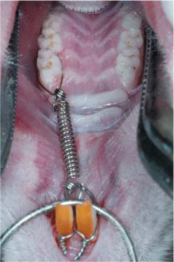
Appliances were placed on the maxillary =/ X 120 days, divided into 7 experimental groups, according to the period of tooth movement (1 to 7 days)13,17. Three animals were analyzed at each period, yet nine animals were evaluated at 3, 5 and 7 days, which are the most representative periods of the biological effects of induced tooth movement. Other 10 animals were taken apart as the control group and were killed without accomplishment of any orthodontic movement. The device was placed as suggested by Heller and Nanda13 (1979) and improved by Martins-Ortiz17 (2005), and was composed of a stainless and anchored to the maxillary incisors, which delivered 75 g of force (Figure 1). To place the device for tooth movement, the animals were anesthetized with a mixture of equal parts of ketamine hydrochloride 100 mg/mL (Dopalen – Vetbrands) and muscle relaxant xylazine hydrochloride 20 mg/mL (Anasedan – Vetbrands), at a dose of 1 mL/Kg. The solution was applied with a 1-mL syringe and 12.7-mm sterile needle by intramuscular injection.
The rats were killed by an overdose of anesthetics (ketamine and xylazine), according to the periods of tooth movement of each group. After that, the maxillae were removed with surgical scissors and the specimens were +$\ ] the specimens were decalcified in Morse’s L longitudinally sectioned of 6-Pm thickness. The sections were placed on glass slabs and stained with hematoxylin-eosin. Ten slabs with 5 sections each were prepared for each animal.
Small hyaline areas and frontal bone resorption were interpreted as the result of biologically acceptable forces.
At the apical third, more specifically at the apical periodontal ligament, microscopic evaluation addressed the detection of circulatory
Figure 1- Orthodontic appliance used to achieve mesial maxillary incisors, delivering a force of 75 g
Periodontal changes Groups
No ITM 1 day 2 days 3 days 4 days 5 days 6 days 7 days
n=10 n=3 n=3 n=9 n=3 n=9 n=3 n=9
Focal hyalinization 0 2 0 2 0 2 0 3
Segmental hyalinization 0 1 3 7* 3 7* 3 6
Frontal bone resorption 0 0 1 3 1 2 0 3
Undermining resorption 0 0 0 6** 2 7* 3 6**
Root resorption 0 0 0 0 1 8* 3 9*
Table 1- Frequency of periodontal phenomena microscopically observed in the control and experimental groups, with
! " # $ %&! " # $
changes such as blood vessel congestion, hemorrhage and thrombosis.
The main changes investigated in the dental pulp comprised cell vacuolization, pulp hyalinization, pulp nodules, dystrophic calcifications, reactive dentin, blood vessel congestion, areas of hemorrhage and thrombosis. The analysis also addressed the disorganization of the odontoblast layer and areas of pulp necrosis.
Statistical analysis
Statistical evaluation was performed between control and experimental groups and between periods of observation using non-parametric chi-square, Kruskal-Wallis and Dunn tests.
] 8$ 30 specimens were randomly selected and re-^ analyses were compared for investigation of intra-examiner agreement by Kappa analysis, considering values above 0.80 as good level of agreement.
RESULTS
Evaluation of intra-examiner agreement revealed a kappa value of 0.83, demonstrating the effectiveness of the analysis of microscopic sections conducted by the examiner.
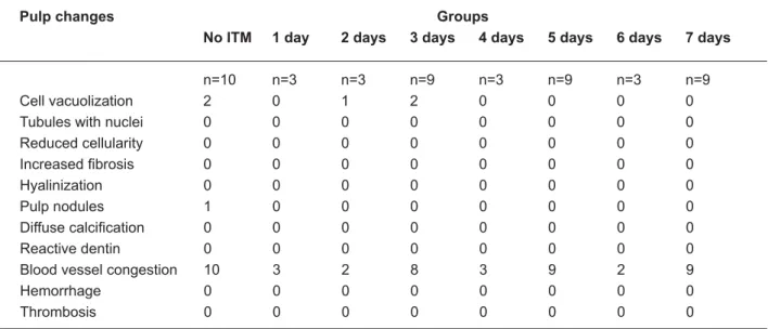
Pulp changes Groups
No ITM 1 day 2 days 3 days 4 days 5 days 6 days 7 days
n=10 n=3 n=3 n=9 n=3 n=9 n=3 n=9
Cell vacuolization 2 0 1 2 0 0 0 0
Tubules with nuclei 0 0 0 0 0 0 0 0
Reduced cellularity 0 0 0 0 0 0 0 0
' & & & & & & & &
Hyalinization 0 0 0 0 0 0 0 0
Pulp nodules 1 0 0 0 0 0 0 0
; & & & & & & & &
Reactive dentin 0 0 0 0 0 0 0 0
Blood vessel congestion 10 3 2 8 3 9 2 9
Hemorrhage 0 0 0 0 0 0 0 0
Thrombosis 0 0 0 0 0 0 0 0
! " # $ %&! " # $
Table 2- Frequency of pulp phenomena microscopically observed in the control and experimental groups, with induced
Figure 2- Periodontal morphological changes induced on the cervical region of the distobuccal root submitted to intense < >? E# # G ? < I # ## J? <K L N >K Q&
groups compared to the Control group. This difference was found for the presence of segmental hyalinization at 3 and 5 days, at the 5% level, between control and experimental groups. The analysis of frequency of frontal bone
Figure 3- Periodontal morphological changes induced on the cervical region of the mesiobuccal root (A and B) submitted to moderate forces with frontal bone resorption. Experimental group, after 3 days of induced tooth movement on the maxillary ? <K L N >K Q&
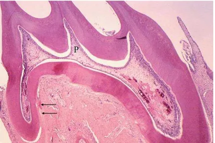
Figure 4- Microscopic aspects of the pulp and periodontal morphological changes in the periodontal ligament, after 4 days ? I X # the distal root (arrows) and the normal aspect of the pulp (P). Pulp vascular congestion was due to operatory procedures #? %&
was greater at 3, 5 and 7 days compared to the { ! observed for the presence of root resorption at 5 and 7 days compared to the Control group (Table 1).
Evaluation of the Control and Experimental
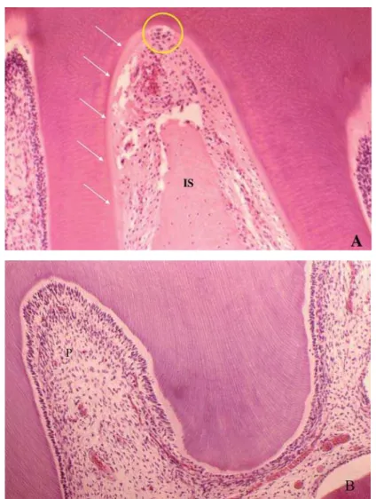
Figure 5- Microscopic aspects of the root pulp (P) in A, and coronal pulp in B, compatible with normal morphology of maxillary ' Y ? Z # ' the cementoblast layer was extensively lost (arrows), with presence of several clasts and areas of root resorption (circle). <K L N >K L
yet associated with artifacts. These observations on morphological normality were valid for the entire dental pulp, from the coronal to the apical region. In the internal dentinal wall, turned toward the dental pulp, there was neither thickening of the pre-dentin layer nor morphological signs of reactive dentin that could indicate any pulpal effect from the force applied throughout this experiment of induced tooth movement in rats (Table 2).
DISCUSSION
The forces applied to the maxillary first molars of rats were intense on the mesial interseptal surface of the distobuccal root (Figure 2) and moderate on the mesial region of the mesiobuccal root, both analyzed on the cervical
third (Figure 3).
Analysis of the distobuccal root microscopically demonstrated the biological effects of application of intense forces, with presence of hyaline areas of periodontal ligament at 3 to 5 days of induced tooth movement, especially on the cervical region at the pressure side (Figure 2 and 5). At 5 days and especially at 7 days, on the same region, there was higher frequency of root resorption (Figure 5). The forces applied to the mesiobuccal root had moderate intensity, with absence of hyalinization of the periodontal ligament and root resorption at the most advanced periods of induced tooth movement (Figure 3).
up to 7 days (Table 2, Figure 2, 4 and 5). The experimental model of induced tooth movement adequately reproduces the biological situation found in humans. However, the metabolic rate of connective tissues of rats should be considered when using this experimental model. A period of 21 days is required for the observation of all stages of induced tooth movement in humans, whereas in rats this period is reduced to 7 days11.
Previous studies in the literature1,4,19,25,27,28 indicate that orthodontic movement does not induce pulp changes such as increased number of pulp modules, calcific metamorphosis of the pulp, permanent vascular changes, or early pulp aging represented by hyalinization ! changes are generically called regressive and/ or degenerative8,24.
Investigations reveal the occurrence of several biological reactions during induced tooth movement, including increased15 or reduced14,18 blood flow; reduced12,31 or increased16 cell respiration; and increased angiogenesis9,10. However, changes in the local level of enzymes and mediators observed in cultures and laboratory biochemical tests on the pulp tissue23,32 indicate possible changes in the metabolic rates of the pulp tissue, rather than loss of biological viability or vitality. These metabolic changes did not morphologically changes the pulp tissue.
Clinical cases8,30 should properly include all records, from photographs to periapical radiographs, before and during orthodontic treatment, or rule out the possibility of dental trauma5,6,19.
For the endodontist and orthodontist, the pulp status should be precisely and safely determined before the onset of orthodontic treatment. After diagnosis of pulp status of teeth which will be submitted to orthodontic movement, treatment may be performed in a safe manner and with good prognosis. In teeth without caries, restorations or periodontal disease, trauma should be investigated as the main suspected cause of pulp changes during and after orthodontic treatment.
Pulp necrosis induced by trauma may be diagnosed during orthodontic treatment. The
tissue changes inherent to tooth movement may exacerbate the clinical and radiographic signs of chronic pericementitis and periapical granuloma, not diagnosed on the initial orthodontic records. The initial correct diagnosis of the teeth should be done with periapical radiographic.
When compared to traumatisms or mechanic forces of different kinds, orthodontic forces can be considered little or small, even when called heavy or intense5,6. Studies comprising experimental intrusion and tooth movement with forces considered intense did not produce pulp necrosis in a similar context as the orthodontic treatment4,25.
In the present study, the effectiveness of the force applied was demonstrated by the microscopically observable changes in the periodontal tissue, including significant loss of segmental hyalinization at 3 to 5 days, undermining resorption at 5 days and root resorption at 7 days; however, the pulps of moved teeth did not present any degenerative * "| > = %& The odontoblast layer occasionally presented cell vacuolization and loss of continuity, yet associated to artifacts.
Therefore, according to the present results and previous reports in the literature, it is suggested that clinical cases of pulp necrosis occurring during and after orthodontic treatment should be managed with greater emphasis on the dental history of the patient and possibility of trauma, rather than on the orthodontic force applied.
CONCLUSION
REFERENCES
1- Anstendig HS, Kronman JH. A histologic study of pulpal reaction to orthodontic tooth movement in dogs. Angle Orthod. 1972;42:50-5.
2- Barwick PJ, Ramsay DS. Effect of brief intrusive force on human * ] ~ +//<^++$>'8(/ 3- Butcher EO, Taylor C. The effects of denervation and ischemia upon the teeth of the monkey. J Dent Res. 1951;30:265-75. 4- Butcher EO, Taylor C. The vascularity of the incisor pulp of the monkey and its alteration by tooth retraction. J Dent Res. 1952;31:239-47.
5- Consolaro A. Movimento ortodôntico não promove necrose pulpar. R Clin Ortodon Dental Press. 2002;1:99-100.
6- Consolaro A. Reabsorções dentárias nas especialidades clínicas. Maringá: Dental Press; 2005.
7- Davidovitch Z. Tooth movement. Crit Rev Oral Biol Med. 1991;2:411-50.
( ~ ) orthodontic patient. Am J Orthod Dentofac Orthop. 1982;82:58-61.
9- Derringer KA, Jaggers DC, Linden RWA. Angiogenesis in human dental pulp following orthodontic tooth movement. J Dent Res. 1996;75:1761-6.
10- Derringer KA, Liden RW. Enhance angiogenesis growth induced by diffusible angiogenic growth factors release from human dental pulp explants of orthodontically moved teeth. Eur J Orthod. 1998;20:357-67.
++( | ! ) | ! rat in laboratory investigation. New York: Hafner Publication; 1963. p.120.
12- Hamersky PA, Weimer AD, Taintor JF. The effect of orthodontic force application on the pulpal tissue respiration rate in the human premolar. Am J Orthod Dentofac Orthop. 1980;77:368-78. +8( )~ Q on orthodontic tooth movement. Am J Orthod. 1979;75:239-58. 14- Ikawa M, Fujiwara M, Horiuchi H, Shimauchi H. The effect of ( * * ] J >$$+^=<'+(' 15- Kvinnsland S, Heyeraas K, Ofjord ES. Effect of experimental * Q ~ Orthod. 1989;11:200-5.
16- Labart WA, Taintor JF, Dyer JK, Weimer, AD. The effect of orthodontic forces on pulp respiration in the rat incisor. J Endod. 1980;6:724-7.
+'( ( | { ] )* na movimentação dentária induzida e nas reabsorções dentárias associadas. In: Consolaro A. Reabsorções dentárias nas especialidades clínicas. 2nd ed. Maringá: Dental Press International; 2005. p. 523-70.
+( | | ! J * maxillary canines during retraction. Eur J Orthod. 1994;16:1-9. 19- Mjör IA, Stenvik A. Microradiography and histology of ^ emphasis on resorption. Arch Oral Biol. 1969;14:1355-64. 20- Oppenheim A. Biologic orthodontic therapy and reality. Angle Orthod. 1936;6:153.
21- Oppenheim A. Biologic orthodontic therapy and reality. Angle Orthod. 1937;7:58-9.
22- Oppenheim A. Human tissue response to orthodontic intervention of short and long duration. Am J Orthod. 1942;28:263-301.
23- Perinetti G, Varvara G, Festa F, Esposito P. Aspartate aminotransferase activity in pulp of orthodontic tooth movement in adolescents: a radiographic study. Am J Orthod Dentofac Orthop. 2004;125:88-92.
24- Popp TW, Artun J, Linge L. Pulpal response to orthodontic tooth movement in adolescents: a radiographic study. Am J Orthod Dentofac Orthop. 1992;101:228-33.
>%( J] ]] N, Jahanbin A, Shakeri MT. Histological changes in human dental pulp following application of intrusive and extrusive orthodontic forces. J Oral Sci. 2009;51:109-15.
26- Sano Y, Ikawa M, Sugawara J, Horiuchi H, Mitani H. The effect * Q ~ Orthod. 2002;24:159-66.
27- Santamaria M Jr, Milagres D, Iyomasa MM, Stuani MBS, Ruellas ACO. Initial pulp changes during orthodontic movement: histomorphological evaluation. Braz Dent J. 2007;18(1):34-9. 28- Santamaria M Jr, Milagres D, Stuani AS, Stuani MBS, Ruellas ACO. Pulpal vasculature changes in tooth movement. Eur J Orthod. 2006;28:217-20.
29- Strang RHW. A textbook of orthodontia. Philadelphia: Lea and Febiger; 1943.
30- Stuteville OH. Injuries caused by orthodontic forces and the ultimate results of these injuries. Am J Orthod Oral Surg. 1938;24:103-19.
31- Unterseher RE, Neiberg LG, Weimer AD, Dyer JK. The response of human pulp tissue after orthodontic force application. Am J Orthod Dentofac Orthop. 1987;92:220-4.