Introduction
Diabetic peripheral neuropathy (DPN) is the most common chronic complication of diabetes mellitus (DM), affecting ap-proximately 50% of patients1. When associated with vascular impairment of the lower limbs, this generates a set of changes in the feet, characterized as diabetic foot2.
Over the course of the disease, there may be deforma-tions, decreased mobility, and muscle hypotrophism of the foot and ankle, resulting in changes in plantar pressure and thus predisposing the individual to the development of ulcers and the risk of falls3-6. This, added to deicits in the motor control system, intensiies the imbalances and alterations in posture and gait3.
Investigations in this population indicate a reduction in gait velocity and step length increasing the duration of the support phase, a decrease in the mobility of the ankle joint, a redistribu-tion of joint moments, and changes in kinematic lower limb and muscle activation, compared with the normal range of subjects without diabetes7-10.
According to the International Working Group on Diabetic Foot (IWGDF), based on the aggravation of lower limb com-plications and thus, the severity of peripheral neurovascular aflictions, the feet of diabetic patients can be classiied into categories11. This is easy to apply in a clinical setting and could be important to aid the understanding of the clinical condition of diabetic patients.
Accordingly, early studies suggest possible relationships between the degree of incapacity in the diabetic population and the disability in gait variables10,12; however, without con-sidering the presence of diabetic peripheral vascular disease (PVD), creating a gap in the scientiic knowledge since the population with this level of impairment presents a challenge due to the limited ability to walk without the need for auxiliary gait devices. In this sense, the present study aimed to analyze the inluence of peripheral neurovascular impairment on gait variables, believing that the conirmation or dismissal of this relationship could contribute to awareness of the risks of the chronic complications of diabetes, as well as highlight the im-portance of early intervention.
Thus, this study aimed to analyze the behavior of gait param-eters of non-diabetic and diabetic individuals with neuropathy and/or peripheral diabetic vasculopathy.
Method
This was an observational, cross-sectional controlled study, de-veloped at the Clinical Studies Laboratory in Physical Therapy (LECFisio) at the School of Science and Technology – Universidade Estadual Paulista (UNESP), Presidente Prudente – SP.
All participants, in accordance with the means and purposes of the research, signed the informed consent form. All proce-dures complied with the ethical principles for clinical research Original article (short paper)
Vasculopathy associated with peripheral
neuropathy in gait parameters of diabetic people
Alessandra Madia Mantovani
Universidade Estadual Paulista “Júlio de Mesquita Filho”, Rio Claro, SP, Brasil
Nathalia Ulices Savian Mariana Romanholi Palma Claudia Regina Sgobbi de Faria Cristina Elena Prado Teles Fregonesi
Universidade Estadual Paulista “Júlio de Mesquita Filho”, Presidente Prudente, SP, Brasil
ABSTRACT — Diabetic peripheral neuropathy (DPN) is a common complication of diabetes mellitus when glycemic levels are poorly controlled. Sometimes DPN is accompanied by vasculopathy (DPV), which can worsen the clinical prognosis. The aim of this study wasto analyze the gait parameters of nondiabetic individuals and diabetic individuals with DPN with or without DPV. Method: The study included 68 individuals (50 to 65 years old) divided into three groups: people without diabetes mellitus (n = 33), diabetic patients with DPN (n = 18), and diabetic patients with both DPN and DVP (n = 17). The participants underwent a gait evaluation using electronic baropodometry to obtain the single and double support, velocity, and pressure-time integral. Results: The pressure-time integral, velocity, and single support variables were lower, and the double support and double support/single support ratio were higher in the diabetic neuropathy and vasculopathy group. The velocity was lower the greater the degree of impairment of the diabetic foot. Some correlations were identiied with velocity. Conclusion: In diabetic individuals, there was a signiicant worsening
of the gait parameters analyzed according to increasing degree of clinical impairment.
involving human beings and were approved by the Research Ethics Committee of the proponent university (protocol number: 10/2011).
Sample
Initially, a sample size calculation was carried out based on a sampling error of 0.2, from which it was determined that the groups required a minimum of 18 subjects; however, some groups exceeded this value. The inal sample consisted of 68 subjects divided into three groups: CG – control group (n = 33), 30% males and 70% females, NG – diabetic neuropathy group (n = 18), 40% males and 60% females, and NVG–diabetic neuropathy and vasculopathy group (n = 17), 60% males and 40% females. The CG contained a higher number of individuals than expected due to the high number of eligible volunteers, whereas there was some dificulty composing the NVG as individuals with these levels of complication rarely walk independently.
Sample Selection Criteria
Inclusion in the CG: an absence of diagnosis of DM and/or other diseases of possible neural damage, sensitivity to 2 g monoilament (described in the procedure section), preserved feet conirmed by somatosensory sensitivity, ankle/upper arm index above 0.90, and normalcy of blood perfusion, with dif-ferences in this index value between the upper and lower limbs of less than two points and grade 0 of impairment of diabetic foot according to the IWGDF11,13,14.
Inclusion in the NG: medical conirmation of type 2 DM, DPN conirmed by insensitivity to the 10 g Semmes–Weinstein monoilament in nine points in the plantar region and two in the dorsal region13 and the Scale for Diagnosis of Diabetic Distal Polyneuropathy15, without a medical diagnosis of peripheral vas-cular disease, ankle/upper arm index greater than 0.9 (obtained by analysis and Doppler sphygmomanometer in the upper and lower members), and blood perfusion with a difference of up to two points between the upper and lower limbs measured by the oximetry test on the ingers and toes and grade 1 diabetic foot impairment11.
Inclusion in the NVG: medical conirmation of type 2 DM, DPN, and PVD. DPN conirmed by insensitivity to the 10 g Semmes–Weinstein monoilament at least one of the 11 points tested in the plantar and dorsal regions of the foot13 and the Scale for Diagnosis of Diabetic Distal Polyneuropathy15. Change in the circulation and peripheral blood perfusion, detected respectively by an ankle/arm index of <0.90 (obtained by analysis and Doppler sphygmomanometer in the upper and lower members) and by values that differ by two or more points between the upper and lower limbs in the oximetry test14. Medical conirmation of pe -ripheral vascular disease and grade 3 diabetic foot involvement11. In all groups, the absence of other neurological or neuropathic diseases and the ability to understand the tests were mandatory.
Participants who presented a history of alcohol abuse and/or nephropathy of multiple causes, smokers, and former smokers were excluded.
Procedures
The same researcher performed the evaluations with expertise in diabetic patient evaluation and the subjects underwent an initial assessment to obtain personal and anthropometric data (body weight and height) using an electronic scale (Welmy, W110H, Brazil), which was used to calculate the body mass index (BMI) in kg/m2. The following aspects related to diabetes were evaluated: presence and type of diabetes, time of medical diagnosis, and postprandial blood glucose. A foot inspection was performed to verify skin condition and the presence of ulceration and/or amputation.
To conirm the DPN, the Scale for Diagnosis of Diabetic Distal Polyneuropathy was applied, composed of the Neuropathy Symptom Score (NSS) and the Neuropathy Disability Score (NDS)15. For the NSS, a score of 3–4 implies mild symptoms, 5–6 moderate symptoms, and 7–9 severe symptoms and for NDS, scores of 3–5 are considered mild neuropathic signs, 6–8 as moderate, and a score of 9–10 as severe neuropathic signs. In addition, the somatosensory sensitivity test was ap-plied using Semmes–Weinstein monoilaments (Sorri-Bauru, Brazil). This test consisted of stimuli with nylon monoilaments to 11 points on the feet (nine of them in the plantar region and two in the dorsal region), which stimulate sensory ibers; each participant was instructed to inform the evaluator the moment they felt the percussion wire. Graduation was recorded by the smallest diameter felt. For the feet, the irst three monoilaments (0.05 g, 0.2 g, and 2 g) correspond to normal sensitivity values; therefore, 2 g was considered the limit of normal sensitivity. A lack of response to a 10 g monoilament was essential for the diagnosis of peripheral neuropathy13. As an additional means of conirming the validity of the response to the test, the volunteers were requested to inform the investigator in which region they had felt the stimulus.
Vasculopathy was conirmed by evaluating the peripheral circulation using the ankle/brachial index (ABI) and the as-sessment of blood perfusion using a inger oximeter, with the individual in the supine position, having previously rested for 5 min14,16. To assess the ABI between the upper and lower limbs, three recordings of systolic pressure of the brachial arteries (proximal pressure) and posterior tibial (distal pressure) were collected in the right and left limbs, using a Doppler ultrasonic device (DV-2001, Medpej®, Brazil) and sphygmomanometer (Becton Dickinson, USA). The ABI was calculated from the ratio between the greatest brachial systolic pressure value and systolic pressure at the ankle. When the result was <0.90, a diagnosis of peripheral obstructive arterial disease was consid-ered. The assessment of blood perfusion was performed in the hallux of both feet and compared with values obtained from the foreingers of both hands by means of a inger oximeter (Nonin Onyx®, USA). The result was considered altered when the oxygen saturation was less than the index inger by more than two percentage points.
and deceleration of motion in the three initial and inal meters. Analyses were performed using FootWork Pro® software, version 3.7.0.1 (IST Informatique – Intelligence Service et Tecnique, France).All participants underwent a period of adaptation to the equipment prior to data collection. Each subject was instructed to walk barefoot on the gait track three times. Data from six complete cycles of gait for the two intermediate meters of the track were captured automatically by the software.
The values of the pressure-time integral (kgf/cm2/ms) (ratio between the area of the foot that sustained higher weight for a longer period of time during the gait cycle) and some gait spa-tiotemporal parameters such as velocity (m/s) (ratio of length and total time of the stride), the double support period (%), (the sum of the time when both feet are in contact with the ground during a step), single support period (%) (the time at which a foot is loading the weight on the ground while the other oscillates) and double support/single support ratio (division between the value of the double support and single support, wherein values greater than one indicate a higher value of double support and values lower than 1 account for a higher value of single sup-port) were analyzed.
The velocity was calculated by the division between the stride length and cycle time variables, generated by the software and the mean of these values was used in the analysis. The values of double support and full support were extracted from the graph, which presents completed cycles of the gait for both lower limbs, generated by the software.
To obtain the single support period the double support time (start and end) was subtracted from the value of full support.
Statistical Analysis
Statistical analysis was performed initially by applying the Komolgorov–Smirnov test to check for normality of the distri-bution of all study variables. Then, the comparisons between groups were analyzed using ANOVA with Tukey’s post-test when the distribution was normal and the Kruskal–Wallis test with Dunn’s post-test in cases of non-normal distribution of data; only the time-pressure integral presented a non-normal distribution. Subsequently, ANCOVA was used to analyze gait parameters adjusted by velocity. In addition, Pearson correlation tests were applied to analyze the possible relationship between the double support/single support ratio and pressure-time integral variables with the gait velocity and, as complementary analysis, these variables were tested in a linear regression model. Spearman correlation was used to verify possible relationships between foot impairment with gait parameters. Analyses were performed using SPSS 13.0 software and the signiicance level was 5%.
Results
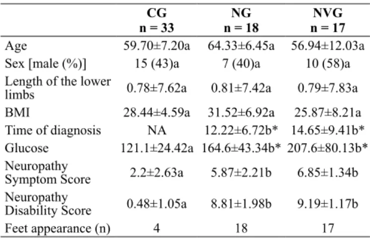
The sample characteristics are described in Table 1. The gender variable was dichotomized and compared between groups by means of categorical statistical analysis that identiied no sig -niicant difference between groups (p > 0.05).
Table 1. Characteristics of the sample, mean ± standard deviation of the control group (CG), neuropathy group (NG) and neuropathy vas-culopathy group (NVG) for the variables age (years), sex, length of the lower limbs (m), body mass index (BMI) in kg/m2, time of
diag-nosis (years), and postprandial blood glucose (mg/dl). CG
n = 33
NG n = 18
NVG n = 17 Age 59.70±7.20a 64.33±6.45a 56.94±12.03a Sex [male (%)] 15 (43)a 7 (40)a 10 (58)a Length of the lower
limbs 0.78±7.62a 0.81±7.42a 0.79±7.83a
BMI 28.44±4.59a 31.52±6.92a 25.87±8.21a Time of diagnosis NA 12.22±6.72b* 14.65±9.41b* Glucose 121.1±24.42a 164.6±43.34b* 207.6±80.13b* Neuropathy
Symptom Score 2.2±2.63a 5.87±2.21b 6.85±1.34b Neuropathy
Disability Score 0.48±1.05a 8.81±1.98b 9.19±1.17b
Feet appearance (n) 4 18 17
Note: Different letters on the same line indicate signiicant differences
between the groups, equal letters on the same line indicate no difference between the groups. †some physical aspects of the feet such as dryness,
blemishes, skin tears, and ulcers. *p < 0.05; NA: not applicable.
We chose to present the average of the right and left lower limbs for the pressure-time integral values and for the spatio-temporal gait variables when no statistical differences were detected between them (p > 0.05).
Table 2 presents comparisons between the groups regarding the pressure-time integral, velocity, double support, single sup-port, and double support/single support ratio variables.
Table 2. Mean ± standard deviation of the control group (CG), neu-ropathy group (NG), and neuneu-ropathy vasculopathy group (NVG) for the pressure-time integral (P-T Integral) (kgf/cm2/ms), velocity (m/s),
double support (DS) (%), single support (SS) (%), and double sup-port/single support ratio (DS/SS) variables.
CG n=33
NG n=18
NVG n=17
P-T Integral 6.77±2.32ª 5.45±1.60a 9.74±4.37b* Velocity 1.05±0.15ª 0.84±0.13b* 0.56±0.22c* DS (%) 39.68±5.7ª 42.42±5.02a 50.45±4.54b* SS (%) 59.94±6.2ª 57.58±5.02a 51.07±5.18b*
DS/SS 0.67±0.14ª 0.75±0.15a 1.0±0.18b*
Note: Different letters in the same line indicate signiicant differences
between the groups. *p < 0.05.
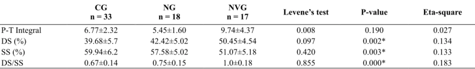
In addition, ANCOVA analysis adjusted by velocity was performed in which the behavior of the other gait variables could be observed, regardless of velocity. It can be seen that only the pressure-time integral lost signiicance during this adjustment.
In the Spearman correlation test, the degree of impairment according to the IWGDF was related to the pressure-time integral (r = 0.381, p < 0.0001), double support (r = 0.646, p < 0.0001) single support (r = −0.544, p < 0.0001), gait velocity (r = −0.776, p < 0.0001), and double support single/support ratio (r = 0.669,
p < 0.0001). The values from the linear regression model are also displayed in Table 4, considering the relationship between the severity of the disease and baropodometric variables.
Table 4. Linear regression between foot impairment according to the International Working Group on Diabetic Foot and gait variables.
Degree of involvement according to the IWGDF
Variables Β CI (95%) P R2
Pressure-Time Integral 0.14 0.05; 0.23 0.001 0,145 Double Support 0.11 0.08; 0.15 ≤0.001 0,417 Single Support -0.10 -0.13; – 0.06 ≤0.001 0,296 Velocity -3.65 -4.38; – 2.92 ≤0.001 0,602
DS/SS 4.07 2.95; 5.18 ≤0.001 0,448
IWGDF = International Working Group on the Diabetic Foot;
CI = conidence interval DS/SS = double support/single support ratio;
R2 = coeficient of determination.
Discussion
This study aimed to analyze the behavior of gait parameters according to the severity of the disease complications of individuals with and without diabetes, enabling, through the results, the inference that the higher the degree of impairment, represented by the association of peripheral vascular disease, the worse the behavior of the gait which, in turn, relects on the functionality and independence of this population in basic day-to-day activities.
The double support, single support, double support/single support ratio, and pressure-time integral variables behaved simi-larly. Summarized, they demonstrated a signiicant difference between the CG and NG groups compared with the NVG, with no difference between the CG and NG groups. Therefore, it is believed that alterations in these variables are strongly related to the presence of peripheral vascular disease and a history of ulcer or amputation associated with DPN, present in the NVG group, according to a previous study17 in which the danger of an association between vasculopathy and DPN in aggravating the chronic complications of diabetes is pointed out, as it is
considered an important predictor for the occurrence of plantar ulceration in the feet of diabetic patients. Thus, the prevention of DPN and clinical evolution to a more impaired condition with vasculopathy in diabetic individuals may be the key to the prevention of disability of gait parameters.
An increase in the double support/single support ratio was also proportional to the complications of DM, as evidenced by the positive correlation between these variables. This fact can be explained by increased double support values associated with decreased single support values, conirming the dificulty in maintaining the gait swing phase, which may indicate a balance deicit and hence, a greater risk of falling18,19. A previous study found a longer period of support in the gait of patients with DPN, suggesting that this was due to the generation of compensatory mechanisms of a musculoskeletal nature, as well as inding aggravations of other variables related to gait4. However, the present data showed that this condition was signiicant only in the NVG; therefore, it is probable that the individuals in the NG group have developed a compensatory mechanism during gait that favors the space-time variables, among them, velocity that may represent a considerable tool; however, not the NVG individuals. This is another point that highlights the importance of not having controlled the pace of the participants to a single speed, as it is important for clinical research to capture these functional artifacts.
Similarly, due to the positive correlation detected, the ag-gravation of the pressure-time integral was proportional to the complexity of the complication of DM. This inding could be one possible explanation for the fact that increases in peak plantar pressure10, associated with other factors, precede the appear-ance of ulcerations and foot amputations in this population20, 21 consistent with the indings of this study, which indicated that points receiving abusive overload to structures frequently bore signs of existing injuries.
Velocity decreased according to the increase in the degree of complicacy, with even lower values in the diabetic foot clinical state (NVG), in agreement with previous indings by other authors who studied the diabetic population with DPN, who considered DPN as a precursor to diabetic foot22. The causes could be re-lated to the fact that diabetic individuals, especially those with greater levels of complications, present a shorter stride length and a longer cycle time when compared with healthy subjects, thus agreeing with other studies4, 20,22.
Furthermore, with respect to velocity, it was noted that the lower values found in the NG and NVG groups were inversely Table 3. ANCOVA analyses presented as mean ± standard deviation in which each model was adjusted by velocity. Control group (CG), neuropa-thy group (NG), and neuropaneuropa-thy vasculopaneuropa-thy group (NVG), pressure-time integral (P-T Integral) (kgf/cm2/ms), double support (DS) (%), single
support (SS) (%) and double support/single support ratio (DS/SS) (n = 68).
CG n = 33
NG n = 18
NVG
n = 17 Levene’s test P-value Eta-square
P-T Integral 6.77±2.32 5.45±1.60 9.74±4.37 0.008 0.190 0.027
DS (%) 39.68±5.7 42.42±5.02 50.45±4.54 0.097 0.002* 0.134
SS (%) 59.94±6.2 57.58±5.02 51.07±5.18 0.420 0.003* 0.133
DS/SS 0.67±0.14 0.75±0.15 1.0±0.18 0.855 0.000* 0.183
related to the double support/single support ratio. Therefore, it can be suggested that velocity reduction is a strategy used by this population to confront their musculoskeletal deicits as walking more slowly enables more precise movements and safer steps. This is not enough to avoid the risk of falling, veriied by the behavior of the other variables, especially the double support/ single support ratio in which the phases of gait oscillation (single support) require good motor control and balance; however, there was compensation for higher double support values and this relationship relects a compensation in the development of normal gait. All of these aspects, in conjunction with the lower values of the pressure-time integral when the velocity was greater (detected in the CG), could justify the higher tendency to plantar ulcer in the NVG, in which the decreased velocity resulted in higher values of the pressure-time integral, which, in turn, corresponded to the ratio between the speciic area of the foot that sustained higher weight bearing for longer periods.
Gait analysis represents much more than the simple fact of observation of walking, but transcends the evaluation of a complex motor act able to represent the basic functionality of the individual and, in the case of the elderly, may also indicate their level of independence. Given the relationship shown in this study between gait parameters and the clinical worsening of neuropathy and peripheral vascular disease, it is important to highlight the ongoing care of diabetic patients in particular, with regard to the prevention and/or control of peripheral vas-cular disease, which arises as a cause or consequence of more serious cases.
However, these relationships, although strong in this study, have not been investigated by other researchers, thus hampering the theoretical basis of these indings; it is suggested that further studies investigate the relationship between these variables so that in future it will be possible to explain the complicacy of the gait of people with diabetes and diabetic peripheral neuropathy, with or without peripheral vascular disease, more precisely.
Study limitations include the need for another sample group with diabetic peripheral neuropathy and vasculopathy, with no history of ulcers or amputations and it is expected that in the future, the present study will be reproduced taking into con-sideration standardization for velocity and analysis by regions of the foot. In addition, the use of postprandial blood glucose cannot exclude cases of acute neuropathy and therefore, it is suggested that future studies give preference to the examination of glycated hemoglobin.
In conclusion, DPN represents damage to the integrity of the gait and, for the majority of the variables evaluated, the association with diabetic peripheral vascular disease and the presence of ulcers and/or amputations is strongly related to the worsening of the conditions for good ambulation. Neurovascular compromise was found to be positively related to the values as-signed to the pressure-time integral, double support, and double support/single support ratio and negatively related to the values of velocity and single support.
Thus, we highlight the importance of early intervention supported by vasculopathy and DPN veriication tests as well as eficient diabetes education as soon as the diagnosis is es -tablished. In particular, for a professional with close contact
with diabetic patients, tests of tactile sensitivity, perfusion, and blood low integrity, in addition to questionnaires on neuropathic symptoms and commitment and an assessment of gait parameters should be included in the medical routine. In situations where the professional does not perform the above-mentioned tests, it is of the utmost importance to perceive the need for them and, as a minimum, refer the individual to other professionals.
References
1. Bona SF, Barbosa MAR, Ferraz CLH, Guarita LKS, Nina RVAH, Barbosa N et al. Prevalência do pé diabético nos pacientes atendi-dos na emergência de um hospital público terciário de Fortaleza. Rev Soc Bras Clin Med. 2010; 8(1): 1-5.
2. Monteiro-Soares M, Boyko EJ, Ribeiro J, Ribeiro I, Dinis-Ribeiro M. Risk stratiication systems for diabetic foot ulcers: a systematic review. Diabetologia. 2011; 54:1190–1199.
3. Sacco ICN, Sartor CD, Gomes AA, João SMA, Cronfli R. Avaliação das perdas sensório-motoras do pé e tornozelo decor-rentes da neuropatia diabética. BJPT. 2007; 11 (1):27-33. 4. Martinelli AR, Mantovani AM, Nozabieli AJL, Ferreira DMA,
Barela JA, Camargo MRD, et al. Muscle strength and ankle mo-bility for the gait parameters in diabetic neuropathies.The Foot. 2013; 23(1):17-21.
5. Fortaleza ACS, Fortaleza ACDS, Chagas EF, Ferreira DMA, Mantovani AM, Barela JA, et al. Postural control and func-tional balance in individuals with diabetic peripheral neuropathy. Rev. bras. cineantropom. desempenho hum. (Online). 2013; 15(3):305-314.
6. Macedo A, Campos C, Correia J, Gomes P. Pé em risco aumentado de ulceração em doentes com diabetes mellitus tipo 2. Rev Port Clin Geral. 2010; 26:159-68.
7. Andersen H. Diabetes/metabolism research and reviews. Diabetes Metab Res Rev. 2012; 28(Suppl 1):89–92.
8. Sawacha Z, Spolaor F, Guarneri G, Contessa P, Carraro E, Venturin A, et al. Abnormal muscle activation during gait in diabetes patients with and without neuropathy. Gait & posture. 2012; 35(1):101-105.
9. Allet L, Armand S, De Bie RA, Golay A, Monnin D, Aminian K, et al. The gait and balance of patients with diabetes can be improved: a randomised controlled trial. Diabetologia. 2010; 53(3):458-466.
10. Hohne A, Ali S, Stark C, Bruggemann G. Reduced plantar cuta-neous sensation modiies gait dynamics, lower-limb kinematics and muscle activity during walking. Eur J Appl Physiol. 2012; 112:3829–3838.
11. Bakker K, Schaper NC. The development of global consensus guidelines on the management and prevention of the diabetic foot 2011. Diabetes Metab Res Rev. 2012, 28(S1):116-118.
12. Saura V, Santos ALG, Ortiz RT, Parisi MC, Fernandes TD, Nery M. Fatores preditivos da marcha em pacientes diabé-ticos neuropádiabé-ticos e não neuropádiabé-ticos. Acta Ortop Bras. 2010; 18(3):148-51.
14. Nozabieli AJL, Martinelli AR, de Camargo MR, de Souza Fortaleza AC, de Faria CRS, et al. Diabetic peripheral neuropathy in ankles and feet: muscle strength and plantar pressure. Int J Diabetes Develop Countries. 2013; 34(2):82-88.
15. Moreira RO, Castro AP, Papelbaum M, Appolinario JC, Ellinger VCM, Coutinho WF, et al. Tradução para o português e avaliação da coniabilidade de uma escala para diagnostico da polineuropa -tia. Arq Bras Endocrinol Metab. 2005; 49 (6):944-950.
16. ADA. American Diabetes Association. Diagnosis and Classiication of Diabetes Mellitus. Diabetes Care. 2010; 33:62-69.
17. Monteiro-Soares M, Boyko EJ, Ribeiro J, Ribeiro I, Dinis-Ribeiro M. Predictive factors for diabetic foot ulceration: a systematic review. Diabetes Metab Res Rev. 2012; 28(7):574-600. 18. MacGilchrist C, Paul L, Ellis BM, Howe TE, Kennon B, Godwin
J. Lower-limb risk factors for falls in people with diabetes mel-litus. Diabet Med. 2010; 27:162-158.
19. Wrobel JS, Najai B. Diabetic Foot Biomechanics and Gait Dysfunction. J Diabetes Sci Technol. 2010; 4 (4):833-45. 20. Bennetts CJ, Owings TM, Erdemir A, Botek G, Cavanagh PR.
Clustering and classiication of regional peak plantar pressures of diabetic feet. J Biomechanics. 2013; 46(1):19-25.
21. Ledoux WR, Shofer JB, Cowley MS, Ahroni JH, Cohen V, Boyko EJ. Diabetic foot ulcer incidence in relation to plantar pressure
magnitude and measurement location. J Diabetes its complica-tions. 2013; 27(6):621-626.
22. Sawacha Z, Gabriella G, Cristoferi G, Guiotto A, Avogaro A, Cobelli C. Diabetic gait and posture abnormalities: A biomechani-cal investigation through three dimensional gait analysis. Clin Biomechanics. 2009; 24:722–728.
Corresponding author
Alessandra Madia Mantovani
Roberto Simonsen Avenue, 305. Centro Educacional, Presidente Prudente, São Paulo, Brazil.
Email: [email protected]
Manuscript received on February 29, 2016 Manuscript accepted on March 03, 2016