Mariana Coelho Correia da Silva
centromere inheritance
Dissertation presented to obtain the Ph.D degree in
Cell Biology
' ( ) *
This dissertation was sponsored by
Fundação para a Ciência e Tecnologia.
Apoio financeiro da FCT e do FSE no
âmbito do Quadro Comunitário de apoio,
Declaração
Declaro que esta dissertação de candidatura ao grau de Doutor é da minha autoria e que os dados aqui incluídos são o resultado de trabalho original por mim desenvolvido entre Outubro de 2007 e Outubro de 2011 no laboratório do Dr Lars Jansen, Instituto Gulbenkian de Ciência em Oeiras, Portugal. Este doutoramento foi realizado no âmbito do Programa Doutoral do Instituto Gulbenkian de Ciência PGD 2007. Todas as colaborações estão indicadas em cada capítulo, na secção de Acknowledgements. Esta dissertação teve o apoio financeiro da FCT BD nº SFRH/BD/33219/2007 e dos projectos PTDC/BIA-BCM/100557/2008, Marie Curie Reintegration grant (FP7-PEOPLE-2007-4-3-IRG) e EMBO installation grant.
Do trabalho desenvolvido durante este período resultaram as seguintes publicações:
Christopher W. Carroll, Mariana C.C. Silva, Kristina M. Godek, Lars E.T. Jansen, and Aaron F. Straight (2009). Centromere assembly requires the direct recognition of CENP-A nucleosomes by CENP-N. Nature Cell Biol, 11(7):896-902
Mariana C.C. Silva and Lars E.T. Jansen. (2009) “At the right place at the right time: Novel CENP-A binding proteins shed light on centromere assembly”.
Chromosoma, 118(5):567-74.
Mariana C.C. Silva, Dani L. Bodor, Madison E. Stellfox, Nuno M. C. Martins, Helfrid Hochegger, Daniel R. Foltz, and Lars E.T. Jansen. (2012). Cdk Activity Couples Epigenetic Centromere Inheritance to Cell Cycle Progression.
Acknowledgements
First, I would like to thank my supervisor Lars Jansen for his guidance, support and motivation during these 4 years. Thank you for the confidence and for all the great opportunities you gave me. A sincere and big thank you for helping me develop in the scientist I am today. It was really great being your first PhD student. Thank you! I would like to thanks the present and former members of Epilab: João, Dani, Luis, Mariluz and Nuno. In special, I thank Dani for his friendship, for always finding time to help, for the great science discussions and for helping me with some experiments. Um obrigada especial também ao João, pelo apoio nos momentos mais difíceis, pela amizade e ajuda constantes.
I would like to thank Don Cleveland and the members of Cleveland lab, for having me as a visitor for 3 months in San Diego and for treating me as an actual member of the lab. I am grateful to Dan Foltz and Madison Stellfox for helping with experiments. I also thank Dan for reagents, fruitful discussions and ongoing collaborations. I thank Helfrid Hochegger and the members of Hochegger lab, for having me as a visitor for some weeks. I am grateful to Aaron Straight and Christopher Carroll for our collaboration on the CENP-N story. I hope I will meet you in some meeting in the near future.
that discussed my work and gave me burst of motivation and ideas. Thanks also to the ones that made meetings so much fun. Agradeço à FCT pelo financiamento deste projecto. Agradeço às duas grandes amigas que fiz no IGC as minhas Marias Catarina e Fátima. Tem sido fantástico ter a vossa amizade ao longo destes 5 anos. I thank to my sis Baixinha, we started PhD together and we will finish it together. It has been great to share my joy and tears during these 4 years with you my polish friend. Agradeço aos amigos que fiz no IGC pelo apoio e carinho, à Clara, Chica, Ana, Sónia e em especial à Bá por ouvir os meus desabafos quando mais precisei. Ao Gil pelos abraços grátis e pela amizade ao longo de quase 10 anos. À Inês, minha amiga desde que nasci, pela ajuda e pela inspiração nesta vida de cientista. Às Joanas, Sofis, Telma, Ricardo e Meguie pela amizade. Às minhas gajas Raquel, Mafalda, Sol, B e Sara por todos os nossos bons momentos, pelos nossos jantares e sobretudo pela vossa amizade. Adoro-vos! À Filipa, em especial, por estares sempre sempre lá miuda e por nestes últimos meses teres-me ajudado a esquecer a tese quando mais precisei. Não sei o que seria de mim sem ti, adoro-te minha linda. Aos meus surfer budies, Stefan, Nuno, Gaston, Maria e Francis. Agradeço a todos os meus amigos e familiares que me têm apoiado sempre e que fazem com que eu seja assim como me disseram no outro dia: “és sempre assim tão feliz?”. Em especial aos primos Catarina, Zé e Teresa. E à família Ramos, Zé, Bé e Isaura pela grande amizade ao longo destes mais de 20 anos.
Abstract
Cell division is a fundamental process of all living organisms by which a parental cell divides into two genetically identical daughter cells. Faithful cell division requires duplication and subsequent equal distribution of the parental genetic information, the genome, between daughter cells. In eukaryotes, genomic information is organized in chromosomes, which consist of linear DNA sequences packaged into histone protein-DNA complexes called nucleosomes. Chromosomes comprise a defined region, the centromere, which is responsible for delivering the correct number of chromosomal copies to daughter cells during cell division. The centromere directs the formation of the kinetochore, a proteinaceous structure that is responsible for connecting chromosomes to spindle microtubules during mitosis, allowing accurate segregation of chromosomes across generations. Centromere identity in most eukaryotes is not specified by any particular DNA sequence, but rather by an epigenetic chromatin-based mechanism. Key to the epigenetic propagation of the centromere is the histone H3 variant CENP-A that is uniquely incorporated into centromeric chromatin. Consequently, replication and inheritance of this epigenetic mark is crucial to epigenetically maintain centromere identity across cell divisions. Consistently, CENP-A nucleosomes are stably maintained throughout the cell cycle, being turned over only by redistribution between the two sister chromatids during DNA replication. Unlike assembly of the canonical histone H3.1, CENP-A assembly is uncoupled from DNA replication and occurs during late telophase/early G1 in metazoans. Gaining insight into how CENP-A chromatin is propagated throughout cell divisions has been a major research focus in recent years and is essential for our broad understanding of the mechanisms of cell division.
mechanism underlying centromere propagation. Using siRNA-mediated depletion in combination with a unique method to visualize centromere assembly of nascent CENP-A (SNAP-tagging), we show that CENP-N and CENP-C (two proteins that bind directly to CENP-A nucleosomes), and also CENP-T, contribute to CENP-A incorporation into centromeres. This result reveals that these structural components of the centromere are part of an epigenetic feedback loop responsible for propagation of centromeric chromatin.
Although many proteins have been identified to play a role in CENP-A targeting to centromeres, it was still unclear how centromere propagation is restricted to late telophase/early G1 phase. In Chapter 3 we describe the identification of the molecular signal that initiates CENP-A assembly exclusively upon mitotic exit. This phase of the cell cycle is marked by extensive reorganization of the centromere, kinetochore, chromatin, chromosomes and the nucleus as a whole. In addition, a large number of proteins are either selectively destroyed or post-translationally modified at this stage. Using pharmacological and genetic experiments, we show that although CENP-A assembly initiation is not directly dependent on any of these aspects of mitosis, it does require the down regulation of the major mitotic master regulator cyclin-dependent kinase 1 (Cdk1). Additionally, we show that specific inhibition of both Cdk1 and Cdk2 in any phase of the cell cycle is sufficient to trigger rapid CENP-A assembly without passage through mitosis. Neither de novo synthesis nor protein destruction are
required to trigger CENP-A assembly indicating that the CENP-A assembly machinery is already present in an inactive state prior to mitosis.
In Chapter 4, we sought to characterize the inhibitory mechanism mediated by Cdk activity that regulates the timing of CENP-A assembly and restricts it to late telophase/early G1 phase of the cell cycle. We show that high Cdk1 or Cdk2 activity prevents centromere targeting of well-characterized CENP-A assembly factors such as Mis18 , Mis18BP1HsKNL2, along with the CENP-A specific
Mis18BP1HsKNL2 is phosphorylated in a cell cycle dependent manner, and that
phosphorylation prevents its centromere localization. In addition, we identify a domain in HJURP, conserved among vertebrates, as a regulatory domain required for timely control of CENP-A assembly. Together these results suggest that Cdk1 and Cdk2 control the cell cycle timing of CENP-A assembly by inhibiting Mis18BP1HsKNL2 and potentially other assembly factors through
phosphorylation and consequent delocalization.
Sumário
O processo de divisão celular, pelo qual uma célula mãe se divide para dar
origem a duas células filhas, é fundamental para todos os organismos vivos.
Para que uma célula se divida correctamente é necessário a duplicação de toda
a sua informação genética, que posteriormente é distribuída igualmente por duas
células filhas. Nas células eucariotas, a informação genética está organizada em
cromossomas. Os cromossomas são constituídos por sequências lineares de
ADN (ácido desoxirribonucleico) enroladas em torno de um conjunto de
proteínas chamadas histonas. Os complexos formados por histonas e ADN
denominam-se de nucleossomas e são as unidades básicas do cromossoma. Os
cromossomas possuem uma estrutura designada centrómero, essencial para a
distribuição do número correcto de cromossomas pelas células filhas durante o
processo de divisão celular. O centrómero é responsável pela formação do
cinetocoro, um complexo multiproteico que liga os cromossomas aos
microtúbulos do fuso mitótico durante a mitose. Deste modo, o centrómero
permite que os cromossomas sejam distribuídos igual e correctamente pelas
duas células filhas. Na maioria dos eucariotas, o centrómero é herdado de célula
para célula por mecanismos epigenéticos, isto é, independentemente de
qualquer sequência especifica de ADN. A proteína CENP-A, uma variante da
histona H3, é essencial para a propagação do centrómero. Esta proteína, é
incorporada apenas na cromatina centromérica. Logo, a duplicação e herança
desta marca epigenética é essencial para a correcta transmissão do centrómero
ao longo de diversas gerações. Consistentemente, os nucleossomas que contêm
CENP-A são extremamente estáveis, e durante a replicação do ADN são
reutilizados e distribuídos pelos dois cromatídeos recentemente formados. Ao
contrário do que acontece com a histona H3, a inclusão de CENP-A nos
nucleosomas centroméricos ocorre desfasada e independentemente da
cromatina tem início após as células terminarem a mitose e continua durante a
fase G1 do ciclo celular. Perceber como a cromatina centromérica é propagada
durante a divisão celular é uma área de investigação que tem ganho destaque
nos últimos anos e é essencial para o conhecimento geral dos mecanismos da
divisão celular.
No capítulo 2 desta tese, descrevemos a identificação de novas proteínas
envolvidas no processo de incorporação de CENP-A no centrómero, de modo a
perceber detalhadamente o mecanismo molecular responsável pela propagação
desta estrutura. Focámos a nossa atenção particularmente em proteínas
capazes de se associarem directamente com os nucleossomas de CENP-A.
Utilizando uma técnica que permite visualizar apenas a CENP-A recentemente
sintetizada e que foi incorporada no centrómero (SNAP-tagging) em combinação
com a depleção de proteínas utilizando oligos de siRNA, identificámos três novas
proteínas que participam no processo de inclusão de CENP-A no centrómero.
Com esta metodologia, demonstrámos que duas proteínas que se ligam
directamente aos nucleossomas de CENP-A, CENP-C e CENP-N, e também a
proteína CENP-T, participam no processo de propagação da cromatina
centromérica. A localização destas proteínas no centrómero é, por sua vez,
dependente da presença de CENP-A. Este resultado indica que a CENP-A e
estes componentes estruturais do centrómero se regulam mutuamente de modo
a permitir a transmissão do centrómero através de gerações.
Apesar da recente identificação de várias proteínas que fazem parte do
processo de incorporação de CENP-A, permanece por descobrir o mecanismo
responsável por limitar a propagação do centrómero até que o processo de
mitose seja concluído. No capítulo 3, descrevemos o mecanismo responsável
pela activação da transmissão do centrómero após a conclusão do processo de
mitose. Esta fase do ciclo celular é caracterizada pela reorganização do
centrómero, cinetocoro, cromatina, cromossomas e inclusive de todo o núcleo.
Além disso, muitas proteínas são degradadas selectivamente ou são
técnicas genéticas e farmacológicas mostrámos que nenhum destes aspectos da
mitose está directamente envolvido na propagação do centrómero. O único
factor comum necessário é a inactivação de uma proteína extremamente
importante para o controlo da divisão celular, a cinase dependente de ciclina 1
(Cdk1). A inactivação das Cdk1 e Cdk2 em qualquer fase do ciclo celular é
suficiente para induzir a incorporação de CENP-A no centrómero sem o
envolvimento da mitose. A propagação do centrómero não depende da síntese
nem da degradação de proteínas, indicando que as proteínas necessárias para
inserir CENP-A na cromatina centromérica estão presentes mas inactivas entre a
fase S e a mitose.
No capítulo 4, explorámos o mecanismo de inibição dependente da Cdk1,
que controla o timing da inclusão da CENP-A no centrómero, e que limita este
timing ao período final da mitose e à fase G1 do ciclo celular. Mostrámos que a
actividade elevada de Cdk1 e Cdk2 impede a localização centromérica de
proteínas necessárias à propagação de CENP-A, tais como Mis18 ,
Mis18BP1HsKNL2 e HJURP, a chaperone específica da CENP-A. Estas proteínas
são essenciais tanto para a propagação natural do centrómero na fase G1 como
para a propagação induzida nas fases anteriores à mitose. Mostrámos também
que Mis18BP1HsKNL2 é fosforilada em períodos específicos do ciclo celular e que
esta fosforilação impede a localização desta proteína no centrómero.
Adicionalmente, identificámos um domínio regulatório na proteína HJURP,
necessário para controlar o timing de propagação de CENP-A. De notar que este
domínio é conservado em todos os vertebrados. Em conjunto, estes resultados
indicam que Cdk1 e Cdk2 controlam o timing da transmissão do centrómero
através da inactivação da Mis18BP1HsKNL2 e potencialmente de outras proteínas.
Esta inactivação depende da fosforilação destas proteínas e, consequente, da
sua deslocalização.
Resumidamente, identificámos novos participantes do processo de
propagação de CENP-A e revelámos o mecanismo que limita a propagação do
entre o processo de divisão celular e a herança epigenética do centrómero.
Deste modo, este trabalho contribui para o conhecimento dos mecanismos de
List of Abbreviations
(A) Ala Alanine
APC/C Anaphase Promoting Complex/Cyclosome
ATP Adenosine triphosphate
Borax Tetraborato de sódio (Na2B4O7·10H2O)
bp Base pair
BrdU Bromodeoxyuridine
BTP Bromothenylpteridine
CAF1 Chromatin Assembly Factor 1
CAK Cdk-Activating Kinase
CATD CENP-A Targeting Domain
CCAN Constitutive Centromere-Associated Network
Cdc Cell Division Cycle
Cdc20 Cell Division Cycle 20 homologue
CDE Centromere DNA Element
Cdh1 Cdc20 Homologue 1
Cdk Cyclin-Dependent Kinase
cDNA Complementary DNA
Cdt1 Chromatin licensing and DNA replication factor 1
CENP Centromere Protein
CENP-A Centromere Protein A
ChIP Chromatin Immunoprecititation
CKI Cdk inhibitor
CMG Cdc45-Mcm2-7-GINS
DAPI 4’,6-diamidino-2-phenylindole
DNMT DNA Methyltransferase
DNA Deoxyribonucleic acid
E (Glu) Glutamate
Emi1 Early Mitotic Inhibitor
FACS Fluorescence-Activated Cell Sorting
FBS Fetal Bovine Serum
FRAP Fluorescence Recovery After Photobleaching
G (Gly) Glycine
GBP GFP binding protein
GEF Nucleotide Exchange Factor
GFP Gree Fluorescent Protein
HA Hemagglutininepitopetag
HCl Hydrochloric acid
HDAC Histone Deacetylase
HeLa Henrietta Lacks Cervical Cancer Cell Line
HFD Histone Fold Domain
HIRA Hir-related protein A
HJURP Holliday Junction Recognizing Protein
HP1 Heterochromatin-Associated Protein 1
HRP Horseradish Peroxidase
hTERT Human Telomerase Reverse Transcriptase
HU Hydroxyurea
ICEN Interphase Centromere Complex
K (Lys) Lysine
kb kilo bases
KMT Lysine Methyltransferase
KNL2 Kinetochore Null 2
L (Leu) Leucine
LINE Long Interspersed Elements
MAP Microtubule-Associated Protein
MCC Mitotic Checkpoint Complex
MCM Minichromosome Maintenece Complex
M Methionine
Mis S. pombe mutants with high loss rate of
minichromosomes
Mis18BP1 Mis18 Binding Protein 1
mRNA Messenger RNA
MTOC Microtubule Organizing Center
NaV Sodium Orthovanadate
NEBD Nuclear Envelope Breadown
NPM/B23 Nucleophosmin
ORC Origin Recognition Complex
ORF Open Reading Frame
P (Pro) Proline
PC Polycomb protein
PcG Polycomb Group
PCM Pericentriolar material
PCNA Proliferating Cell Nuclear Agent
PCR Polymerase Chain Reaction
Plk1 Polo Like Kinase 1
PP1 Protein phosphatase 1
PP2A Protein phosphatase 2A
pRb Retinoblastoma protein
PRC Polycomb Repressive Complex
pre-RC Pre-Replicative Complex
R (Arg) Arginine
RbAp46 Retinoblastoma-associated protein 46kDa
RbAp48 Retinoblastoma-associated protein 48kDa
RPE Retinal Pigment Epithelial Cell Line
RSF Remodeling and Spacing Factor
RT Room Temperature
SCF Skp1/Cul1/F-box
S (Ser) Serine
SINE Short Interspersed Elements
T (Thr) Treonine
TMR Tetramethylrhodamine
Tome-1 Trigger of Mitotic Entry 1
TrxG Trithorax Group
TSA Trichostatin A
Ub Ubiquitin
Table of Contents
Declaração ... i Acknowledgements ... iii Abstract ... v Sumário ... ix List of Abbreviations ... xiii Chapter 1 – General Introduction ... 1
1. Chromosome structure and genetic inheritance ... 3
1.1. Packing of DNA into chromosomes ... 3
1.2. Genetic inheritance across cell divisions ... 5
1.3. Chromosomal elements required for proper genetic inheritance ... 8
2. Organization and function of centromeres and kinetochores ... 10
2.1. Centromeric DNA ... 12
2.2. Centromeric chromatin organization ... 15
2.2.1. CENP-A, a histone H3 variant unique to the centromere ... 15
2.2.2. Patterns of histone modifications at the centromere ... 17
2.3. The Constitutive Centromere-Associated Network (CCAN) ... 19
2.3.1. CENP-C and CENP-N bind directly to CENP-A nucleosomes ... 20
2.3.2. CENP-T, CENP-W, CENP-S, and CENP-X form a nucleosome like structure able to warp centromeric DNA ... 22
2.3.3. CENP-H/I/K complex ... 23
2.4.1. Mitotic checkpoint and metaphase to anaphase transition ... 26
3. Epigenetic inheritance of centromeres ... 29
3.1. The basic concept of Epigenetics... 29
3.1.1. Inheritance of DNA methylation during DNA replication ... 30
3.1.2. Histone modifications and inheritance of chromatin states ... 31
3.1.2. Polycomb and Trithorax family of proteins as key players in epigenetic control of gene expression ... 33
3.1.2. Other players in epigenetic processes ... 34
3.2. Centromere as a show case to study epigenetic inheritance ... 35
3.2.1. CENP-A as a key epigenetic mark ... 37
3.2.2. CENP-A maintenance and assembly during the cell cycle ... 40
4. Cell cycle control ... 45
4.1. Cyclin-dependent kinases and cell cycle control ... 45
4.1.1. Cyclin-dependent kinases and cell cycle transitions ... 46
4.1.2. Evolution of the Cdk protein family ... 47
4.1.3. Substrate specificity of Cdks ... 48
4.1.4. Cdk activity regulates DNA replication ... 49
4.1.5. Cdk activity controls centrosome duplication ... 52
4.2. Ubiquitin-mediated proteolysis and cell cycle control ... 54
4.2.1. Essential functions of the SCF complex during cell division ... 54
4.2.2. Essential functions of the APC/C during cell division ... 55
4.3. Regulation of mitosis by mitotic kinases and phosphatases ... 57
5. Aims of this thesis ... 59
Chapter 2 – Dissecting the CENP-A assembly pathway ... 83
Abstract ... 85
Introduction ... 86
Material and Methods ... 89
Cell lines and constructs ... 89
Cell synchronization ... 89
siRNA transfection ... 90
SNAP quench-chase-pulse labeling ... 91
Immunofluorescence ... 91
Immunoblotting ... 92
Microscopy ... 92
Fluorescence Quantification ... 93
Results ... 94
Mis18 complex is targeted to centromeres prior to CENP-A and is required for
centromere assembly ... 94
Structural components of the centromere affect CENP-A assembly ... 99
Discussion and Conclusions ... 104
Being at right place at the right time: the Mis18 complex licenses centromeric
chromatin for CENP-A assembly ... 104
CCAN forms an epigenetic loop responsible for propagating the centromeric
chromatin ... 106
CENP-A assembly occurs in three distinct steps: licensing, assembly and
stabilization ... 110
References ... 118
Chapter 3 – Cell cycle control of CENP-A assembly is maintained by Cdk activity ... 125
Abstract ... 127
Introduction ... 128
Material and Methods... 130
Cell lines and constructs ... 130
Immunoblotting ... 131
Cell synchronization and drug treatments ... 132
SNAP quench-chase-pulse labeling ... 133
Immunofluorescence ... 133
Microscopy ... 134
Fluorescence Quantification ... 135
Flow cytometry ... 135
Results ... 136
CENP-N loading at centromeres is not restricted to G1 phase of the cell cycle
... 136
Cdk inhibition triggers CENP-A assembly prior to mitosis ... 138
DNA replication and CENP-A assembly are mutually exclusive in human cells
... 144
APC/C mediated proteolysis and protein synthesis are not required for CENP-A
assembly in G2-phase. ... 147
Cdk1 and Cdk2 activities are sufficient to maintain cell cycle control of CENP-A
assembly ... 151
CENP-A assembly can be induced during S phase in DT40 cells ... 159
Discussion and Conclusions ... 163
The CCAN proteins can be divided in distinct classes based on their dynamic
localization and time of assembly ... 163
APC/C-mediated loss of Cdk1 and Cdk2 activities is the unique mitotic feature
required for CENP-A assembly ... 165
DNA replication and centromere propagation: small molecule inhibitors versus
chemical genetics ... 167
Acknowledgements ... 170
References ... 171
Chapter 4 – Molecular mechanism maintaining cell cycle control of CENP-A assembly ... 177
Abstract ... 179
Introduction ... 180
Material and Methods ... 182
Cell lines and constructs ... 182
Site-directed mutagenesis ... 183
Strand exchage PCR ... 184
Cell synchronization, drug treatments and SNAP quench-chase-pulse labeling
... 186
siRNA and DNA transfections ... 186
Phosphatase Treatment ... 187
Immunofluorescence ... 187
Microscopy and fluorescence quantification ... 188
Results ... 189
Cdk inhibition results in unscheduled recruitment of CENP-A assembly factors to
centromeres ... 189
Unscheduled CENP-A assembly requires the canonical CENP-A assembly
factors ... 192
Canonical CENP-A assembly in G1 and Roscovitine-induced assembly in G2 do
not require Aurora A and Aurora B activities ... 194
Expression of HJURP lacking a domain that is conserved among vertebrates
induces CENP-A assembly in G2 phase ... 197
Phosphorylation of Mis18BP1HsKNL2 controls its centromere localization ... 200
Discussion and Conclusions ... 208
The HJURP vertebrate conserved domain is an important regulatory domain
required for cell cycle control of CENP-A assembly ... 209
CENP-A assembly is activated upon mitotic exit by Cdk inactivation and
dephosphorylation of Mis18BP1HsKNL2 ... 211
Acknowledgements ... 213
References ... 214
Chapter 5 – General Discussion ... 217
Epigenetic inheritance of centromeres ... 219
Cdk activity couples epigenetic inheritance of the centromere with cell cycle
progression ... 220
Why is CENP-A assembly cell cycle regulated? ... 227
Cdk activity and cell cycle control of CENP-A assembly in other organisms . 229
CENP-ACnp1 assembly in Schizosaccharomyces pombe ... 229
CENP-ACID assembly in Drosophila melanogaster ... 231
CENP-ACENH3 assembly in Arabidopsis thaliana ... 235
Cdks control CENP-A assembly at two distinct levels ... 236
General importance of the principles of centromere inheritance ... 237
Cdk1 and Cdk2 as general regulators of epigenetic inheritance throughout cell
divisions ... 238
Future prospects for the study of centromere inheritance ... 239
References ... 241
Appendix 1 – Deletion mutants of Mis18 , Mis18 and HJURP and effect on CENP-A assembly ... 247
1. Chromosome structure and genetic inheritance
1.1. Packing of DNA into chromosomes
DNA is the primary carrier of genetic information in nearly all living
organisms. In eukaryotes, the total length of DNA sequences can reach up to a
hundred thousand times the diameter of the cell. Therefore, compartmentalization
of the genetic material inside the nucleus of the cell requires a dramatic packing
of DNA molecules into a protein-DNA complex called chromatin in eukaryotes.
Chromatin is primarily composed of double stranded DNA folded around small
basic proteins called histones (Kornberg, 1974). The fundamental repeating unit
of chromatin is the nucleosome (Oudet et al., 1975). Each nucleosome is
comprised of a DNA segment with 146 base pairs (bp) in length wrapped around
a compact histone protein core (Luger et al., 1997). This histone core is
composed of an octamer containing two copies of each of the canonical histones
H2A, H2B, H3, and H4 (Richmond et al., 1984; Luger et al., 1997). The structure
of histones is strongly conserved across evolution, suggesting that this type of
DNA packaging has evolved very early and is one of the defining features of
eukaryotes.
The packing of DNA into nucleosomes shortens the DNA length about
sevenfold. Further compaction is achieved by folding of nucleosomes into higher
order arrays with multiple levels of packing ultimately resulting in mitotic
chromosomes. The nature of these higher order structures is a matter of debate
but may include the formation of a fiber about 30 nm wide, whose formation is
facilitated by the linker histone H1 [Figure 1.1; (Everid et al., 1970; Thoma et al.,
1979)]. Additional levels of compaction are achieved by looping the chromatin
fibers along a structural scaffold formed by non-histone proteins such as
topoisomerase II and the condensin complex [Figure 1.1; (Moser and Swedlow,
process by which a cell separates its chromosomes into two identical sets and
distributes them between two daughter cells.
Figure 1.1. Model for the packing of chromatin into metaphase chromosomes. The simplest level of organization of the genetic material is the double stranded helical structure of
DNA. DNA is folded around specialized eukaryotic proteins called histones to form
nucleosomes, the primary repeating unit of chromatin. Each nucleosome consists of a core of
eight histone proteins around which the DNA wraps 1,65 times. This first level of compaction
is facilitated by the linker histone H1 that binds to each nucleosome forming the
chromatosome. Nucleosomes further fold around each other to produce a fiber about 30 nm
wide that forms loops averaging 300 nm in length. The 300 nm fibers are compressed and
folded to create a fiber with a width of 250 nm. Tight coiling of these fibers is responsible for
the final level of chromatin compaction into a metaphase chromosome. Adapted from Pierce,
2005.
The multiple packing levels of chromatin serve not only as a way to
compact the DNA within the eukaryotic nucleus, but also play important functional
expression and DNA metabolism, principally by controlling access of regulatory
proteins to DNA. Highly compacted chromatin is not accessible to the enzymes
involved in DNA transcription, replication or repair. Therefore, regions of
chromatin where active transcription takes place (euchromatin) are less
condensed than regions where transcription is inactive or is actively repressed
(constitutive and facultative heterochromatin, respectively) (Horn and Peterson,
2002; de la Serna and Imbalzano, 2002). Alternatively, looping of
nucleosome-containing fibers can bring specific regions of chromatin together, thereby
influencing gene expression (Misteli, 2007).
The state of chromatin can be regulated directly by energy consuming
chromatin remodeling motors that change chromatin conformation, allowing
targeted access of regulatory proteins, for example, to specific genes
(Varga-Weisz and Becker, 2006). Additionally, gene expression is controlled by
modification of histones with small chemical moieties, such as methyl and acetyl
groups on the N-terminal tail that extend from the core particle of the
nucleosomes (Jenuwein and Allis, 2001; Wang et al., 2004b). The importance of
these histone modifications will be further discussed in the context of centromere
function and epigenetics in the following sections.
1.2. Genetic inheritance across cell divisions
The faithful inheritance of genetic information during meiosis and mitosis is
central to the growth and development of all living organisms. Aberrant
chromosome inheritance causes the formation of cells with an abnormal number
of chromosomes, called aneuploid cells (Torres et al., 2008). Aneuploid cells are
unstable and are normally eliminated by apoptosis. However, in rare cases,
aneuploidy can result in tumor formation and in the development of severe
genetic disorders associated with birth defects (Hassold and Hunt, 2001; Kops et
The universal process responsible for the formation of two genetically
identical cells is called the cell cycle. The cell cycle can be divided in four stages.
The two critical phases are S phase (synthesis phase; during which the genetic
material is duplicated) and M phase (mitosis; during which the duplicated material
is distributed between the two daughter cells). These two phases are separated
by two gap phases: G1 phase (first gap phase; preceding S phase) and G2
phase (second gap phase; preceding mitosis) (reviewed in Nurse, 2000; Pollard
et al., 2004). G1, S, and G2 phases are collectively called interphase, the period
between one mitosis and the next (Figure 1.2). Not only the chromosomes, but
also the cytoplasm and the cell organelles are divided between two daughter
cells during mitosis and subsequent cytokinesis.
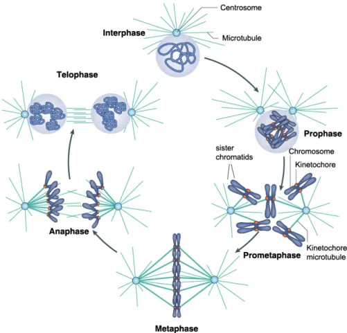
Mitosis is a complex process that is divided into five discrete cell
biologically defined stages: prophase, prometaphase, metaphase, anaphase and
telophase (Figure 1.2). During prophase chromosomes condense and
centrosomes, the organelles that serve as the main microtubule organizing
centers (MTOC) in animal cells, migrate to opposite poles of the cell to facilitate
the assembly of the mitotic spindle (Figure 1.2). At the end of prophase, nuclear
envelope breakdown (NEBD) occurs and cells enter prometaphase. During
prometaphase the formation of the mitotic spindle is completed and
chromosomes start attaching to spindle microtubules (Figure 1.2). The
chromosome-microtubule attachment is mediated by a chromosome-borne
multiprotein complex called the kinetochore. Once all kinetochores are attached
to microtubules that emanate from opposite poles, the chromosomes move to the
middle of the cell. When all chromosomes are properly attached and aligned, the
cell reaches metaphase (Figure 1.2). During anaphase, the chromosomes are
separated into two sister chromatids that migrate to opposite poles of the spindle
(Figure 1.2). When the chromatids approach the spindle poles and nuclear
envelope reformation starts, the cell is in telophase (Figure 1.2). At the end of
masses, leading to separation of the two daughter cells (cytokinesis) (Morgan,
2007).
Figure 1.2. The stages of mitosis in animal cells. The various stages of the cell cycle are depicted. During interphase the cell undergoes growth (G1 phase) and replication of the DNA
(S phase). Upon duplication of the centrosomes and DNA, the cell undergoes a second round
of growth (G2 phase) and subsequently enters mitosis. Mitosis can be divided in five stages:
prophase, prometaphase, metaphase, anaphase and telophase. See the text for details.
Adapted from Verdaasdonk and Bloom, 2011.
Meiosis occurs only in germ cells and is characterized by a single round of
DNA replication followed by two divisions called meiosis I and meiosis II. Unlike
mitosis that leads to the formation of two diploid daughter cells, meiosis produces
germ line. In meiosis I, the replicated homologous chromosomes are segregated
to opposite poles, and in meiosis II, the sister chromatids of each homolog are
segregated (Kleckner, 1996).
1.3. Chromosomal elements required for proper genetic inheritance
Accurate genome duplication and inheritance during mitosis and meiosis
require specific chromosomal elements and protein complexes (Allis et al., 2007).
These chromosomal elements include replication origins, centromeres and
telomeres (Figure 1.3).
Figure 1.3. Key elements of chromosome inheritance. The diagram indicates chromosomal elements or protein complexes essential for
proper inheritance of the genetic material: the
centromere, kinetochores, telomeres and sister
chromatin cohesion. Centromeres include
centromeric chromatin and pericentromeric
heterochromatin. Centromeric chromatin
nucleates the formation of the kinetochore, which
forms the site of attachment for spindle
microtubules. Pericentric heterochromatin is the
site of cohesion accumulation, contributing to
sister chromatid cohesion. Telomeres form the
ends of chromosomes, protecting them from
degradation and fusion. Adapted from Allshire and
Karpen, 2008.
Replication origins are the defined chromosomal regions where DNA
replication is initiated in a eukaryotic cell. At each origin, two replication forks are
formed, and replication proceeds bidirectionally until the replication forks
encounter another fork approaching from the opposite direction. The mechanism
responsible for the formation and regulation of replication origins will be
Several important processes occur at the replication fork, such as assembly
of new histones to form chromatin. Proteins required for sister chromatid
cohesion, called cohesins, are also assembled during this stage, but associate
with chromosomes before DNA replication. In this way, sister chromatid cohesion
is ensured to maintain duplicated sister chromatids together until anaphase onset
(Nasmyth and Haering, 2009).
While cohesion holds newly replicated sister chromatids together,
centromeres are chromosomal elements crucial to drive separation of
chromosomes during mitosis. The centromere is a chromosomal locus composed
of DNA and specialized chromatin proteins that serve as the foundation for
kinetochore formation during mitosis (Allshire and Karpen, 2008; Silva and
Jansen, 2009). The kinetochore is a multi-protein complex that links each
chromosome to spindle microtubules, ensuring chromosome movement and
proper chromosome segregation during mitosis and meiosis (Cleveland et al.,
2003; Cheeseman and Desai, 2008). The detailed function and structure of
centromeres will be discussed in the Section 2 of this Chapter.
Finally, telomeres are important chromosome elements that are located at
the end of the chromosomes to protect them from degradation and to prevent
chromosome rearrangements, such as chromosome fusion (O’Sullivan and
Karlseder, 2010).
Defects in sister chromatid cohesion or loss of telomere and/or centromere
functions result in chromosome instability, which can cause or contribute to the
development of tumors (Bailey and Murnane, 2006; Ricke et al., 2008; Thompson
2. Organization and function of centromeres and
kinetochores
The centromere is a specialized chromosomal region that was originally
identified cytologically as the primary constriction of a metaphase chromosome
[Figure 1.4 A; (Fawcett, 1994)]. The broader centromere region serves two
purposes. The core region nucleates the kinetochore, which in turn ensures
proper chromosome segregation during mitosis and meiosis (Cleveland et al.,
2003). A second centromeric domain that surrounds the kinetochore named
pericentromeric heterochromatin is the site of cohesion accumulation during
mitosis and thereby contributes to sister chromatid cohesion until anaphase onset
(Sullivan, 2001; Cleveland et al., 2003; Ekwall, 2007).
Based on the size and localization of the centromere, eukaryotic
chromosomes can be classified into two distinct types: monocentric and
holocentric (Figure 1.4 C). Monocentric chromosomes assemble the centromere
and kinetochore at a single defined region. In contrast, holocentric chromosomes
have “diffuse” centromeres and kinetochores that are formed along the entire
length of each chromosome. Although most eukaryotes have monocentric
chromosomes (Ekwall, 2007), holocentric chromosomes are found in a wide
variety of species such as in certain plants and in various types of animals
including nematodes, arachnids, and insects (Schvarzstein et al., 2010).
Interestingly, some organisms such as centipedes appear to have both
holocentric and monocentric chromosomes in the same nucleus (White, 1973).
Among common model organisms, only the nematode Caenorhabditis elegans (C. elegans) features holocentric chromosomes (Maddox et al., 2004). Localized centromeres present on monocentric chromosomes can be divided in two
classes: point centromeres and regional centromeres (Figure 1.4 C). Point
centromeres can be found in the budding yeast (Saccharomyces cerevisiae), are located on a small stretch of centromeric DNA, and direct the formation of
Regional centromeres can be found in several model organisms, such as in the
fission yeast (Schizosaccharomyces pombe), the fruit fly (Drosophila melanogaster), the African clawed frog (Xenopus laevis), the chicken (Gallus gallus), the mouse (Mus musculus) as well as in humans (Homo sapiens). Regional centromeres drive the assembly of larger kinetochores on bigger and
more complex chromosomal regions and have multiple microtubule attachment
sites. How and why such an essential chromosomal component has evolved into
markedly different structures is unclear. However, the functional mechanisms
involved in the formation and molecular composition of different centromeres are
conserved between even the most distant centromere types (Sullivan et al., 2001;
Allshire and Karpen, 2008; Joglekar et al., 2008).
Figure 1.4. Centromere/kinetochore structure. (A) Electron micrograph of an entire chromosome. Adapted from Fawcett, 1994. (B) Electron micrograph of a human kinetochore.
The micrograph represents a single slice from a tomographic volume of a high-pressure
frozen mitotic cell. The key structural features of the centromere/kinetochore are labelled.
Scale bar, 100 nm. Adapted from Cheeseman and Desai, 2008. (C) Schematic of the different
types of chromosomes: monocentric, which can contain point or regional centromeres; and
holocentric, in which the centromere occupies the entire chromosome. Black lines represent
DNA, gray lines represent kinetochore microtubules, beige rectangles represent centromeric
2.1. Centromeric DNA
Centromeres are directly associated with chromosomal DNA. Therefore,
initial models proposed that centromere location and function is directed by
specific DNA sequences underlying centromeric chromatin. This occurs in simple
eukaryotic organisms such as S. cerevisiae. The point centromere of S. cerevisiae is defined by a specific DNA sequence found on all chromosomes (Clarke and Carbon, 1983). This characteristic sequence is comprised of three
functional elements termed centromere DNA element I (CDEI), CDEII and CDEIII
(Figure 1.5 A). Together they form a sequence of approximately 125 bp that is
sufficient to confer mitotic stability when introduced into plasmids (Clarke and
Carbon, 1980; Fitzgerald-Hayes et al., 1982; Hieter et al., 1985). The sequences
of CDEI and CDEIII are conserved among all S. cerevisiae chromosomes and are responsible for the recruitment of centromere proteins and kinetochore
formation (Mellor et al., 1990; Lechner and Carbon, 1991). CDEII is a 78-86
AT-rich region that organizes a single centromeric nucleosome, which contains a
specific histone H3 variant called Cse4 (the S. cerevisiae homologue of CENP-A, called CENP-ACse4 throughout this thesis) (Stoler et al., 1995; Meluh et al., 1998).
In fission yeast and metazoans, specific DNA sequences that drive
centromere assembly have not been identified. Instead, the centromere in these
organisms is formed within highly repetitive tandem sequence repeats (Figure 1.5
B-D) (Tyler-Smith and Floridia, 2000; Choo, 2001). A comparison of these
repetitive sequences from different species reveals that centromeric DNA
sequences are extremely divergent among eukaryotes. Consistently, centromeric
DNA was identified as one of the fastest evolving regions in the genome
(Henikoff et al., 2001). In fission yeast, centromeric DNA ranges from 30 to 110
kb in length and contains a central core element that is flanked by various
inverted repeats (Figure 1.5 B) (Pidoux and Allshire, 2004). In multicellular
eukaryotes, centromeres are composed of repetitive DNA sequences that are
monomeric DNA elements that assemble into these repetitive arrays range in
size from 5 or 7 bp (in flies) to 340 bp (in pigs).
In humans, the repeat unit is a 171 bp monomer known as -satellite (or
alphoid) DNA (Willard, 1985, 1990). The arrays formed by -satellite repeats can
range from less than 100 kb up to several megabases (Willard, 1998). Alphoid
DNA repeats are present in two distinct subtypes, type I and type II. Type I
repeats, also called -I satellite DNA contain a 17 bp sequence termed the
CENP-B box that recruits the conserved centromere protein B (CENP-B)
(Earnshaw et al., 1987; Masumoto et al., 1989; Ikeno et al., 1994). -I satellite
repeats are flanked on both sides by divergent repetitive sequences and
retrotransposons, which are referred as -II satellite DNA. These type II satellite
repeats are generally interspersed with other repetitive elements such as long
and short interspersed elements (LINEs and SINEs, respectively).
Despite significant differences in size and DNA sequence, general features
of centromeres across various eukaryotes appear to be highly conserved (Figure
1.5). In general, centromeres are surrounded by heterochromatin regions, are
embedded into AT-rich repetitive sequences, and have a similar protein
composition (Sullivan and Karpen, 2001; Morris and Moazed, 2007). Therefore, it
is likely that, in most eukaryotes, centromere identity and function is mediated by
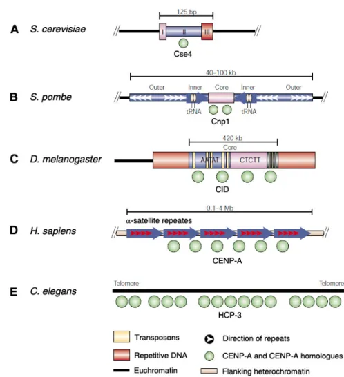
Figure 1.5. Organization of centromeric chromatin in different eukaryotes. The DNA sequence of centromeres differs between species, but the presence and function of CENP-A
and its homologues (shown in green) at centromeres is highly conserved. (A) S. cerevisiae
centromere function depends on conserved DNA elements (I, II, III), to which Cse4 localizes.
(B) S. pombe centromeres contain a unique central core, the site of Cnp1 nucleosome assembly, flanked by conserved inverted inner and outer repeats. (C) The defined D. melanogaster centromere at minichromosome Dp1187 consists of a core of 5 bp satellites and transposons, flanked by other repetitive DNA (red). (D) Human centromeres consist of
-satellite DNA (red arrows) tandemly arranged into higher order repeats (blue arrows), which
extend over megabases. CENPA localizes to a portion of these arrays. (E) C. elegans
centromeres assemble along the length of each chromosome. Adapted from Sullivan et al.,
2.2. Centromeric chromatin organization
2.2.1. CENP-A, a histone H3 variant unique to the centromere
Histone modifications and histone variants serve to define unique
chromosome regions, including the centromere. The centromere differs from the
rest of chromatin primarily by the presence of a specific histone H3 variant, the
centromere protein A (CENP-A), that replaces canonical histone H3.1 in
nucleosomes at active centromeres [Figure 1.6; (Palmer et al., 1987, 1991; Yoda
et al., 2000)]. This protein was one of the first centromere proteins identified as
an antigen recognized by sera from patients suffering from scleroderma spectrum
disease (Earnshaw and Rothfield, 1985; Valdivia and Brinkley, 1985). Homologs
of human CENP-A have been identified in all eukaryotes thus far: mouse
(Cenpa), chicken (CENP-A), plants (CenH3), D. melanogaster (CID), X. laevis (CENP-A), C. elegans (HCP-3), S. pombe (Cnp1) and S. cerevisiae (Cse4) (Wulf and Earnshaw, 2008; Silva and Jansen, 2009).
At point centromeres, such as those in S. cerevisiae, a single CENP-ACse4 -containing nucleosome forms the basis for kinetochore formation and microtubule
attachment (Furuyama and Biggins, 2007). Regional centromeres have multiple
CENP-A nucleosomes interspersed between nucleosomes containing the
canonical histone H3.1 and nucleosomes containing H3.3, another histone H3
variant [Figure 1.6; (Blower et al., 2002; Dunleavy et al., 2011)]. In holocentric
chromosomes, CENP-A and canonical H3 nucleosomes are interspersed and
spread throughout chromosome arms (Buchwitz et al., 1999; Oegema et al.,
2001). Importantly, loss of CENP-A or CENP-A homologs results in a complete
failure in chromosome segregation in all organisms tested so far (Earnshaw and
Migeon, 1985; Palmer et al., 1987; Meluh et al., 1998; Buchwitz et al., 1999;
Howman et al., 2000; Takahashi et al., 2000; Blower and Karpen, 2001; Oegema
et al., 2001; Régnier et al., 2005). This can be explained by the fact that CENP-A
is responsible for nucleating the centromere/kinetochore complex, which is
Foltz et al., 2006; Liu et al., 2006). Loss of CENP-A results in concomitant loss of
several centromere/kinetochore proteins, whereas overexpression of CENP-ACID,
in flies, results in mislocalization of CENP-ACID to noncentromeric regions and in
the formation of ectopic kinetochores (Heun et al., 2006; Olszak et al., 2011).
These observations suggest that CENP-A forms the foundation for kinetochore
assembly.
CENP-A and H3 share sequence homology within their histone fold
domains (approximately 60% in humans), but there is no sequence identity
between N-termini of both proteins (Sullivan et al., 1994). Consistent with the
pattern of sequence homology between CENP-A and H3, the histone fold domain
(HFD) directs CENP-A deposition at centromeres. A stretch of residues
responsible for CENP-A localization are found across the loop L1 and the
adjacent 2-helix of the HFD and is called the CENP-A targeting domain (CATD)
(Sullivan et al., 1994; Shelby et al., 1997; Vermaak et al., 2002; Black et al.,
2004). Replacement of the corresponding region within the HFD of H3 by the
CATD creates a chimeric H3CATD protein that targets to centromeres (Black et al.,
2004, 2007b). Remarkably, this chimeric histone H3CATD rescues the lethality
induced by depletion of CENP-A and sustains the assembly of a functional
kinetochore (Black et al., 2007b). Using hydrogen/deuterium exchange
experiments, the CATD was found to induce conformational rigidity to
(CENP-A/H4)2 tetramers and CENP-A containing nucleosomes relative to the
conventional counterparts containing histone H3.1 (Black et al., 2004, 2007a).
The molecular basis for this conformational rigidity was later provided by the
atomic structure of the subnucleosomal (CENP-A/H4)2 tetramers (Sekulic et al.,
2010). The crystal structure of the entire human CENP-A-containing nucleosome
has only been recently reported (Tachiwana et al., 2011). Overall, the structure of
the CENP-A nucleosome is extremely similar to the structure of canonical H3.1
nucleosome. The major structural differences of CENP-A nucleosomes are: 1)
the extended loop L1 that appears at the surface of the nucleosome and could
enters and exits the nucleosome resulting in a smaller DNA protection footprint in
nuclease assays (Maddox et al., 2011; Tachiwana et al., 2011). These different
DNA angles may affect higher order packing of CENP-A nucleosomes that could
contribute to maintaining a unique centromeric chromatin structure. Despite these
differences, these results indicate that human CENP-A-containing nucleosomes
are octameric and contain two copies of each of CENP-A, H4, H2A and H2B.
However, we cannot exclude that in other organisms or in different cell cycle
stages, CENP-A nucleosomes have alternative structures, such as tetrasomes,
hemisomes, and hexasomes, as several reports have suggested (Dalal et al.,
2007a, 2007b; Mizuguchi et al., 2007; Furuyama and Henikoff, 2009; Williams et
al., 2009).
2.2.2. Patterns of histone modifications at the centromere
The highly homogenous, repetitive nature of centromeric DNA makes
evaluation of the long-range chromatin organization of centromeric regions, for
example by chromatin immunoprecipitation (ChIP), a challenging task (Spence et
al., 2002; Lam et al., 2006). Nevertheless, high resolution cytogenetic techniques
such as chromatin fiber analysis have revealed that centromeric chromatin in
humans and flies is composed of CENP-A nucleosomes interspersed with
nucleosomes containing the canonical H3 dimethylated at Lysine 4 (H3K4me2)
[Figure 1.6; (Blower et al., 2002; Sullivan and Karpen, 2004)]. This modification,
which is a mark of euchromatin (Bernstein et al., 2002; Schneider et al., 2004), is
thought to be important for structural organization of the centromere (Dunleavy et
al., 2005). HJURP (Holliday Junction Recognizing Protein) is a CENP-A specific
chaperone important for CENP-A assembly (Dunleavy et al., 2009; Foltz et al.,
2009). Importantly, loss of H3K4me2 results in failure of centromere targeting of
HJURP, impairing incorporation of CENP-A at centromeres (Bergmann et al.,
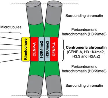
Figure 1.6. The unique organization of centromeric chromatin and pericentromeric heterochromatin. Centromeric chromatin consists of CENP-A nucleosomes interspersed with histone H3 nucleosomes. These H3 nucleosomes feature a unique pattern of histone
modifications rich in H3K4me2 and low in acetylation as well as the histone variant H2A.Z.
Centromeric chromatin also features nucleosomes containing H3.3, which may function as a
CENP-A placeholder during S, G2 and M phases. In metaphase chromosomes, it has been
proposed that these different histone variants are organized in distinct domains (represented
in red and blue). The CENP-A domain is located at the exterior surface, on top of which the
kinetochore is formed. The other histones are mainly located at the inner centromere.
However, it is possible that H3.1 and H3.3 are also part of the kinetochore forming domain
that is in contact with other centromere and kinetochore proteins. Recently a new nucleosome
like complex (CENP-T-W-S-X) was identified that is present within the CENP-A containing
chromatin domain (see Figure 1.7). Adapted from Vos et al., 2006.
CENP-A/H3K4me2 containing chromatin is typically embedded within a
large domain of heterochromatin named pericentromeric heterochromatin [Figure
1.6; (Carroll and Straight, 2006; Verdaasdonk and Bloom, 2011)]. The assembly
part, on the trimethylation of histone H3 on Lysine 9 (H3K9me3) and on the
binding of this histone modification by the chromodomain-containing protein HP1.
H3K9me3 chromatin in turn recruits cohesin that is responsible for establishment
of sister chromatid cohesion (Nonaka et al., 2002). Indeed, cells that lack
Su(var)3-9, the methyltransferase generating the H3K9me3 mark, do not recruit
HP1 to pericentromeric heterochromatin and do not establish sister chromatid
cohesion (Guenatri et al., 2004). In some species, such as in fission yeast, the
RNA interference (RNAi) machinery also plays a critical role in maintaining the
pericentromeric heterochromatin (Grewal and Moazed, 2003).
Another feature of centromeric chromatin is the lack of histone acetylation
that is normally associated with actively transcribed chromatin. The
hypoacetylation of histones at the centromere alter the chromatin structure and
organization, defining a chromatin region distinct from traditional euchromatin and
heterochromatin (Sullivan and Karpen, 2004; Dunleavy et al., 2005). Combined,
these modifications may help maintain the identity of centromeric chromatin and
pericentromeric heterochromatin, which are required for kinetochore assembly,
sister chromatid cohesion and condensation, and for proper chromosome
segregation during mitosis (Hendzel et al., 1997; Bernard et al., 2001).
2.3. The Constitutive Centromere-Associated Network (CCAN)
The core centromere is composed of several proteins that are associated
with CENP-A chromatin throughout the cell cycle. Although many designations
exist in the literature, this protein complex is now commonly called the
constitutive centromere-associated network (CCAN) and is composed of 16
proteins: CENP-C, CENP-H, CENP-I, CENP-K through CENP-U, CENP-W and
CENP-X (Obuse et al., 2004; Foltz et al., 2006; Izuta et al., 2006; Okada et al.,
2006; Cheeseman and Desai, 2008; Hori et al., 2008; Amano et al., 2009;
McAinsh and Meraldi, 2011; Perpelescu and Fukagawa, 2011). Many of these
CENP-C, have not been identified in D. melanogaster or C. elegans (Oegema et al., 2001; Heeger et al., 2005; Orr and Sunkel, 2011). In addition to these 16,
many other proteins have been found to copurify with CENP-A chromatin but
their centromere localization is either not constitutive or not yet verified (Obuse et
al., 2004; Izuta et al., 2006).
The CCAN complex localizes at the inner kinetochore and creates a bridge
between centromeric chromatin and the kinetochore proteins that bind plus ends
of spindle microtubule (McAinsh and Meraldi, 2011; Perpelescu and Fukagawa,
2011). While most proteins within the CCAN are interdependent, detailed
analysis has assigned specific roles to individual components. The specific roles
of the most relevant and best characterized CCAN proteins will be discussed
below.
2.3.1. CENP-C and CENP-N bind directly to CENP-A nucleosomes
In vitro experiments have shown that the CENP-C and CENP-N proteins
bind to distinct domains of CENP-A to direct the assembly of other CCAN and
kinetochore proteins (Carroll et al., 2009, 2010). While CENP-C recognizes the
C-terminal LEEGLG motif of CENP-A, CENP-N recognizes the CATD domain
embedded in the HFD of CENP-A (Carroll et al., 2009, 2010).
CENP-C is a DNA binding protein that localizes to the inner kinetochore
(Saitoh et al., 1992). CENP-C homologues have been identified in virtually all
model organisms, including yeast, flies, plants and mammals, and have been
shown to be required for proper chromosome segregation and mitotic
progression (Tomkiel et al., 1994; Dawe et al., 1999; Fukagawa et al., 2001;
Moore and Roth, 2001; Oegema et al., 2001; Ogura et al., 2004; Schuh et al.,
2007; Erhardt et al., 2008; Orr and Sunkel, 2011). The centromere localization of
CENP-C depends on CENP-A (Oegema et al., 2001; Liu et al., 2006; Hori et al.,
2008), and, at least in flies and to a lesser extend in human cells, centromere
et al., 2010). CENP-C is required for the centromere localization of several
kinetochore proteins, including Knl1, the Mis12 complex and the Ndc80 complex
that together are known as the KMN network, which forms the principal
microtubule binding complex in the kinetochore [(Cheeseman et al., 2006; Milks
et al., 2009; Przewloka et al., 2011; Screpanti et al., 2011); see also Section 2.4].
CENP-C is also required for the recruitment of checkpoint proteins, for mitotic
checkpoint function (Kwon et al., 2007; Przewloka et al., 2011; Screpanti et al.,
2011), and for the centromere localization of other CCAN components such as
CENP-H, CENP-I, CENP-K and CENP-T (Carroll et al., 2010).
CENP-N is also required for proper chromosome segregation and mitotic
progression (Foltz et al., 2006; McClelland et al., 2007; Carroll et al., 2009). This
protein is required for centromere localization of several CCAN components,
including CENP-H, CENP-I, CENP-K, CENP-C and CENP-O (McClelland et al.,
2007; Carroll et al., 2009). However, depletion of CENP-N did not affect the
levels of the Nnf1 component of the Mis12 complex (McClelland et al., 2007).
This observation suggests that CENP-N is not directly involved in recruiting
kinetochore proteins that bind to spindle microtubules during mitosis. In Chapter
2 of this thesis we will analyze and discuss the additional function of CENP-N in
assembling centromeric chromatin.
The fact that both CENP-N and CENP-C bind directly to CENP-A-containing
nucleosomes suggests they play a central role in the early stages of
centromere/kinetochore assembly (Figure 1.7). Indeed, a recent study showed
that C when artificially tethered onto chromosomes together with
CENP-T/W, is sufficient to drive the formation of a functional centromere/kinetochore
2.3.2. CENP-T, CENP-W, CENP-S, and CENP-X form a nucleosome like
structure able to warp centromeric DNA
CENP-T was initially identified as a component that copurified with CENP-A
chromatin (Obuse et al., 2004; Foltz et al., 2006). Those affinity purifications also
identified CENP-M and CENP-U which, when tagged and purified, leaded to the
identification of CENP-S (Foltz et al., 2006), indicating that CENP-S is not directly
associated with CENP-A nucleosomes. Later, CENP-W and CENP-X were
discovered as binding partners of CENP-T and CENP-S, respectively (Hori et al.,
2008; Amano et al., 2009). Strikingly, these four CCAN proteins all carry a
histone fold domain (HFD) and a recent report showed that, in vitro, they form a heterotetrameric complex capable of binding and wrapping centromeric DNA
[Figure 1.7; (Nishino et al., 2012)]. The resolution of the crystal structure of this
heterotetrameric complex revealed that CENP-T/W and CENP-S/X dimerize
through their HFDs. The interface between the CENP-T/W and CENP-S/X
heterodimers occurs in the regions of CENP-T and CENP-S that are similar to
those involved in heterotetramerization of histones (Nishino et al., 2012). An
important difference between these complexes and normal histone complexes is
that the histone H3-H4 heterotetramer is a dimer of heterodimers and is therefore
symmetric across the tetramerization interface. In contrast, the CENP-T/W/S/X
complex is asymmetric. Mutations in the tetramerization interface in either
CENP-T or CENP-S abolish their recruitment to centromeres (Nishino et al., 2012),
suggesting that these complexes are formed in vivo and are important for centromere/kinetochore establishment and function. The discovery that the
CENP-T/W/S/X complex is able to bind DNA in a nucleosome-like fashion
suggests that functional centromeric chromatin is composed by four different
chromatin-binding complexes: H3.1-, H3.3- and CENP-A-containing nucleosomes
as well as CENP-T/W/S/X complexes.
Importantly, a portion of CENP-T that is N-terminal to the HFD extends
(Gascoigne et al., 2011)]. Consistently, CENP-T and CENP-W are loaded and
become enriched at centromeres during late S and G2 phases (Prendergast et
al., 2011), just before the recruitment of the KMN network to the kinetochores.
Another connection of the CCAN to the Ndc80 complex is built through CENP-C,
which links the CENP-A nucleosome to the KMN network through its binding to
the Mis12 complex (Screpanti et al., 2011). All together, these results indicate
that CENP-C and the CENP-T/W/S/X complex form the primary platform that
recruits the KMN network (Figure 1.7).
2.3.3. CENP-H/I/K complex
One CCAN subcomplex that localizes to the centromere downstream of the
CCAN proteins described above is the CENP-H complex, composed of CENP-H,
CENP-I and CENP-K proteins. The centromere localization of this complex has
been shown to be dependent on CENP-A, CENP-N, CENP-M, CENP-C, CENP-T
and CENP-L (Foltz et al., 2006; Okada et al., 2006; McClelland et al., 2007; Hori
et al., 2008). The CENP-H/I/K complex is required for the centromere localization
of the CENP-O/P/Q/R/U complex and of the KNM network (Liu et al., 2003;
Okada et al., 2006; Kwon et al., 2007; Cheeseman et al., 2008; Hori et al., 2008).
Accordingly, depletion of any component of the CENP-H/I/K complex induces
severe defects in kinetochore assembly and chromosome segregation
(Nishihashi et al., 2002; Okada et al., 2006).
Besides their role in recruiting other centromere/kinetochore proteins,
CENP-H and CENP-I interact with the plus end of kinetochore microtubules
modulating their turnover rate and promoting accurate chromosome alignment at
the metaphase plate (Amaro et al., 2010). The CENP-H/I/K complex also has a
role in recruiting CENP-A to the centromere as will be described in the Chapter 2
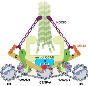
Figure 1.7. The CCAN forms a bridge between centromeric DNA and microtubule plus ends. The CENP-T/W/S/X complex links centromeric chromatin to the mitotic kinetochore through a direct interaction between CENP-T and the NDC80 microtubule-binding complex.
The CENP-A nucleosome is contacted by CENP-C and CENP-N that bind to the Mis12
complex and the remaining CCAN, respectively. In this way, parallel contacts between
centromeric DNA and microtubules are established. Adapted from Foltz and Stukenberg,
2012.
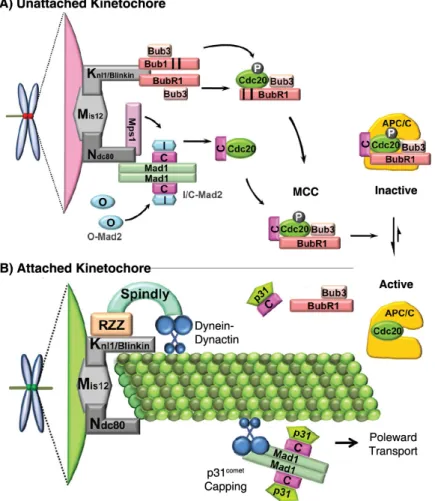
2.4. The Kinetochore
The kinetochore is a highly complex proteinacious structure involved in
several mitotic functions, most importantly in attachment to spindle microtubules
and in mitotic checkpoint signaling (Rieder and Salmon, 1998). Despite a few
minor organism-specific differences, kinetochore composition and organization
appears to be highly conserved among most eukaryotes (Musacchio and
Salmon, 2007; Gascoigne and Cheeseman, 2011). Kinetochore proteins are