Evaluation of neuroprotective potential of
polyphenols derived from Portuguese
native plants:
Juniperus
sp. and
Rubus
sp.
Lucélia Rodrigues Tavares
Dissertation presented to obtain the Ph.D degree in Biochemistry
Instituto de Tecnologia Química e Biológica | Universidade Nova de Lisboa
Oeiras,
Evaluation of
neuroprotective potential of
polyphenols derived from
Portuguese native plants:
Juniperus
sp. and
Rub us
sp.
Lucélia Rodrigues Tavares
Dissertation presented to obtain the Ph.D
degree in Biochemistry
Instituto de Tecnologia Química e Biologica |
Universidade Nova de Lisboa
Oeiras, November, 2012
INSTITUTO DE TECNOLOGIA QUIMICA E BI0L0GICA / UNL
ii
The work presented in this thesis was developed at:
Disease and Stress Biology Laboratory
Instituto de Tecnologia Química e Biológica
Universidade Nova de Lisboa
Av. da República, Apartado 127
2781-901 Oeiras, Portugal
Supervised by:
Doctor Ricardo Boavida Ferreira
Doctor Maria Cláudia Nunes dos Santos
iii
Acknowledgements………..……….
iv
Abreviation
list………..………..….…
.
…
.v
Abstract………..………
...ix
Resumo……….………
..
…
.xi
Thesis outline………..……….…x
iii
Chapter 1:
General introduction………..………..………
...1
Juniperus
sp. .………...…………59
Chapter 2: The neuroprotective potential of phenolic-enriched fractions
from four Juniperus species found in
Portugal ………
...
….
61
Chapter 3: Elucidating phytochemical production in
Juniperus sp.:
seasonality and response to stress situations
……….
.
…………
87
Rubus sp. ………..………..11
3
Chapter 4 : Neuroprotective effect of blackberry (Rubus sp.) polyphenols
is potentiated after simulated gastrointestinal digestion
………..…..
115
Chapter 5: Neuroprotective effects of digested polyphenols from wild
blackberry species………..…..
141
Chapter 6: Differential expression of genes involved in neuroprotection
conferred by blackberry polyphenols after simulated gastrointestinal
digestion………..……
.
…..…
169
Chapter 7: General discussion……….…..…..…
221
Appendix: Comparison of different methods for DNA-free RNA isolation
iv
I dedicate this thesis to my all family, specially to my parents, my sister
and my grandfather “Zé” that always believe on me and supported me.
I also would like to acknowledge to my supervisors for all knowledge,
advices and support. A special thank to Cláudia for being in the last years
my
“
mom
”
, my
“
sister
”
and specially my friend.
To my all lab colleagues. Many thanks for the time we shared together, on
the lab and besides it. They enriched my life in many diverse aspects. A
special thanks to all that help me in “flow cytometry marathon”
and in
thesis revision and to Rui for the amazing plant pictures.
To my all friends not included above, that always support me and make
me laugh, thanks.
To Doctors Helena Vieira, Derek Stewart and Gordon McDougall I would
like to acknowledge all the help and the involvement into the work.
Last but not least, I thank to ITQB for hosting me and all people that make
this work possible providing or helping in the search of plants, namely
Doctors Carlos Aguiar, Pedro Oliveira, Dalila Espírito-Santo and Fátima
v
Abbreviation Termm Mitochondrial transmembrane potential 4CL 4-coumarate:CoA ligase
6-OHDA 6-hydroxydopamine 8-OHdG 8-hydroxydeoxyguanosine
A -amyloid
ABC ATP-binding cassette AC Antioxidant capacity ACC Acetyl-CoA carboxylase
ACh Acetylcholine
AChE Acetylcholinesterase
ACT Actin
AD Alzheimer’s disease
Aha1 ATPase homolog 1 Akt Protein kinase B
ALP Autophagy-lysosome pathway ALS Amyotrophic lateral sclerosis ANR Anthocyanidin reductase ANS Anthocyanidin synthase AP-1 Activating protein-1
APC/CDC20 Anaphase-promoting complex/cell division cycle 20 homolog APP Amyloid precursor protein
Arc Activity-regulated cytoskeleton-associated protein ARE Antioxidant response element
Bcl-2 B-cell lymphoma 2
BDNF Brain-derived neurotrophic factor
bp Base pair
Brca Breast and ovarian cancer susceptibility protein BuChE Butyrulcholinesterase
C3H p-coumarate 3 hydroxylase C4H Cinnamate 4-hydroxylase cAMP Cyclic adenosine monophosphate
CAT Catalase
CBF Cerebral blood flow CE Catechin equivalents
CFTR Cystic fibrosis transmembrane conductance regulator CHI Chalcone isomerise
CHS Chalcone synthase CNS Central nervous system COP Coat protein complex
COX Cyclooxigenase
CREB cAMP response element binding CyDAGlc Cyanidin-3-O-dioxayl-glucoside CyGlc Cyanidin-3-O-glucoside
CyHMGG Cyanidin-3-O-hydroxymethyl-glutaroyl glucoside CyMalG Cyanidin-3-O-malonylglucoside
CyXyl Cyanidin-3-O-xyloside CyXyl Cyanidin-3-O-xyloside DAPI 4',6-diamidino-2-phenylindole
DAVID Database for Annotation, Visualization and Integrated Discovery
DCF 2’,7’-dichlorofluorescein
DFR Dihydroflavonol reductase
DG Dentate gyrus
DiOC6(3) 3,3’-dihexyloxacarbocyanine iodide DSB Double strain break
DW Dry weight
EA Ellagic acid
vi
EGCG Epigallocatechin gallateeIF2 Eukaryotic translation initiation factor 2 EMEM Eagle Minimum Essential Medium ENaC Epithelium sodium channel ER Endoplasmic reticulum
ERK Extracellular-signal-regulated kinase
ET Ellagitannin
F3’5’H Flavonoid 3′5′-hydroxylase
F3’H Flavonoid 3′-hydroxylase F3GT Flavonoid-3-glucosyltransferase F3H Favanone 3-hydroxylase
FA Formic acid
FBS Foetal bovine serum FLS Flavonol synthase
FS Flavone synthase
GA Golgi apparatus
GABAA Gamma-aminobutyric acid GAE Gallic acid equivalents
GAPDH Glyceraldeide 3-phosphate dehydrogenase GCN2 General control nonrepressed 2
GI Gastrointestinal
GITC Guanidine Isothiocyanate GPx Glutathione peroxidase GR Glutathione reductase GRAS Generally Recognized As Safe
Grx Glutaredoxin
GSH Glutathione (reduced form) GSK 3 Glycogen synthase kinase 3 GSSG Glutathione disulphide
H2DCFDA 2’,7’-dichlorofluorescein diacetate
HD Huntington’s disease
HNE 4-hydroxynonenal
HO-1 Heme oxygenase
HSD Honest Significant Difference HSP Heat shock protein
IAPs Inhibitors of apoptosis IL-1 Interleukin-1
iNOS Inducible nitric oxide synthase IVD In vitro digestion
JNK c-Jun N-terminal kinase
KV Voltage-gated K+
Lamb Lambertianin C
LAR Leucoanthocyanidin reductase
LC-MS Liquid chromatography-mass spectrometry LPO Lipid peroxidation
m/z Mass-to-charge ratio
MAPK Mitogen-activated protein kinase MEAD Methyl ellagic acid derivative MeEAGlcA Methyl ellagic acid glucuronide MeEApent Methyl ellagic acid pentose MeJa Methyl jasmonate
MEK Mitogen-activated protein kinase kinase
MMR Mismatch repair
MPP+
1-methyl-4-phenylpyridinium Chk2 Cell cycle checkpoint kinase 2
MPTP 1-methyl-4-phenyl-1,2,3,6-tetrahydropyridine MRE11 Meiotic recombination element 11
MRN MRE11/Rad50/NBS1 complex
vii
NDDs Neurodegenerative diseasesNER Nucleotide excision repair Nf-B Nuclear factor-B
NFT Neutrophin
NHEJ Non-homologous end joining NMDA N-Methyl-D-aspartic acid
NP Natural product
NQO1 NAD(P)H dehydrogenase, quinone 1 Nrf2 Nuclear factor erythroid 2-related factor 2 OMT o-methyltransferase
OPA Orthophthalaldehyde
ORAC Oxygen radical absorbance capacity OST Oligosaccharyltransferase
PAL Phenylalanine ammonia-lyase PBS Phosphate buffer saline PCD Programmed cell death
PCNA Proliferating cell nuclear antigen PCR Polymerase chain reaction
PD Parkinson’s disease
PDA Photo diode array PEF Phenolic-enriched fraction PG Post gastric (digest)
PI Propidium iodide
PI3K . Phosphoinositide 3-kinase PKA cAMP-dependent protein kinase PLA2 Group II phospholipase A2
POLR2A DNA-directed RNA polymerase II subunit A
PP Polyphenol
PP1-cat Protein phosphatase catalytic subunit PP2A Protein phosphatase 2A catalytic subunit PP2C Protein phosphatase 2C
QGlc Quercetin-glucoside QGlcA Quercetin-glucuronide QHMGG Quercetin-HMG-glucoside QHMGGlc Quercetin-HMG-glucoside Qpent Quercetin pentose Qrut Quercetin-rutinoside QXyl Quercetin-xyloside RAN Ras-related nuclear protein RMA Robust Multi-array average RNS Reactive nitrogen species
RONS Reactive oxygen and nitrogen species ROS Radical oxygen species
RT Retention time
RT-PCR Reverse transcription-polymerase chain reaction
Sang Sanguiin
SD Standard deviation SIRT1 Sirtuin 1
SN Substancia nigra
SOD Superoxide dismutase SPE Solid phase extraction STS Stilbene synthase TE Trolox equivalents TFC Total flavonoid content TFIIH Transcription factor II H
TOP Topoisomerase
TPC Total phenolic content TrkB Tyrosin-related kinase B
viii
UPW Ultra pure waterix
Neurodegenerative diseases have become a challenge to modern societies
in industrialized countries due to the high social and economic impacts
and therefore demand intensive research. These diseases present diverse
common features, in which oxidative stress plays a very important role. As
result of oxidative stress, some molecular mechanisms become
dysfunctional and ultimately lead to cell death. Amongst the compounds
displaying neuroprotective activity, phenolics have been identified
amongst the most active substances from natural sources.
The purpose of the work presented in this thesis was to address the
neuroprotective capacity of phenolic compounds from juniper leaves
(Juniperus sp.) and blackberry fruits (Rubus sp.) from Portuguese native
plants that display high phenolic content. Two different approaches in
the prevention of neurodegenerative diseases could be foreseen:
nutritional and pharmacological approaches.
Concerning the pharmacological approach, leaves from four junipers were
evaluated and all displayed characteristics for promising neuroprotective
products. Among these species,
Juniperus oxycedrus badia was
distinguished by the capacity of its phenolic compounds to protect
neuronal viability and functionality. Junipers are evergreen plants with
leaves available all the year, although their phenolic composition could be
affected by environmental factors. Therefore, it is important to
understand the seasonal dynamics of their phytochemical content and
variation due to stress. In periods when active growth is retarded,
particularly in winter, junipers had higher phenolic content. Juniper
species displayed different susceptibilities to stresses, and salt stress
enhanced the phenolic content of J. oxycedrus badia. The increase in total
content was due to a general rise in all classes of compounds with a few
alterations in the relative content of specific compounds. This increase
was concomitant with the enhancement of transcripts encoding
x
yield can be modulated/maximized.
Related to the nutritional perspective, the impact of gastrointestinal
digestion on blackberry polyphenols was accessed to ensure that the
physiological conditions affecting foods were taken into account. This
approach was then used to evaluate the neuroprotective potential of
polyphenols from Portuguese wild blackberries compared to commercial
blackberries. Although they presented few quantitative chemical
differences, differential cytoprotective activities were discovered. Wild
blackberry species were revealed as more effective in neuroprotection
possibly through the mediation of molecular adaptive responses.
Therefore, a transcriptomic approach was used to compare the differential
molecular mechanisms mediating neuroprotection between commercial
species and
R. vagabundus. Blackberry metabolites were able to
differentially prevent transcript alterations induced by oxidative stress,
with wild blackberry more effective in reducing the number of genes
altered. The main pathways differentially affected between stressed and
blackberry pre-treated cells were related to transport, cell cycle and DNA
repair. A common regulation by small GTP-binding proteins and/or
associated
pathways could be foreseen,
although confirmatory
biochemical assays are required. In conclusion, metabolites from
J. oxycedrus badia and
R. vagabundus show neuroprotection potential
xi
Resumo
As doenças neurodegenerativas têm-se tornado um desafio das
sociedades modernas nos países industrializados, devido ao elevado
impacto económico e social, requerendo por isso de uma investigação
intensiva. Estas doenças apresentam diversas características em comum
das quais se destaca o stress oxidativo. Como resultado deste, alguns
mecanismos moleculares tornam-se disfuncionais podendo conduzir à
morte celular. Entre os compostos apresentando atividade neuroprotetora,
os compostos fenólicos têm sido identificados como entre as substâncias
mais ativas obtidas da Natureza.
O objetivo do trabalho apresentado na presente tese foi de determinar a
capacidade neuroprotetora de compostos fenólicos de folhas de zimbro
(Juniperus sp.) e amoras de silva (Rubus sp.), obtidos de plantas nativas
portuguesas com um elevado conteúdo em compostos fenólicos. Duas
abordagens diferentes podem ser antecipadas na prevenção das doenças
neurodegenerativas: farmacológica e nutricional.
Relativamente à abordagem farmacológica, folhas de quatro espécies de
zimbro foram avaliadas e todas demonstraram características importantes
para a obtenção de produtos neuroprotetores promissores.
J. oxycedrus
badia foi distinguido pela capacidade dos seus compostos fenólicos
protegerem a viabilidade e funcionalidade neuronal. Sendo os zimbros
plantas perenes, cuja composição fenólica das folhas pode ser afetada
pelos fatores ambientais, revelou-se importante perceber a dinâmica
sazonal dos fitoquímicos bem como as suas variações devido a stresses.
Em períodos em que o crescimento ativo é retardado, particularmente no
Inverno, os zimbros apresentaram um elevado conteúdo em compostos
fenólicos. As diferentes espécies de zimbro apresentaram diferentes
suscetibilidades aos stresses, tendo o stresse salino aumentado o
conteúdo em polifenóis de
J. oxycedrus badia. O aumento do conteúdo
total deveu-se ao aumento geral de compostos das várias classes,
xii
particular. Este aumento foi concomitante com o aumento de transcritos
que codificam fenilalanina amónia liase, um enzima chave da via
responsável pela produção de polifenóis. Verificou-se portanto que a
produção de fitoquímicos nos zimbros pode ser modulada/maximizada.
Quanto à perspetiva nutricional, de forma a garantir que as condições
fisiológicas que afetam os alimentos eram comtempladas, foi
determinado o impacto da digestão gastrointestinal sobre os polifenóis de
amoras. Esta abordagem foi posteriormente usada na avaliação do
potencial neuroprotetor dos polifenóis de amoras silvestres portuguesas
comparativamente com amoras comerciais. Apesar da composição
química das amoras apresentar poucas diferenças quantitativas entre
espécies, as atividades citoprotetoras foram diferenciadas. As espécies de
amora silvestres mostraram-se mais efetivas na neuroprotecção,
possivelmente devido à mediação de respostas adaptativas moleculares.
Consequentemente, foi usada uma abordagem transcritómica para
comparar os mecanismos moleculares que intervêm na neuroprotecção e
que são diferentes entre as amoras comerciais e selvagens,
particularmente
R. vagabundus. Os metabolitos de ambas as amoras
conseguiram prevenir as alterações nos transcritos induzidas pelo stresse
oxidativo, apresentando-se a amora silvestre mais efetiva na redução do
número de genes alterados. As principais vias diferencialmente alteradas
entre células stressadas e pré-tratadas com amoras encontram-se
relacionadas com funções de transporte, ciclo celular e reparação de DNA.
Uma regulação comum pelas pequenas proteínas de ligação ao GTP e/ou
vias associadas pode ser antecipada, apesar de ser necessário a realização
de ensaios bioquímicos confirmatórios. Em conclusão, os metabolitos de
J. oxycedrus badia e R. vagabundus mostram um potencial neuroprotetor
xiii
Thesis outline
This thesis is organized in seven chapters.
The first chapter (General introduction) brings in the main topics related
to the work developed and necessary to its comprehension, namely
neurodegenerative diseases, polyphenols and the possible role of
polyphenols
activities
in
protecting
human
body
against
neurodegeneration.
Presentation of experimental work is divided in two parts:
Juniperus sp.
(A) and
Rubus sp. (B), where is presented work developed applied to
species of these two genus.
In the second and third chapters are presented the results concerning the
first type of strategy, the role of phenolic extracts from
Juniperus sp.
leaves as natural products. The second chapter rely on the potential
neuroprotective activity of the extracts and the third chapter on
understanding polyphenols dynamics in plants due to seasonality and
stress induction.
The fourth, fifth and sixth chapters rely on a nutritional approach, where
the neuroprotective potential of blackberries polyphenols are determined.
In the fourth chapter it is highlighted the importance of using a more
physiological model to unravel the real potential of polyphenols that are
submitted to gastrointestinal digestion, by using a commercial variety of
blackberry. In the fifth chapter it is evaluated the neuroprotective
potential of two Portuguese wild blackberries. In the sixth chapter the
potential molecular mechanisms underlying the protective effect
previously verified by blackberries are studied using transcriptomics
approach.
In the seventh chapter (General discussion) it is presented a general
discussion of findings obtained and the main conclusions as well as future
2
1. Neurodegeneratives diseases (NDDs)
NDDs are a heterogeneous debilitating and until now an incurable group
of degenerative disorders. They are characterized by a slow and
progressive loss of neuronal cells, leading to gradual and progressive
impairments of selective functions of central nervous system (CNS),
depending upon the involved type of neuronal cells 1. The increase in life
expectancy observed over the last century has led to the emergence of
these age-related disorders that pose novel challenges to modern
societies in industrialized countries and demand intensive research 2.
1.1.Social and Economic impact
These diseases cause devastating effects on the patient and are currently
only treated palliatively. Despite the profound impact on patients and on
their families, the combination of expensive formal care services,
increasing number of affected people and major reliance on informal care,
remains a great challenge for society. It was estimated in 2004 by WHO
that NDDs (Alzheimer’s disease (AD), other dementias and Parkinson’s
disease (PD)) reached 29.4 million people all around the world 3.
Focusing just on dementia, a more recent survey estimated that there
were about 10.11 million people affected in Europe 4. The total costs of
illness for these disorders in the whole Europe were €177 billion, being
the cost per demented person about €22,000 per year 4.
1.2.The main NDDs
Alzheimer’s disease
AD is the most common NDD, where patients experience progressive
cognitive deterioration, behavioral changes, and neuropsychiatric
changes 5. AD is characterized by the loss of neurons, synapses, and
neurotransmitters throughout the brain, especially in the hippocampus
and cerebral cortex 6. The pathological hallmarks of AD are neuronal loss,
extracellular senile plaques containing the peptide -amyloid (A) and
neurofibrillary tangles, composed of a hyperphosphorylated form of the
3
Parkinson’s diseasePD is a slowly progressive NDD, the second most common, characterized
by loss of catecholaminergic neurons from the substantia nigra (SN) that
eventually leads to bradykinesia, muscle rigidity, resting tremor, and
postural instability 8. The motor symptoms of PD are directly related to the
loss of pigmented cells in the SN and to the reduction of the
neurotransmitter dopamine in the striatum. The characteristic hallmark is
also the presence of the Lewy body, aggregates of proteins containing
ubiquitin and alpha-synuclein, within the cytoplasm of dying nerve cells.
Huntington’s disease (HD)
HD is characterized by personality changes as well as motor and cognitive
symptoms, including the hallmark feature, chorea, psychiatric
disturbances and dementia 9. Pathologically it is characterized by loss of
long projection neurons in the cortex and striatum and premature
death 10,11. HD is an autosomal dominant disease that causes degeneration
of medium spiny GABAergic neurons in the striatum. This
neurodegeneration causes a progressive atrophy of the caudate nucleus,
putamen, and globus pallidus 5. HD is a genetic disease caused by a CAG
repeat expansion of exon 1 in the gene that encodes huntingtin protein.
Amyotrophic lateral sclerosis (ALS)
ALS is a devastating disease resulting in progressive skeletal muscle
weakness, muscle atrophy, paralysis, and ultimately death within 2–5
years of onset. ALS results from a progressive degeneration of motor
neurons within the ventral horn of the spinal cord, brainstem, and motor
cortex. Degeneration also occurs within the corticospinal tract.
Approximately 20 % of patients have a mutation in the superoxide
dismutase 1 (SOD1) gene, however, the majority of patients have normal
SOD1 activity 12.
1.3.Common features of NDDs
The various NDDs have different symptoms, affect different parts of the
brain and have different physiological causes 13. NDDs constitute a
4
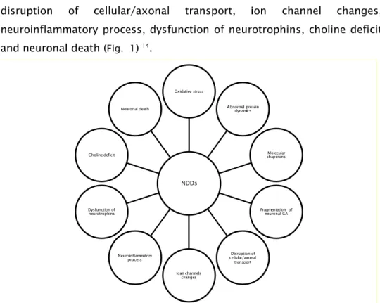
pathogenesis. Cellular features that have been thought to contribute to
neuronal loss include oxidative stress, abnormal protein dynamics,
molecular chaperones, fragmentation of neuronal Golgi apparatus (GA),
disruption of cellular/axonal transport, ion channel changes,
neuroinflammatory process, dysfunction of neurotrophins, choline deficit
and neuronal death (Fig. 1) 14.
1.3.1. Oxidative stress
Oxidative stress has been implicated in the pathogenesis of several
disease processes, including, ischemia/reperfusion injury, AD, PD, among
others 15. It is basically a pathologic metabolic condition arising upon
imbalance between the production of potentially toxic reactive oxygen
species (ROS; e.g. hydrogen peroxide, superoxide and hydroxyl radical)
and the antioxidants, in favor of the former 16. ROS include oxygen free
radicals and non-radical derivatives of O2 that participate in free radical
production.
ROS have been shown to play both beneficial and deleterious roles. At
very low concentration, in non-pathological conditions, it may act as a
Fig. 1- Cellular features that contribute to neuronal loss. NDDs
Oxidative stress
Abnormal protein dynamics
Molecular chaperons
Fragmentation of neuronal GA
Disruption of cellular/axonal transport
Ioan channels changes Neuroinflammatory
process Dysfunction of neurotrophins Choline deficit
5
second messenger in some signal transduction pathways 17. However,
overproduction of ROS can damage cells through free radicals generation
that leads to a progressive decline in physiological function 18. ROS can
attack proteins 19, oxidize lipids and damage DNA 20.
Protein oxidation
ROS may damage the proteins via nitration or oxidation 21,22. Protein
oxidation contributes to the inhibition or impairment of multiple enzymes,
thus affecting multiple cellular functions, ranging from protein synthesis,
energy production, and cytoskeleton dynamics to signal transduction 23.
Additionally, protein oxidative modification could contribute to the
formation of intracellular protein aggregates, which may have additional
effects on intracellular homeostasis 24. Once protein aggregation begins,
other proteins may be sequestered into the protein aggregates, resulting
in their loss of cellular function 22,25.
Lipid peroxidation
All cellular membranes are especially vulnerable to oxidation due to their
high concentrations of unsaturated fatty acid 21. Thus lipid peroxidation
(LPO) is the consequence of ROS attack. Their role is well established in
the pathogenesis of a wide range of NDDs 26. LPO causes changes in
fluidity and permeability of the cell membranes leading to the production
of conjugated dienic hydroperoxides 27,28. These unstable substances
decompose either into various aldehydes, such as malondialdehyde and
4-hydroxynonenal (HNE) 28. HNE in primary neurons and tissues leads to
the activation of cell repair or cell death programs, being its adducts used
as biomarkers of oxidative damage within cells 29. HNE accumulation has
been observed in NDDs, including AD, PD, and ALS 30.
DNA oxidation
Although DNA is a stable and well-protected molecule, ROS can promote
extensive damages. The damage of nuclear DNA can be caused by two
different mechanisms: oxidative modification and endonuclease-mediated
DNA fragmentation 31. Several types of DNA damages could be caused by
6
purines, damage to the deoxyribose sugar, DNA–protein cross-linkage and
damage of the DNA repair system 21. All these chemical damages impair
function and leads to cellular debilitation and possibly premature death 32.
The 8-hydroxydeoxyguanosine (8-OHdG), an oxidized form of guanine, is
a major oxidative DNA-damage product that can produce mutations.
The consequences of oxidative stress increase may result in imbalance
calcium homeostasis, alterations in cellular signaling cascades and
changes in gene expression 33,34. These effects are detected as more
elevated in the aging population 35,36, AD 37-39 and PD 40,41.
Cells are also equipped to counteract these oxidative attacks with
numerous cellular antioxidant defenses, such as glutathione (GSH),
glutathione peroxidase (GPx), SOD, and catalase (CAT) 42. However, during
aging and various disease states, these antioxidant defense systems can
be altered leading to progressive oxidative damage and subsequent cell
death and/or significant loss of function 43.
Brain sensitivity to oxidative stress
The brain is particularly sensitive to oxidative stress since it presents:
high content of peroxidizable unsaturated fatty acids, high oxygen
consumption per unit of weight, high content in iron and ascorbate (LPO
key ingredients) and a scarcity of antioxidant defense systems(e.g. GSH,
GPx, CAT and vitamin E) 44-49.
In fact, neurons have high metabolic demand. In humans, the brain
accounts for only a few percent of the body weight, but it processes 20 %
of basal oxygen consumption. A neuron uses much of oxygen via
mitochondrial respiratory chain to make ATP for maintaining low
gradients (high intracellular K+, low Na+, very low and free Ca2+) 50,51.
Mitochondria role
Mitochondria are the ‘‘powerhouses’’ of the cells. The main function is the
production of ATP 52. It has been estimated that about 90 % of mammalian
oxygen consumption is mitochondrial, which primarily serves to
7
neurons, owing to their high demands for energy because of their
specialized functions, complex morphology, and synaptic activity 53. It has
been increasing attention for the recognition of NDDs as mitochondrial
diseases 54. Although each of the NDDs has its distinct etiological
processes and different affected brain regions, neurodegenerative
disorders as a whole, share similar mitochondrial dysfunctions 54. Since
neurons in the brain strongly depend on mitochondrial driven aerobic
respiration, when mitochondria become dysfunctional, it starts a vicious
and detrimental cycle in which neurons become much more susceptible to
oxidative stress. Mitochondria have already a high level of oxidative stress
and, therefore, any increase in internal or external ROS leads to
dysfunctional mitochondria, which in turn produces more ROS.
Additionally, mitochondrial DNA is also a target of oxidation which can
mediate impairment of mitochondrial functions, leading to more oxidative
stress and eventual cell death (Fig. 2).
Role of oxidative stress on the main NDDs
Oxidative stress seems to be involved in the neuronal loss that occurs in
AD although its cause is unclear 55. Oxidative damage occurs early in the
AD brain, before the onset of significant plaque pathology, and seems
also to precede the A deposition in animal models 56. Increased protein
oxidation, protein nitration, and LPO occur in neurofibrillary tangles and
neuritic plaques of AD patients and also levels of oxidation products are
increased in cerebrospinal fluid of AD patients 57. Increased amounts of
Fig. 2- Vicious cycle of mitochondria promoted by oxidative stress.
Higher ROS levels
Mitochondrial DNA damage Mitochondria
dysfunction
8
glutathione reductase (GR) glutaredoxin (Grx) and GPx were found in
postmortem samples from AD patients, suggesting the activation of an
antioxidant defense response 58,59.
Oxidative stress is also involved in the pathology of PD. Although the
disease could be attributed to genetic or environmental factors, both lead
to oxidative stress, mitochondrial dysfunction and protein aggregation 60.
The pathogenesis of PD has been reported to include increased dopamine
turnover, diminished GSH content, and increased iron levels in the SN 61.
Dopaminergic neurons of the SN from brains of PD patients also exhibit
hallmarks of oxidative stress, including LPO, nucleic acid and protein
oxidation, and changes in some antioxidant molecules 62. Mitochondrial
defects are observed in PD 63; specifically, reduced activity of the
mitochondrial respiratory complex I (NAD(P)H: quinone oxidoreductase
(NQO)) in the SN 64 as well as the frontal cortex 65,66. It is well known that
PD pathology includes the selective loss of dopamine neurons 67, and it is
hypothesized that the main cause of this loss is the high levels of ROS in
dopamine neurons due to dopamine metabolism and their high iron
content 68.
Oxidative stress is also present in HD as represented by increased DNA
oxidative products and elevated DNA strand breaks 5. Oxidative
challenges present with HD are thought to arise from poor mitochondrial
membrane integrity. Impaired function of complex II and complex III of
the electron transport chain 69, should contribute to the decreased ATP
levels detected in HD patients.
In ALS like disease, onset and progression is also associated with
enhanced production of ROS in the spinal cords 70. Oxidative stress has
been suggested as favoring neuronal damage 71. Protein carbonyls and
protein nitration have been found to be elevated in human ALS
patients 72,73. Markers for LPO and higher levels of 8-OHdG were detected
in spinal cord from ALS patients 74,75. Other cellular characteristics of ALS
include a loss of mitochondrial membrane potential and increased
9
1.3.2. Abnormal protein dynamics
Aberrant self-aggregating misfolded proteins with formation of intra- or
extra-cellular high-ordered insoluble fibrils deposits are common
pathological hallmarks of many NDDs 76,77. A causative link between the
formation of protein aggregates and NDD has been established, which
may occur as a result of the toxic action of substances produced during
early phases, where soluble oligomers and protofibrillar derivatives of
misfolded proteins may play a pathogenic role 78-86.
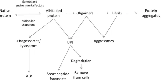
Fig. 3- Abnormal protein model of protein misfolding and fibrillation leading to the deposition of aggregated proteins via misfolded proteins, formation of
aggresomes or to degradation via autophagy–lysosome pathway (ALP) or
ubiquitin–proteasome system (UPS) (adapted from Jellinger 14).
Progressive intracellular protein accumulation can result from various
pathological processes: abnormal synthesis and folding, abnormal
interaction with other proteins, overproduction of protein constituents,
impaired degradation and turnover, altered post-translational
modifications of newly synthesized proteins, abnormal proteolytic
cleavage, improper expression or altered gene splicing, insufficient
molecular chaperone activity, impaired intracellular transport of proteins
and protein folding/misfolding modified on surfaces such as lipid
membranes 87.
Aberrant proteins often cannot fold correctly and will be trapped in
misfolded conformations. To get rid of the misfolded proteins, the living
Native protein
Misfolded
protein Oligomers Fibrils
Protein aggregates
Phagossomes/ lysosomes
ALP
UPS
Short peptide fragments
Degradation
Remove from cells
Genetic and environmental factors
Molecular chaperons
10
cell contains a large number of intracellular proteases, which, together
with the chaperones, comprise the cellular protein quality-control systems
in the endoplasmic reticulum (ER) 88. The two principal routes of
intracellular protein catabolism are the UPS and the ALP (autophagy). Both
were implicated to play important roles in the pathogenesis of NDDs 89,
but also to collaborate in neuroprotection 90. When the capacity of UPS to
degrade misfolded proteins is overwhelmed, aggregation occurs and
proteins are moved to ubiquitin-rich structures termed aggresome 91. The
formation of these structures has been associated with activation of
caspases and apoptosis 92. However, is not clear if the formation of
aggresomes is causative or protective, although data suggest that they
facilitate the degradation of toxic proteins 93. Not all aberrant proteins
can be eliminated and the misfolded protein may accumulate and form
toxic oligomeric and/or aggregated inclusions. By this way, the loss of
protein function may be accompanied by a gain of pathogenic function,
culminating in death of affected cells 94,95.
1.3.3. Molecular chaperones defective function
Molecular chaperones have essential roles in many cellular processes,
including protein folding, targeting, transport, degradation, and signal
transduction. Chaperones have been found to be effective in preventing
misfolding of different disease-causing proteins, reducing the severity of
several NDDs and many other protein-misfolding diseases. They are
ubiquitous in cells and are stress-induced or administered
pharmacologically. Mutations on chaperones, such as heat shock proteins
(HSP) have been increasingly associated to diseases 96. It is likely that
molecular chaperones facilitate neuroprotection by functioning at various
levels. They could prevent protein aggregation, interfere with oxidative
stress (eg. HSP27 through GSH uphold) and block apoptotic signaling
pathways, thus promoting survival. Thus, molecular chaperones may
11
1.3.4. Fragmentation of neuronal GA
Fragmentation of the neuronal GA was reported in ALS, AD, PD,
Creutzfeldt–Jakob disease and in spinocerebellar ataxia type 2 97. Since the
GA is involved in numerous important functions, such as the transport,
processing, and targeting of proteins synthesized in the ER, fragmentation
of Golgi might have detrimental effects and lead to dysfunction of the
cytoplasmic machinery in neurons. In ALS cell model expressing mutant
SOD1, fragmentation of the GA is associated with dysfunction of the
secretory pathway 98. Several types of neuronal insult induce Golgi
fragmentation, including excitotoxicity (nerve cells are damaged and
killed by excessive stimulation by neurotransmitters), ROS, reactive
nitrogen species (RNS) and ER stresses. GA has been found as a sensor for
controlling entry into apoptosis 99. Evidences from Nakagomi et al. work
have suggested that GA fragmentation/dispersal is a process downstream
mitochondria and ER cell death pathway in response to apoptotic effectors
99. Moreover, this process seems to be an early event in neuronal cell
death, initiated prior to tubulin degeneration, which is observed in the
middle stages of apoptosis and not a consequence of cytoskeletal
degradation during cell death. Finally, it has been suggested that GA
fragmentation aberrant entry into the cell cycle can trigger neuronal
apoptosis. Since mature neurons cannot undergo mitosis, the initiation of
cell cycle events, such as GA fragmentation, may contribute to an
apoptotic pathway 99,100.
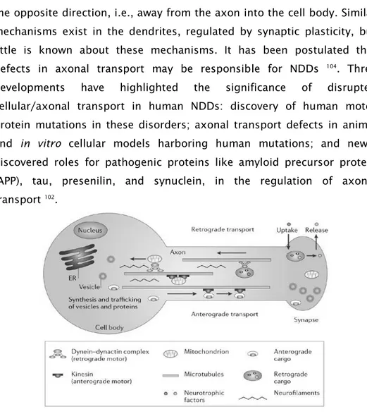
1.3.5. Disruption of cellular/axonal transport
Defective neuronal and axonal transport also plays a mechanistic role in
several NDDs 101-103. Most of the transport uses the microtubule system,
which is proposed to form a network of trafficking highways, and also
active proteins (Fig. 4). All axonal components are synthesized in the cell
body and transported from there into the axonal processes. Axonal
transport is essential for the movement of vital proteins, vesicles,
organelles, signaling molecules, and other materials to the axon, and
12
the opposite direction, i.e., away from the axon into the cell body. Similar
mechanisms exist in the dendrites, regulated by synaptic plasticity, but
little is known about these mechanisms. It has been postulated that
defects in axonal transport may be responsible for NDDs 104. Three
developments have highlighted the significance of disrupted
cellular/axonal transport in human NDDs: discovery of human motor
protein mutations in these disorders; axonal transport defects in animal
and in vitro cellular models harboring human mutations; and newly
discovered roles for pathogenic proteins like amyloid precursor protein
(APP), tau, presenilin, and synuclein, in the regulation of axonal
transport 102.
Fig. 4- Axonal transport. The motors for anterograde and retrograde fast axonal transport are the kinesins and dynactin complex proteins, respectively; microtubules provide the tracks for these motors. Vesicles for transport are sorted and loaded onto transport motors both in the cell body and the distal nerve terminal. The former are transported not only into the axon but also into dendrites. Those in the distal nerve terminal permit uptake and axosomatic movement of substances such as trophic proteins. Figure from Pasinelli et al. 105
1.3.6. Ion channels changes
Ion channels have a critical role in maintaining proper CNS function, and
13
Ca2+ homeostasis can promote alterations of Ca2+ buffering capacities,
deregulation of Ca2+ channel activities, or excitotoxicity and were
observed in several neurodegenerative disorders including AD, PD, HD
and ALS 107-110. In AD, accumulation of A peptides in neurons has been
shown to activate ion channels, causing an influx of Ca2+ that disrupts
homeostasis, leading to mitochondria dysfunction, oxidative stress and
apoptosis of neurons 111. Moreover, reduction of K+ in cells can cause
neuronal apoptosis 112. Voltage-gated K+ (KV) channels are responsible for
the electrical activity in neurons. The drop in cytosolic K+ relieves
inhibition of an array of pro-apoptotic enzymes such as caspases and
nucleases 113,114. Blocking KV channels has been known to prevent neuronal
apoptosis by preventing K+ efflux.
1.3.7. Neuroinflammatory process
Chronic inflammatory reactions and signs of immune activation in the CNS,
involving major histocompatibility complex class II expression, glial
reaction, T-cell infiltration and blood–brain-barrier (BBB) dysfunction are
well-known features in the pathogenesis and progression of NDDs 115-120.
Microglial cells are the primary immune cells in the CNS. Their primary
functions are to promote defense by destroying pathogens, removing
deleterious debris, promoting tissue repair and facilitating tissue
homeostasis, through their influence on surrounding astrocytes and
neurons 121. In most cases, neuroinflammatory process promoted by
microglial activation stops once homeostasis has been restored. However,
continued, uncontrolled activation of microglia can lead to an excess
production of various factors that contribute to neuronal injury 122-124 . The
insufficient clearance by microglia is also present in several NDDs and in
14
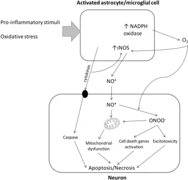
Fig. 5- Neuroinflammatory process. Microglia activation induce increased expression of inducible nitric oxide synthase (iNOS) 126 that promotes excessive
nitric oxide production that can lead to protein S-nitrosylation and nitration 127 and
disruption of neuronal mitochondrial electron transport chain function 128. The
activation of NADPH oxidase 129 also promoted by microglia activation mediates
superoxide production and the release of pro-inflammatory molecules 130.
Superoxide itself can increased iNOS expression 131 and react with nitric oxide
generating peroxynitrite 132. This highly reactive oxygen and nitrogen species
(RONS) can inhibit mitochondrial respiration, induce caspase-dependent neuronal apoptosis, and to induce glutamate release resulting in excitotoxicity and neuronal death 132,133. Additionally, longer lived cytokines are produced which
enhance the expression of iNOS, increasing nitric oxide production and stimulating the release of additional cytokines that activate neuronal death signaling cascades. 134,135. Glial cytokine may also bind to specific cell surface
receptors in neurons that activate pro-apoptotic pathways 136,137.
1.3.8. Dysfunctions of neurotrophins (NTFs)
Reduced neurotrophic support is a significant factor in the pathogenesis
of NDDs 138,139. NTFs regulate development and maintenance of the CNS.
They affect neuronal survival, influence synaptic function and plasticity
and are central to many aspects of the CNS. Since NTFs in neurons are
subject to transport from and to targeting neurons, their effects may be
related to synthesis or to changes in axonal transport. NTFs activate
signaling pathways through binding of tropomyosin-receptor-kinase (Trk)
Activated astrocyte/microglial cell
↑ NADPH
oxidase
↑iNOS
Neuron
NO•
NO•
ONOO
-O2
-Cell death genes activation Mitochondrial
dysfunction Caspase
Excitotoxicity
Apoptosis/Necrosis Pro-inflammatory stimuli
15
receptors, receptors dimerization and subsequent phosphorylation of the
intracellular kinase domain 139. Neurotrophic support is inversely
correlated with oxidative stress 140. In NDDs, such as AD, PD and HD,
oxidative stress appears linked to the loss of neurotrophic support,
causing down-regulation of NTFs which, in turn, can up-regulate
antioxidant enzymes and promote the expression of antioxidant proteins.
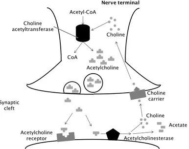
1.3.9. Choline deficit
Acetylcholine (ACh) is a neurotransmitter widely distributed in the nervous
system and has been implicated in cerebral cortical development, cortical
activity, controlling of cerebral blood flow, sleep–wake cycle as well as in
modulating cognitive performances and learning and memory
processes 141. It plays an essential role in structural and functional
remodelling of cortical circuits by establishing synaptic contacts in
networks of cells that will support complex cognitive functions in
adulthood 142. However, in NDDs it has been registered a cholinergic
deficit 143. To the cholinergic dysfunction may contribute imbalances in the
expression of neurotrophins, their precursors or in neurotrophin
receptors, changes in ACh release, high-affinity choline uptake, as well as
alterations in muscarinic and nicotinic ACh receptor expression. As a
consequence of cholinergic hypofunction, cell atrophy may be mediated
by decrements in gene expression, impairments in intracellular signaling
and cytoskeletal transport that lead to functional decline in the brain.
Malfunction of the cholinergic system may be tackled pharmacologically
by intervening in cholinergic as well as neurotrophic signaling cascades
that have been shown to ameliorate the cholinergic deficit at early stages
of the disease, and slow-down its progression. Acetylcholinesterase
(AChE), catalyzes the hydrolysis of the neurotransmitter ACh to choline
(Fig. 6). This process is necessary to return an activated cholinergic
neuron to a resting state. AChE inhibition has been reported to ameliorate
16
Fig. 6- Acetylcoline cycle. Cholinergic nerve transmission is terminated by the enzyme AChE, that is found both on the post-synaptic membrane of cholinergic synapses and in other tissues. ACh binds to AChE and is hydrolysed to acetate and choline. This inactivates the ACh and the nerve impulse is halted. AChE inhibitors prevent the hydrolysis of ACh, increasing its concentration of in the synaptic cleft.
1.3.10.Neuronal death
Neuronal death is the most critical hallmark of many NDDs 14. The nature,
time course, molecular causes of cell death in NDDs and their relations to
basic processes are still a matter of discussion. Based on distinct
morphologic criteria and biochemical features, three major mechanisms
of neuronal failure could be found: apoptosis, a specific form of
gene-directed programmed cell death (PCD); necrosis, a passive killing of the
cell; autophagic degeneration 147,148.
Morphologically, apoptotic cell death is characterized by chromatin
condensation (pyknosis), nuclear fragmentation, cell shrinkage, and
plasma membrane blabbing. Eventually, the cell breaks into small
membrane surrounded fragments (apoptotic bodies), which are cleared by
phagocytosis without inciting an inflammatory response 149
. Apoptosis can
occur locally, without damaging healthy adjacent cells 150.
In contrast, necrotic cell death, an accidental and uncontrolled mode of
cell death, exhibits rapid cell swelling and subsequent rupture of the
Post-synaptic membrane
Synaptic cleft
Nerve terminal
Choline carrier Choline
Choline acetyltransferase
Acetyl-CoA
Acetylcholine CoA
Acetylcholine
receptor Acetylcholinesterase
17
plasma membrane that due to an inflammatory response, usually induces
substantial secondary cell damage in the surrounding tissue 151.
Cell death with autophagy is a normal physiological process active in both
homeostasis and atrophy, probably representing a failure of
neuroprotective mechanisms 152. It is characterized by the formation of
numerous autophagic vacuoles, endocytosis, enlargement of the GA for
the vacuolization of the ER and moderate condensation of nuclear
chromatin that may ultimately leave the pyknotic nuclei and to be
destroyed by autophagosomes 153.
Increasing evidences suggests that regulation of neuronal cell death is
complex, utilizing multiple pathways that are dependent on the damaging
insult 154.
2. Polyphenols
2.1.General aspects
2.1.1. Occurrence and function
Polyphenols (PPs) are among the most widespread class of metabolites in
nature, and their distribution is almost ubiquitous. It is estimated that
exist 100,000 to 200,000 secondary metabolites in plants 155 and 20 % of
the carbon fixed by photosynthesis is channeled into the phenylpropanoid
pathway, thus generating the majority of the natural-occurring
phenolics 156. PPs are plant secondary metabolites, i.e. substances that in
plants have little or no role in primary metabolism: photosynthesis,
respiration, growth and development, but which may accumulate in
surprisingly high concentrations 157.
PPs appear to have many diverse functions in plants, e.g. colour of leaves,
flowers and fruit, anti-microbial function, anti-fungal function, insect
feeding deterrence, screening from damage by solar UV radiation,
chelation of toxic heavy metals and antioxidant protection from free
radicals generated during the photosynthetic process 158.
One of the most obvious roles for colored phenolics is in making flowers
18
Rhizobium spp., are positively chemotactic towards plant phenolics 159,
which is advantageous to the plant in the development of a favourable
rhizosphere environment and in the establishment of symbiotic
relationships, such as with nitrogen-fixing bacteria. Phenolics also protect
plants against damage produced by UV light, by enhancing their
accumulation or shifting towards more hydroxylated PPs 160. Flavonoids, a
particular group of phenolic compounds, are major components of this
screen, that occur in highest levels in plant tissues exposed to strong
light, such as flowers and leaves. 161.
One of the most important roles of phenolics is the defense from
pathogens and herbivorous predators. This protective role results from
PPs activities such as antibacterial, antiviral, damage tissues seal and
insect feeding deterrents. Moreover, the higher-molecular-weight PPs (also
known as tannins) precipitate proteins in the mammalian digestive system
and impair digestion.
2.1.2. Chemistry
PPs are characterized structurally by the presence of one or more six-
-carbon aromatic rings and two or more phenolic (i.e. linked directly to the
aromatic ring) hydroxyl groups. Some compounds are usually integrated
in the group of PPs as ”honorary” PPs, however not sharing the
characteristic chemical properties, such as phenolic acids, stilbenes, etc.
Phenolic compounds display diverse chemical aspects relevant to their
biological actions, such as: (a) interaction of the hydroxyl groups with the
p-electrons of the benzene ring make them able to scavenging reactive
radicals and generate more stable radicals; (b) chelation of pro-oxidants
metal ions, that requires vicinal hydroxyl groups; (c) readily ionized,
acting as weak acids and thus influencing their chemical reactivity;
(d) good hydrogen donors, forming complexes with other molecules
extremely stable and with tendency to precipitate; (e) cis-trans isomerism.
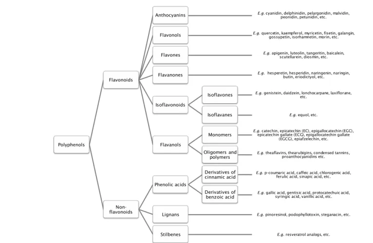
PPs are a diverse class of plant secondary metabolites (Fig. 7). They
19
molecules such as catechol, through to complicated macromolecular
polymers 162.
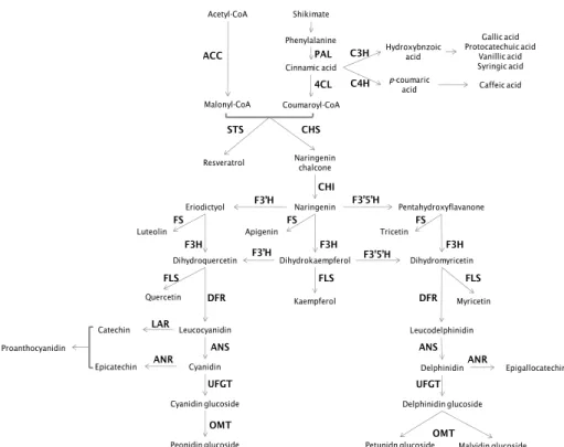
2.1.2.1. Biosynthesis
Although a large variety of plant phenols exists, PPs are mostly
synthesized from phenylalanine, from shikimic acid pathway. Biosynthesis
in then conducted via phenylpropanoids pathway, producing the large
variety of plant phenols (Fig. 8).
The flux into phenolic biosynthesis in plants is known to be highly
sensitive to environmental conditions and also biotic and abiotic
stresses 158. Detailed examination by molecular biological approaches has
indicated that the phenomenon is largely due to enhanced transcription of
the phenolic biosynthetic genes following exposure to the inducing
stimulus 163. Genes for the initial enzymes in the phenylpropanoid pathway
appear to be induced very early on, typically in a co-ordinated, “block-wise”
manner 158. Mineral nutrition, such as a limited nitrogen supply and boron
deficiency, lower temperatures, water availability and light are
20
Fig. 7- Polyphenols classification. Schematic representation of polyphenols groups, including main examples of molecules.
Polyphenols
Flavonoids
Anthocyanins E.g. cyanidin, delphinidin, pelargonidin, malvidin, peonidin, petunidin, etc.
Flavonols E.g. quercetin, kaempferol, myricetin, fisetin, galangin, gossypetin, isorhamnetin, morin, etc.
Flavones E.g. apigenin, luteolin, tangeritin, baicalein, scutellarein, diosmin, etc.
Flavanones E.g. hesperetin, hesperidin, naringenin, naringin, butin, eriodictyol, etc.
Isoflavonoids
Isoflavones E.g. genistein, daidzein, lonchocarpane, laxiflorane, etc.
Isoflavanes E.g. equol, etc.
Flavanols
Monomers E.g. epicatechin gallate (ECG), epigallocatechin gallatecatechin, epicatechin (EC), epigallocatechin (EGC),
(EGCG), epiafzelechin, etc.
Oligomers and
polymers E.g. theaflavins, thearubigins, condensed tannins, proanthocyanidins etc.
Non-flavonoids
Phenolic acids
Derivatives of
cinnamic acid E.g. p-coumaric acid, caffeic acid, chlorogenic acid, ferulic acid, sinapic acid, etc.
Derivatives of
benzoic acid E.g. gallic acid, gentisic acid, protocatechuic acid, syringic acid, vanillic acid, etc.
Lignans E.g. pinoresinol, podophyllotoxin, steganacin, etc.
21
Fig. 8- Biosynthetic pathway of phenylpropanoids. Adapted from Berli et al. 165.
Legend: 4CL- 4-coumarate:CoA ligase (EC 6.2.1.12); ACC- acetyl-CoA carboxylase (EC 6.4.1.2); ANR- anthocyanidin reductase (EC 1.3.1.77); ANS- anthocyanidin synthase (EC 1.14.11.19 );C3H- p-coumarate 3 hydroxylase (EC 1.14._._); C4H- cinnamate 4-hydroxylase (EC 1.14.13.11); CHI- chalcone isomerase (EC 5.5.1.6); CHS- chalcone synthase (EC 2.3.1.74); DFR- dihydroflavonol reductase (EC 1.1.1.219); F3H- flavanone 3-hydroxylase (EC 1.14.11.9); F3′H- flavonoid 3′-hydroxylase (EC 1.14.13.21); F3′5′H- flavonoid 3′5′-hydroxylase (EC 1.14.13.88); FLS- flavonol synthase (EC 1.14.11.23); FS- flavone synthase (EC 1.14.11.22); LAR- leucoanthocyanidin reductase (EC 1.17.1.3); OMT- o-methyltransferase (EC 2.1.1._); PAL- phenylalanine ammonia-lyase (EC 4.3.1.5); STS- stilbene synthase (EC 2.3.1.95); UFGT- UDP glucose-flavonoid 3-glucosyltransferase (EC 2.4.1.115 ). The pathway starts with PAL that catalyzes the conversion of phenylalanine to cinnamate. The C4H catalyzes the synthesis of p-hydroxycinnamate from cinnamate and 4CL converts p-coumarate to its coenzyme-A ester, activating it for reaction with malonyl CoA. The flavonoid biosynthetic pathway starts with the condensation of one molecule of 4-coumaroyl-CoA and three molecules of malonyl-CoA, yielding naringenin chalcone. This reaction is carried out by the CHS. Chalcone is isomerised to a flavanone by the CHI. From these central intermediates, the pathway diverges into several side branches, each resulting in a different class of flavonoids. F3H catalyzes the stereospecific 3ß-hydroxylation of (2S)-flavanones to dihydroflavonols. For the biosynthesis of anthocyanins, DFR catalyzes the reduction of dihydroflavonols to flavan-3,4-diols (leucoanthocyanins), which are converted to anthocyanidins by ANS. Leucoanthocyanins can also be catalyzed by LAR to flavan-3-ol. ANR catalyzes the reduction of anthocyanidins to epi-flavan-3-ol. The formation of glucosides is catalyzed by UFGT, which stabilize the anthocyanidins by 3-O-glucosylation.
Shikimate Phenylalanine Cinnamic acid Hydroxybnzoic acid p-coumaric acid Acetyl-CoA Malonyl-CoA Coumaroyl-CoA Naringenin chalcone Naringenin Eriodictyol Pentahydroxyflavanone
Quercetin Kaempferol Myricetin
Leucodelphinidin Leucocyanidin Cyanidin Cyanidin glucoside Peonidin glucoside Catechin Epicatechin Delphinidin Delphinidin glucoside
Petunidn glucoside Malvidin glucoside Epigallocatechin Gallic acid Protocatechuic acid Vanillic acid Syringic acid Caffeic acid Resveratrol PAL CHS STS CHI F3H
Dihydroquercetin Dihydrokaempferol Dihydromyricetin
F3H F3H F3’H F3’H F3’5’H F3’5’H FLS FLS FLS DFR DFR UFGT UFGT LAR ANR ANR OMT OMT ANS ANS ACC C3H C4H 4CL Proanthocyanidin
Luteolin Apigenin Tricetin
22
2.1.Neuroprotective potential
It has been suggested that phenolics are among the most active
substances from natural sources, displaying a variety of health-promoting
properties 166,167. PPs and PP-rich extracts have been implicated as
beneficial agents in a multitude of disease states 168,169, most commonly
cancer, cardiovascular disease, and neurodegenerative disorders.
It is now believed that the activity of these low-molecular-weight, non
nutrient components and their metabolites on neurological processes can
be defined through a number of distinct biological processes: (a) neuronal
signaling pathways, leading to inhibition of apoptosis, neuronal survival
and differentiation 170,171; (b) inhibition of neuropathological processes in
specific brain regions 172,173; (c) changes in brain blood flow 174-176.
2.1.1. Interactions with neuronal signaling pathways
There is extensive evidence indicating that some native flavonoids 171,177,
their small intestinal metabolites 178,179 and also large intestinal
derivatives 180-182 are capable of exerting beneficial effects on neurological
processes and that this is linked to their interactions with neuronal
signaling pathways. Receptor binding by flavonoids and their metabolites
may underlie changes in the activation status of various downstream
kinases, including various members of both the mitogen-activated protein
kinase (MAPK) and the phosphoinositide 3-kinase (PI3K)
pathways 170,171,183,184 and the nuclear factor-B (Nf-B) pathway 185. Various
flavonoid-binding sites on neurons have now been described, whose
include adenosine 186, gamma-aminobutyric acid (GABA
A)
187,188, delta-
-opioid 189,190, nicotinic 191,192, TrkB 193, estrogen 194, and testosterone
receptors 195. Also a specific brain plasma membrane binding site for PPs
has been proposed 196.
Neuronal survival and differentiation
Modulation of protein kinases and lipid kinases signaling cascades by PPs
allows changes in caspase activity and/or alterations on gene expression,
23
Signaling pathways changes promoted by PPs are highly likely to underpin
enhancements in spatial memory through the facilitation of changes in
synaptic strength and the induction of morphological changes 197, in
particular changes in neuronal spine density and morphology that are
considered vital for learning and memory 198. Compounds or drugs that
are capable of enhancing the activity of cAMP response element binding
(CREB) may be capable of facilitating memory consolidation by promoting
gene expression related with synaptic morphology and associated
changes that are critical for long-term memory 199,200. Moreover, molecules
that are capable of entering the brain and activate upstream regulators of
CREB (e.g., extracellular-signal-regulated kinase (ERK) and protein kinase B
(Akt)) may also be considered excellent candidates for memory-enhancing
drugs. Flavonoids, due to their ability to localize in the brain 201 and their
capacity to activate ERK– CREB and Akt–CREB circuitry 170,202 may be
regarded as promising candidates for the enhancement of memory and
aid patients suffering from AD, PD, stroke, or even normal age-associated
memory deficits.
2.1.2. Inhibition of neuropathological processes
Flavonoids are also able to counteract the neuronal injury underlying
these disorders and thus slowing the disease progression 203,204. Recent
evidences suggests that nonsteroidal anti-inflammatory drugs are
effective at delaying the onset of neurodegenerative disorders, particularly
PD 205. As such, there has been an interest in the development of new
compounds with an ability to counteract neuroinflammatory injury to the
brain. Likewise, flavonoids have been shown to be effective at blocking
oxidant-induced neuronal injury 206, although not via direct radical or
oxidant-scavenging activity 170,178,179,207-209.
‘Direct’ versus‘indirect’ antioxidant activity
Until recently the conventional wisdom was that the direct antioxidant
activity of phytochemicals was the responsible to confer their health
benefits 210. Phytochemicals have been considered able to modulate