* Corresponding author. Mailing address: Departamento de Microbiologia Geral, Instituto de Microbiologia Prof. Paulo de Goés, UFRJ, CCS, Bl. I, CEP 21949-900, Rio de Janeiro, RJ, Brasil. Fax: (+5521) 5608344. E-mail: [email protected]
IDENTIFICATION AND PROPERTIES OF TWO EXTRACELLULAR PROTEASES FROM BREVUNDIMONASDIMINUTA
André Adriano Chaia1; Salvatore Giovanni-De-Simone2,3; Simone Dias Gonçalves Petinate1; Ana Paula Cabral de
Araújo Lima4; Marta Helena Branquinha1; Alane Beatriz Vermelho1*
1Departamento de Microbiologia Geral, Instituto de Microbiologia Professor Paulo de Góes, Universidade Federal do Rio de
Janeiro, Rio de Janeiro, RJ, Brasil. 2Departamento de Bioquímica e Biologia Molecular, FIOCRUZ, Rio de Janeiro, RJ, Brasil. 3Departamento de Biologia Celular e Molecular, Instituto de Biologia, Universidade Federal Fluminense, Rio de Janeiro, RJ,
Brasil. 4Instituto de Biofísica Carlos Chagas Filho, Universidade Federal do Rio de Janeiro, Rio de Janeiro, RJ, Brasil
Submited: March 14, 1998; Returned to authors for corrections: March 25, 1999; Approved: January 16, 2000
ABSTRACT
Extracellular proteases from Brevundimonas diminuta (syn. Pseudomonas diminuta) were studied in sodium dodecyl sulfate polyacrylamide gel electrophoresis (SDS-PAGE) containing a copolymerized substrate. Two proteases were detected migrating at 67 kDa and 50 kDa: both of them hydrolysed preferentially gelatin, but casein was also degraded and a slight hydrolysis was observed with hemoglobin. No detectable extracellular proteolytic activity was found in bovine serum albumin-containing gels. The optima temperature and pH for proteolytic activity were between 40ºC and 50ºC in a pH ranging from 7.0 to 11.0, respectively. These enzymes were isolated by analytical high performance liquid chromatography (HPLC). Protease assays with the synthetic substrate Z-Phe-Arg-MCA and the inhibitors EGTA, EDTA and 1, 10 phenanthroline point out that these enzymes are metalloproteases.
Key words : metalloproteases, Brevundimonas diminuta, Pseudomonadaceae, extracellular proteases
INTRODUCTION
Brevundimonas diminuta is the new nomenclature for former Pseudomonas diminuta based on a new genus name, proposed for the species of Pseudomonadaceae family rRNA group IV, often referred to as the diminuta group. Members of the genus Brevundimonas are found in water and differ from the authentic pseudomonads by having short wavelenght polar flagella, restricted biochemical activity, different polyamine and ubiquinone patterns as well as different fatty acid composition (15).
B. diminuta has been a target of several biochemical studies due to a zinc-dependent phosphotriesterase which catalyses the hydrolysis of several toxic organophosphates triesters (5). The catalytic role of zinc is similar to the mechanisms proposed for many metalloenzymes including proteases such as
carboxypeptidases and thermolysin (6). This group of bacterium is also able to degrade aerobically isoquinoline, a toxic compound used in pesticides, medicaments, antioxidants and corrosion inhibitors (14). As in the family Pseudomonadaceae several species produce metallo and serine proteases (2, 9, 13, 16) and because there is no information about the type and function of these enzymes in B. diminuta, it was decided to study the occurrence and type of secreted proteases from this microrganism.
M ATERIALS AND METHODS
Preparation of cellular extracts and extracellular concentrates. 1.2 x 10⁹ cells were processed as previously
described (4). For protease secretion studies, 30 ml daily aliquots of culture medium were collected during 10 days and concentrated by dialysis against polyethyleneglycol overnight at 4ºC. For substrate-containing SDS-PAGE, the extracellular concentrate was mixed with sample buffer (0.125 mM Tris-HCl, pH 6.8, 4% SDS, 20% glycerol and 2% bromophenol blue) in a proportion of 7:3 (v/v).
Substrate-containing SDS-PAGE. Proteases in cellular and extracellular extracts (100 µg of protein, according to Lowry et al. (11) method) were assayed and characterized by electrophoresis in 7.5% SDS-PAGE (10) with 0.1% copolymerized substrate (7). Gelatin, casein, hemoglobin and bovine serum albumin (BSA) were used as substrates. Following electrophoresis, gels were incubated for 24 h at 37ºC under two different conditions: 50 mM phosphate buffer at pH 5.5, or 100 mM glycine-NaOH buffer at pH 10.0. To detect the presence of proteases, gels were stained with 0.1% amido black in methanol-acetic acid-water (3:1:6) and destained with water until the appearance of clear zones.
Effect of temperature and pH on proteolytic activity. Substrate hydrolysis on SDS-PAGE-gelatin was assayed at different temperatures (28ºC, 40ºC, 50ºC, 60ºC and 70ºC) for 24 h at pH 10.0. The effect of pH was determined with the following buffers: 200 mM acetate (pH 4.0 and 5.0); 200 mM phosphate (pH 6.0 to 8.0); 200 mM carbonate-bicarbonate (pH 9.0 and 10.0); and 100 mM glycine-NaOH (pH 11.0 and 12.0). The assays for optimal pH were carried out at 37ºC for 24 h.
Gel-filtration high performance liquid chromatography (HPLC). The cell-free culture supernatant (1 L) was concentrated and injected in a Shinpack Diol-150 (50 cm x 7.9 cm, I.D.) HPLC column (Shimadzu, Kyoto, Japan) previously equilibrated in 50 mM phosphate buffer (pH 7.2). The proteins were fractionated on an automatic HPLC system (Shimadzu, 6A model) at a flow-rate of 1 ml/min, for 30 min at 25ºC. For molecular mass determination the column was calibrated with the same buffer with the following markers: β-galactosidase (Mr 105 kDa), bovine serum albumin (BSA, Mr 66 kDa), ovalbumin (Mr 45 kDa) and carbonic anhydrase (Mr 29 kDa). Protein determinations were made by the method of Lowry et al. (11), with bovine serum albumin as a standard.
Detection of proteolytic activity using fluorogenic substrate. Proteolytic activity was determined by measuring the rate of hydrolysis in fluorimetric continuous assays. The release of product was observed in 100 mM glycine-NaOH buffer, pH 10.0, containing 0.12 µg of purified proteases and 10 µM Z-Phe-Arg-MCA (Sigma Chem. Co., St. Louis, MO)
as substrate, using a F-4500 fluorescence spectrophotometer (Hitachi, Japan). Table 1 shows the proteolytic inhibitors used.
RESULT S
B. diminuta proteases hydrolysed preferentially gelatin, however distinct profiles were obtained for cellular and extracellular proteases (Fig. 1). A 80 kDa band was only detected in cellular extracts, while 67 kDa and 50 kDa proteases were observed in both extracts. A 67 kDa protease was found in B. diminuta cells and was released to culture medium, being able to hydrolyse casein, gelatin and hemoglobin. No detectable extracellular proteolytic activity was found in BSA-containing gels. However, in cellular extracts, a slight hydrolysis was observed at pH 5.5, for the 80 kDa protease (Fig. 1). Extracellular proteases yield low proteolytic activity in hemoglobin-containing gels, and only at pH 5.5 (Fig. 1). This activity was more proeminent in cellular proteases.
Protease secretion studies showed that only the 67 kDa protease was released to culture medium during the first five days of culture (Fig. 2). After 6 days of cultivation, the 50 kDa enzyme was also detected (Fig. 2). No qualitative differences were observed until the tenth day.
Fig. 3 shows the extracellular proteases pH activity profile. The optimum pH for extracellular proteases activity was found in a 7.0 to 11.0 pH range. Low activity was found below pH 7.0. The effect of temperature on the activity of these enzymes was determined incubating the gels at various temperatures. The optimum temperature for extracellular proteolytic activity was between 40ºC and 50ºC (Fig. 4).
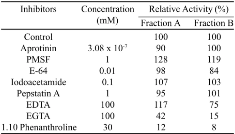
Table 1. Effect of protease inhibitors on Brevundimonas diminuta extracellular proteolytic activity.
Inhibitors Concentration Relative Activity (%) (mM) Fraction A Fraction B
Control 100 100
Aprotinin 3.08 x 10⁻⁷ 90 100
PMSF 1 128 119
E-64 0.01 98 84
Iodoacetamide 0.1 107 103
Pepstatin A 1 95 101
EDTA 100 117 75
EGTA 100 42 15
1.10 Phenanthroline 30 12 8
B (50-52 kDa) and Fraction C (below 30 kDa). This procedure resulted in partial purification of the enzyme activities in fractions A and B, as demonstrated by SDS-PAGE-gelatin. The fractions A, B and C obtained from 2.3 mg of protein yielded 0.29, 0.32 and 1.68 mg of proteins with a percentage recovery of 12.8, 14.2 and 73% (related to the HPLC column), respectively.
The inhibition profile of the extracellular proteases (obtained
in Fractions A and B, respectively) with various protease inhibitors is shown in Table 1. Inhibitors to cysteine-proteases (E-64, iodoacetamide), serine proteases (PMSF, aprotinin) and aspartic proteases (pepstatin A) failed to inhibit the enzymes. The proteases were inhibited by typical metal chelators such as EGTA and 1,10-phenanthroline, which indicates that both enzymes are metalloproteases.
Figure 2. Secretion of proteases during Brevundimonas diminuta growth. Aliquots of the culture medium were collected from the first (1) to the tenth (10) day of culture and analysed on SDS-PAGE-gelatin.
Figure 3. Effect of pH on Brevundimonas diminuta extracellular proteolytic activities. Extracts were analysed on SDS-PAGE-gelatin in a 4.0 to 12.0 pH range.
DISCUSSION
While there have been several metallo and serine-proteases reported in the literature in the Pseudomonadaceae family, the detection of proteases in B. diminuta has never been described (2, 9, 13, 16).
The present study indicates that Brevundimonas diminuta, a member of the Pseudomonadaceae family, is able to release into the culture medium two metalloproteases with molecular masses of 50 kDa and 67 kDa. By means of a SDS-PAGE-gelatin analysis, cell-associated proteases of 50 kDa, 67 kDa and 80 kDa were detected. Our findings point to the possibility that the 50 kDa cellular and extracellular proteases are different enzymes. The cell-associated enzyme hydrolysed gelatin only, but the extracellular one also displayed proteolytic activity with casein and hemoglobin at pH 5.5 and 10.0. The 67 kDa protease showed the same pattern of degradation of substrates, being able to hydrolyse casein, gelatin and hemoglobin, and probably being released to culture medium. These extracellular enzymes typically have low substrate specificity and function in the degradation of exogenous proteins for assimilation into the catabolic pathways, differing from intracellular proteases that have high substrate specificities and are associated with metabolic regulation mechanisms such as protein turnover and modification of regulatory enzymes (3). In addition, extracellular metalloproteases have been shown to contribute to the bacterial pathogenesis of P. aeruginosa (8).
In the growth conditions employed in this study, the proteases were secreted following a 72 h interval. The production of these
enzymes is heterogenous in Pseudomonas spp. It has been described that distinct amounts of enzymes are detected in different strains of P. aeruginosa (12).
The effect of metal chelators on B. diminuta extracellular proteolytic activity suggests that these enzymes are calcium-activated zinc-metalloproteases. The loss of activity in the presence of EGTA, which has affinity for calcium, is probably due to the capture of Ca+2 ions that are critical to the tertiary
structure of the protein, resulting in an enzyme more susceptible to autoproteolysis and to difficulties in refolding. Previous reports showed that P. aeruginosa alkaline protease contains one Zn+2 atom in the catalytic center and requires several Ca+2
ions for stabilization of its folding (13).
Studies with B. diminuta are interesting since this microorganism was already isolated from clinical specimens, sewage and soil (1). As far as we can ascertain from the literature, this is the first example of proteolytic activity described in B. diminuta. The extracellular metalloproteases described in this work are probably involved in the nutrition and/or in the decomposition of organic matter in soil.
ACKNOWLEDGMENTS
This work was supported by CNPq, CEPG/UFRJ, FINEP-BID, FAPERJ and FUJB.
R E S U M O
Identificação e propriedades de duas proteases extracelulares de Brevundimonas diminuta
Proteases extracelulares de Brevundimonas diminuta (Pseudomonas diminuta) foram identificadas e caracterizadas por eletroforese em gel de poliacrilamida com dodecilsulfato de sódio, contendo um substrato co-polimerizado. Duas proteases foram detectadas migrando em 67 kDa e 50 kDa: ambas hidrolisaram preferencialmente a gelatina, embora a caseína também tenha sido degradada e uma pequena hidrólise tenha sido observada com hemoglobina. Nenhuma atividade proteolítica extracelular foi detectada nos géis contendo soro albumina bovina. Condições ótimas de temperatura e pH para a atividade proteolítica foram observadas entre 40ºC e 50ºC e numa faixa de pH que variou de 7,0 a 11,0, respectivamente. Essas enzimas foram isoladas por cromatografia líquida de alta resolução. Os ensaios enzimáticos com o substrato sintético Z-Phe-Arg-MCA e com os inibidores EGTA, EDTA e 1, 10 fenantrolina indicam que essas enzimas são metaloproteases.
Palavras-chave: metaloproteases, Brevundimonas diminuta, Pseudomonadaceae, proteases extracelulares
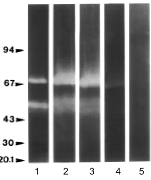
Figure 4. Effect of temperature on Brevundimonas diminuta extracellular proteolytic activities. Extracts were analysed on SDS-PAGE-gelatin at 28ºC (1), 40ºC (2), 50ºC (3), 60ºC (4) and 70ºC (5).
REFERENCES
1. Ballard, R.W.; Doudoroff. M.; Stanier, R.; Mandel, M. Taxonomy of the aerobic Pseudomonads: Pseudomonas diminuta and P. vesiculare.J. Gen. Microbiol., 53:349-361, 1968.
2. Boethling, R.S. Purification and properties of a serine protease from
Pseudomonas maltophilia. J. Bacteriol., 121:933-941, 1975.
3. Bono, F.; Savi, P.; Tuong, A.; Maftouh, M.; Pereillo, J.M.; Capdevielle, J.; Guillemot, J.C.; Maffrand, J.P.; Herbert, J.M. Purification and characterization of a novel protease from culture filtrates of a Streptomyces
sp. FEMS Microbiol. Lett., 141:213-220, 1996.
4. Branquinha, M.H.; Vermelho, A.B.; Goldenberg, S.; Bonaldo, M.C. Ubiquity of cysteine- and metalloproteinase activities in a wide range of trypanosomatids. J. Euk. Microbiol., 43:131-135, 1996.
5. Dumas, D.P.; Caldwell, S.R.; Wild, J.R.; Raushel, F.M. Purification and properties of the phosphotriesterase from Pseudomonas diminuta. J. Biol. Chem., 264:19659-19665, 1989.
6. Dumas, D.P.; Raushel, F.M. Chemical and kinetic evidence for an essential histidine in the phosphotriesterase from Pseudomonas diminuta. J. Biol. Chem., 265:21498-21503, 1990.
7. Heussen, C.; Dowdle, E.B. Electrophoretic analysis of plasminogen activators in polyacrylamide gels containing sodium dodecyl sulfate and copolymerized substrates. Anal. Biochem., 102:196-202, 1980. 8. Holder, I.A.; Neely, N.A. Pseudomonas Elastase Acts as a Virulence Factor
in Burned Hosts by Hageman Factor-Dependent Activation of the Host Kinin Cascade. Infect. Immun., 57:3345-3348, 1989.
9. Kim, H.J.; Tamanoue, Y.; Jeohn, G.H.; Iwamatsu, A.; Yokota, A.; Kim, Y.T.; Takahadhi, T.; Takahashi, K. Purification and characterization of an extracellular metalloprotease from Pseudomonas fluorescens. J. Biol. Chem., 121:82-88, 1997.
10. Laemmli, U.K. Cleavage of structural proteins during the assembly of the head of bacteriophage T4. Nature, 227:680-685, 1970.
11. Lowry, O.H.; Rosebrough, N.J.; Farr, A.L.; Randall, R.J. Protein measurement with the Folin phenol reagent. J. Biol. Chem., 193:265-275, 1951.
12. Mizukane, R.; Hirakata, Y.; Kaku, M.; Ishii, Y.; Furuya, N.; Ishida, K.; Koga, H.; Kohno, S.; Yamaguchi, K. Comparative in vitroexoenzyme-suppressing activities of azithromycin and other macrolide antibiotics.
Antimicrob. Agents Chemother., 38:528-533, 1994.
13. Morihara, K.; Tsuzuki, H.; Oka, T. On the specificity of Pseudomonas aeruginosa alkaline proteinase with synthetic peptides. Biochim. Biophys. Acta, 309:414-429, 1973.
14. Röger, P.; Erben, A.; Lingens, F. Microbial metabolism of quinoline and related compounds. IV. Degradation of isoquinoline by Alcaligenes faecalis
Pa and Pseudomonas diminuta 7. Biol.Chem. Hoppe-Seyler, 371:511-513, 1990.
15. Segers, P.; Vancanneyt, M.; Pot, B.; Torck, U.; Hoste, B.; Dewettinck, D.; Falsen, E.; Kersters, K.; De Vos, P. Classification of Pseudomonas diminuta
Leifson and Hugh 1954 and Pseudomonas vesiculares Büsing, Döll, and Freytag 1953 in Brevundimonas diminuta comb. nov. and Brevundimonas vesiculares comb. nov., respectively. Int. J. Sys. Bacteriol., 44:499-510, 1994.