Pedro M.F. Sousa
Dissertation presented to obtain the Ph.D degree in Biochemistry
Instituto de Tecnologia Química e Biológica | Universidade Nova de Lisboa
Instituto de Investigação Científica Tropical
Aerobic Respiratory Chains
Escherichia coli
and
Bacillus subtilis
supramolecular assemblies
Oeiras,
Pedro M.F. Sousa
Dissertation presented to obtain the Ph.D degree in Biochemistry
Instituto de Tecnologia Química e Biológica | Universidade Nova de Lisboa
Instituto de Investigação Científica Tropical
Oeiras, December, 2013
Aerobic Respiratory Chains
Escherichia coli
and
Bacillus subtilis
supramolecular assemblies
Instituto de Tecnologia Química e Biológica (ITQB)
Av. da República
Estação Agronómica Nacional
2780-157 Oeiras
Portugal
Tel. (+351) 214 469 323
Instituto de Investigação Científica Tropical (IICT)
Rua da Junqueira Nº86, 1º
1300-344 Lisboa
Portugal
Tel. (+351) 213 616 340
reductionist “eyes
-
down” molecular perspective with a new and genuinely
holistic “eyes
-
up” view of the living world, one whose primary focus is on
evolution, emergence, and biology’s innate complexity
.
Carl R.Woese (1928
–
2012)
The work summarized in this thesis is the product of several collaborations established
during my research grant. It is with great appreciation that I would like to acknowledge the following groups and people:Ana Melo, my supervisor, for her endless support on my work. I’m truly honored of being her first PhD student and I have learned with Ana how to be constantly motivated, focused, and above all, how to critically analyze scientific results. I appreciate her confidence in my work, friendship and her ability to promote useful discussions.
Miguel Teixeira, my co-supervisor, for his support on my work, useful discussions, and for the opportunity conceded.
Lígia Saraiva and Célia Romão, for following the work developed these years and promoting fruitful discussions.
Marco Videira, Filipe Santos, Sara Silva and Levi Bolacha, for the outstanding contribution they have provided to the work reported in this thesis. Furthermore, I would like to acknowledge Sara for supporting me all the times I needed, with unconditional love and friendship.
For providing a friendly environment and a stimulating workplace, I would like to thank all my colleagues from IICT, namely, José Ramalho, Ana Ribeiro, António Leitão, Luís Goulão, Ana Fortunato, Paula Batista-Santos, Inês Graça, Elisabete Lopes, Paula Alves, Isabel Palos, Micaela Sousa and Tiago David.
II
João Carita from ITQB is acknowledged for the large scale growth of Escherichia coli wild-type and mutant strains.
Natalya Dudkina and Egbert Boekema from Groningen University, Netherlands, for valuable insights on the methodology used in this work regarding the electrophoretic analysis of supercomplexes.
Julia Steuber and Thomas Vorburger from Stuttgart University, for allowing me to work on their laboratory and for their useful discussions.
My family and friends, for providing me the knowledge and environment I needed to become a better person. I thank Luís Sousa, Maria Sousa, Ana Sousa, Sara Silva, Ricardo Alves, Mário Coelho and Bruno Pais.
III
Pedro M.F. Sousa, Sara T.N. Silva, Brian L. Hood, Nuno Charro, João N. Carita, Fátima Vaz, Deborah Penque, Thomas P. Conrads, Ana M.P. Melo (2011), “Supramolecular organizations in the aerobic respiratory chain of Escherichia coli”, Biochimie 93, 418-425.
Pedro M.F. Sousa, Marco A.M. Videira, Andreas Bohn, Brian L. Hood, Thomas P. Conrads, Luis F. Goulao, Ana M.P. Melo (2012), “The aerobic respiratory chain of Escherichia coli: from genes to supercomplexes”, Microbiology 158, 2408-2418.
Pedro M.F. Sousa, Marco A.M. Videira, Ana M.P. Melo (2013), “The formate:oxygen oxidoreductase supercomplex of Escherichia coli aerobic respiratory chain”, FEBS Lett. 587, 2559-2564.
Pedro M.F. Sousa, Marco A.M. Videira, Filipe A.S. Santos, Brian L. Hood, Thomas P. Conrads, Ana M.P. Melo (2013), “The bc:caa3 supercomplexes from
the gram positive bacterium Bacillus subtilis respiratory chain: a megacomplex organization?”, Arch. Biochem. Biophys. 537, 153-160.
Other publications not included in this thesis
Pedro M.F. Sousa, Marco A.M. Videira, Thomas Vorburger, Sara T.N. Silva, James W. Moir, Julia Steuber, Ana M.P. Melo (2012), “The novel NhaE-type Na+/H+ antiporter of the pathogenic bacterium
V
Aerobic respiratory chains are composed of a series of membrane complexes that catalyze the electron transfer from reducing substrates to oxygen. The energy released through this process is used to translocate protons across the membranes, thus generating a proton motive force that activates F1FO-ATP
synthase to synthesize ATP. The arrangement of these enzymes in the inner mitochondrial membrane is well characterized in mammalian mitochondria, where different sets of supramolecular assemblies, or supercomplexes, involving the majority of the respiratory complexes were described, reinforcing the idea of an operational solid state model wherein the oxidative phosphorylation processes are optimized.
A broader perspective of the ubiquity of such assemblies is obtained when analyzing the prokaryotic components that lead to oxygen utilization. The aerobic respiratory chain of these organisms is more flexible than the mitochondrial one, bearing alternative pathways in response to the metabolic needs of the cell, which reflect the ability of these organisms to cope with extreme environmental conditions.
VI
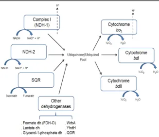
succinate:quinone oxidoreductase (SQR), cytochrome bo3 oxygen reductase and
type I and II cytochromes bd oxygen reductases, that accomplish the transfer of electrons from the reducing substrates NADH and succinate to O2, with proton
translocation through NDH-1 and cytochrome bo3 oxygen reductase. Other
enzymes such as lactate, formate (FDH-O) and glycerol-1-phosphate dehydrogenases may also participate in this process.
In this thesis, the supercomplexes of E. coli aerobic respiratory chain were reported for the first time. Prior to supercomplex detection, a thorough spectroscopic and kinetic characterization was performed in E. coli membranes, allowing the detection of the respiratory chain components operating in this Gram negative bacterium.
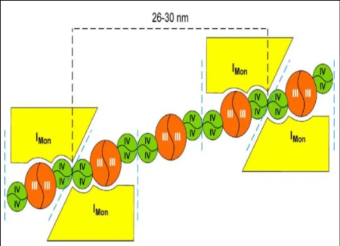
The analysis of supercomplex composition in this bacterium revealed at least three distinct supercomplexes, one resulting from the association of NADH:quinone oxidoreductases NDH-1 and NDH-2, one composed of FDH-O and cytochromes bo3 and bdI oxygen reductases, and another comprising SQR and
cytochrome bdII oxygen reductase. In addition, two homo-oligomers were described in this respiratory chain, namely the trimeric assembly of SQR and the dimeric form of cytochrome bo3 oxygen reductase.
VII
NADH: and formate:NBT oxidoreductase activities in E. coli wild type digitonin solubilized membranes, was nearly absent in mutant strains devoid of FDH-O or cytochromes bo3 or bdI oxygen reductases, although maintaining the same
intensity in other respiratory chain mutants. Its composition was further confirmed by the mass spectrometry identification of several peptides belonging to subunits of these complexes. Moreover, a molecular mass of 432 ± 7 kDa is in agreement with the sum of the individual complexes present in FdOx, which was shown to dissociate in a smaller subcomplex when harsher solubilization conditions were applied. FdOx was capable of cyanide sensitive quinol: and formate:oxygen oxidoreductase activities. The kinetic parameters of the latter activity were established, the KM for formate oxidation being 169 ± 21 µM and the
corresponding VMAX 117 ± 15 nmol O2.min-1.mg-1. As expected, this activity was
severely impaired in the above mentioned mutant strains, in agreement with the composition of FdOx.
In the scope of these results, an investigation of the prevalence of these assemblies during the bacterial growth was performed, complemented by a gene transcription analysis and enzyme activities of the respiratory chain components. The obtained results were globally analyzed and correlations between gene transcription, enzyme activities and bacterial growth were established, providing a multi-level perspective of the E. coli respiratory chain. Interestingly, both supercomplexes were detected throughout growth, namely from the mid-logarithmic to the late-stationary phase. The positive correlations observed between the transcription of NDH-1 and NDH-2 encoding genes were in agreement with the identified NADH:ubiquinone oxidoreductase supercomplex. A similar correlation was established between the transcription of cytochromes bo3
VIII
complexes in a supramolecular assembly. In fact, a polarographic analysis of wild type E. coli digitonin solubilized membranes resolved by sucrose gradient ultracentrifugation also suggested the presence of supercomplexes with cyanide sensitive succinate:oxygen oxidoreductase activities. The same activity was observed in a similar sucrose gradient analysis, although using E. coli solubilized membranes where cytochrome bdII was the sole oxygen reductase expressed, confirming the identification of a third supercomplex in the E. coli aerobic respiratory chain, composed of SQR and cytochrome bdII oxygen reductase. The B. subtilis aerobic respiratory chain comprises at least seven enzymes. NDH-2 and SQR respectively promote the electron transfer from the reducing substrates NADH and succinate to O2 via four different pathways. One involves a
cytochrome c pathway where bc complex and caa3 oxygen reductase operate,
while the other pathways consist on bd, aa3, or bb’ menaquinol:oxygen
oxidoreductases.
Similarly to what was described for the E. coli aerobic respiratory chain studies, a full kinetic and spectroscopic characterization was performed in B. subtilis membranes, prior to supercomplex detection, allowing the identification of the respiratory chain components operating in this Gram positive bacterium.
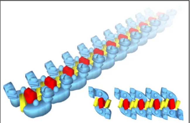
The B. subtilis respiratory chain was shown to contain at least four different supramolecular organizations, in addition to the dimeric assemblies of SQR and
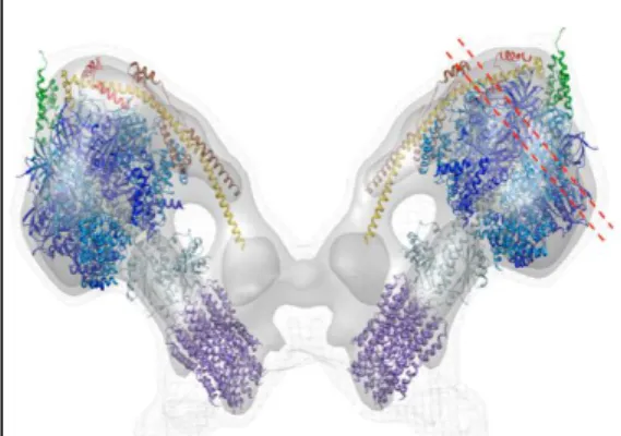
caa3 oxygen reductase. The bc complex and caa3 oxygen reductase were shown
to be the only components of three identified supercomplexes with different stoichiometries, namely (bc)4:(caa3)2, (bc)2:(caa3)4 and 2[(bc)2(caa3)4]. The fourth
supercomplex was shown to comprise a dimeric SQR and nitrate reductase (NAR).
In detail, (SQR)2:NAR supramolecular assembly was detected by the
IX
B. subtilis membranes devoid of SQR. Moreover, the dimeric form of SQR dissociated from the supercomplex upon solubilization with different detergents in a 2D-BN-PAGE analysis.
The same techniques were applied for the identification of the B. subtilis respiratory chain bc:caa3 supercomplexes. Cytochrome c oxidoreductase in gel
activity and heme staining produced three high molecular mass bands in wild type B. subtilis digitonin solubilized membranes resolved by BN-PAGE, which were absent from B. subtilis membranes devoid of bc complex or caa3 oxygen
reductase. In addition, the dissociation of these supercomplex-containing bands with harsher solubilization conditions allowed the resolution of several spots with similar or smaller molecular masses in a 2D-BN-PAGE analysis, which were assigned to bc:caa3 subcomplexes after identification by mass spectrometry
analysis. Guided by this and the molecular masses of bc and caa3 complexes,
each band was assigned to (bc)4:(caa3)2, (bc)2:(caa3)4 and 2[(bc)2(caa3)4]
supercomplexes. In agreement, bc:caa3 supercomplex bands were capable of
cyanide sensitive TMPD/ascorbate:oxygen oxidoreductase activity. The identification of bc:caa3 supercomplexes different stoichiometries allowed us to
XI
As cadeias respiratórias aeróbias são compostas por diferentes complexos membranares que catalisam a transferência de electrões da oxidação de substratos à redução do oxigénio. A energia transferida neste processo é aproveitada para promover a translocação de protões através das membranas, gerando uma força protomotriz que é utilizada pelo complexo F1FO-ATP sintase
na síntese de ATP. Embora o estudo das cadeias respiratórias esteja bem documentado, a organização nativa dos complexos enzimáticos que as constituem é ainda discutida. Diferentes estruturas supramoleculares, ou supercomplexos, envolvendo a maioria dos complexos respiratórios foram descritas em mitocôndrias de mamíferos, reforçando a ideia de que esta organização, suportada pelo modelo do “estado sólido” das cadeias respiratórias, seja fisiologicamente relevante e contribua para a optimização dos processos de fosforilação oxidativa.
A cadeia respiratória aeróbia de procariotas distingue-se da mitocondrial pela sua maior flexibilidade, albergando múltiplas vias alternativas compostas por diferentes complexos enzimáticos cuja expressão está dependente das necessidades metabólicas da célula, assegurando a proliferação destes organismos em condições ambientais extremas. Não obstante, diversos supercomplexos foram descritos nas cadeias respiratórias aeróbias de procariotas, evidenciando a ubiquidade deste tipo de organizações.
XII
massas moleculares superiores às dos componentes individuais da cadeia respiratória de cada organismo.
A cadeia respiratória de E. coli é composta por seis enzimas, nomeadamente NADH:quinona oxidoreductases do tipo I (NDH-1) e II (NDH-2), succinato:quinona oxidoreductase (SQR), citocromo bo3 reductase de oxigénio e citocromo bd
reductase de oxigénio do tipo I e II, que catalisam a transferência de electrões da oxidação dos substratos NADH e succinato à redução do oxigénio, com concomitante translocação protónica através dos complexos NDH-1 e citocromo
bo3 reductase de oxigénio. Outros enzimas como as desidrogenases do lactato,
formato (FDH-O) e glicerol-1-fosfato participam igualmente neste processo.
Nesta tese é descrita, pela primeira vez, a organização supramolecular da cadeia respiratória aeróbia de E. coli. Uma completa caracterização espectroscópica e cinética foi efectuada à cadeia respiratória deste organismo, em membranas obtidas no início da fase estacionária do crescimento, antes de se proceder à sua caracterização supramolecular permitindo a identificação dos complexos enzimáticos presentes na cadeia respiratória aeróbia deste organismo. A cadeia respiratória aeróbia desta bactéria Gram negativa revelou conter, pelo menos, três supercomplexos diferentes, um resultante da associação das NADH:quinona oxidoreductases NDH-1 e NDH-2, outro composto pelos complexos FDH-O e citocromos bo3 e bdI reductases de oxigénio e um terceiro
contendo os complexos SQR e citocromo bdII reductase de oxigénio. Foram também identificados dois homo-oligómeros nesta cadeia respiratória, o trímero do complexo SQR e o dímero do citocromo bo3 reductase de oxigénio.
XIII
NDH-1 através de análises de MALDI-TOF/TOF e LC-MS/MS. Curiosamente, a estabilidade de NDH-1 parece ser influenciada pela presença da proteína NDH-2, uma vez que não foram detectados quaisquer complexos NDH-1 no seu estado monomérico, nas análises electroforéticas efectuadas.
No que diz respeito à composição do supercomplexo formato:oxigénio oxidoreductase (FdOx) os mesmos requisitos foram verificados. A banda de BN-PAGE contendo este supercomplexo foi detectada através das actividades NADH: e formato:NBT oxidoreductase em membranas da estirpe selvagem de E. coli solubilizadas com digitonina, tendo desaparecido quase totalmente nas estirpes mutantes deste organismo em que a expressão dos complexos FDH-O e citocromos bo3 e bdI reductases de oxigénio foi abolida, ao contrário do que foi
verificado na análise de outros mutantes da cadeia respiratória. A composição deste supercomplexo foi confirmada pela identificação de vários péptidos das subunidades dos complexos FDH-O e citocromos bo3 e bdI reductases de
oxigénio através de espectrometria de massa. A massa molecular de 432 ± 7 kDa está de acordo com a soma das massas dos complexos individuais presentes em FdOx, sendo que a dissociação deste supercomplexo em subcomplexos de menor massa molecular foi obtida após solubilização com outros detergentes. A caracterização cinética deste supercomplexo revelou que o mesmo possui actividades quinol: e formato:oxigénio oxidoreductase inibidas por KCN. Os parâmetros cinéticos da actividade formato:oxigénio oxidoreductase foram calculados, tendo sido obtido um KM de 169 ± 21 µM para a oxidação do formato e
a correspondente VMAX de 117 ± 15 nmol O2.min-1.mg-1. Como esperado, de
acordo com a composição do supercomplexo, esta actividade foi severamente reduzida nas estirpes mutantes acima referidas.
XIV
crescimento, desde a fase de crescimento “mid-logarithmic” até à fase “late -stationary”. As correlações positivas observadas entre o perfil de transcrição dos genes codificantes de NDH-1 e NDH-2 estão de acordo com a composição do supercomplexo NADH:ubiquinona oxidoreductase identificado. Uma correlação semelhante foi verificada entre a transcrição dos genes codificantes dos citocromos bo3 e bdI reductases de oxigénio e a actividade formato:oxigénio
oxidoreductase ao longo do crescimento, confirmando a identificação dos complexos na interacção com o FDH-O no supercomplexo FdOx. Uma terceira correlação positiva foi registada entre o perfil de transcrição dos genes codificantes de SQR e citocromo bdII reductase de oxigénio, sugerindo a associação destes complexos numa organização supramolecular. De facto, análises polarográficas a fracções de um gradiente de sacarose contendo membranas da estirpe selvagem de E. coli solubilizadas com digitonina sugeriram a presença de supercomplexos com actividade succinato:oxigénio oxidoreductase inibida por KCN. Esta actividade foi igualmente observada num gradiente de sacarose contendo membranas solubilizadas de uma estirpe de E. coli expressando apenas o citocromo bdII como único reductase de oxigénio.
A cadeia respiratória de B. subtilis é composta por sete enzimas. A proteína NDH-2 e o complexo SQR catalisam a transferência de electrões da oxidação dos substratos NADH e succinato, respectivamente, à redução de oxigénio através de quatro vias distintas. Uma consiste na via do citocromo c, da qual fazem parte os complexos bc e citocromo caa3 reductase de oxigénio, ao passo que as outras
vias consistem nos citocromos bd, aa3, ou bb’ menaquinol:oxigénio
oxidoreductases.
XV
A cadeia respiratória aeróbia de B. subtilis revelou conter, pelo menos, quatro supercomplexos, para além das organizações diméricas dos complexos SQR e citocromo caa3 reductase de oxigénio. Três dos supercomplexos são constituídos
pelos complexos bc e citocromo caa3 reductase de oxigénio com diferentes
estequeometrias, nomeadamente (bc)4:(caa3)2, (bc)2:(caa3)4 and 2[(bc)2(caa3)4]. O
quarto supercomplexo é constituído pelos complexos SQR dimérico e nitrato reductase (NAR).
Em detalhe, a organização supramolecular (SQR)2:NAR foi detectada em
BN-PAGE através das actividades succinato:NBT e metil viologénio:nitrato oxidoreductase em membranas da estirpe selvagem de B. subtilis solubilizadas com digitonina. As bandas produzidas, com massa molecular de 301-337 kDa, não foram detectadas após solubilização de membranas de B. subtilis em que a expressão de SQR foi abolida. A composição deste supercomplexo foi confirmada pela identificação de vários péptidos das subunidades dos complexos SQR e NAR, sendo que a dissociação deste supercomplexo foi promovida após solubilização com diferentes detergentes e confirmada por 2D-BN-PAGE.
As mesmas técnicas foram aplicadas na identificação dos supercomplexos bc:caa3. Três bandas de BN-PAGE contendo supercomplexos de elevada massa
molecular foram detectadas através da actividade citocromo c oxidoreductase e coloração de hemos em membranas da estirpe selvagem de B. subtilis solubilizadas com digitonina, tendo desaparecido nas estirpes mutantes deste organismo em que a expressão dos complexos bc e citocromo caa3 reductase de
oxigénio foi abolida. A dissociação destes supercomplexos analisada por 2D-BN-PAGE após solubilização com diferentes detergentes e subsequente análise por espectrometria de massa permitiu identificar subcomplexos bc:caa3 de igual ou
menor massa molecular. De acordo com esta informação e com as massas moleculares dos complexos bc e citocromo caa3 reductase de oxigénio, as três
XVI
oxidoreductase inibida por KCN. A identificação de diferentes composições estequiométricas destes supercomplexos permitiu-nos sugerir a organização de um megacomplexo na cadeia respiratória aeróbia de B. subtilis. Esta foi a primeira vez que este tipo de organização supramolecular foi proposta nas cadeias respiratórias de procariotas.
XVII
Abbreviations
Å Angstrom
ADP Adenosine diphosphate nucleotide AOX Alternative oxygen reductase ATP Adenosine triphosphate nucleotide AU Arbitrary units
BCA Bicinchoninic acid
BN-PAGE Blue native polyacrylamide gel electrophoresis
CO Carbon monoxide Complex III Ubiquinol:cytochrome c
oxidoreductase Complex IV Cytochrome c:oxygen
oxidoreductase Cyt c Cytochrome c
DCPIP Dichlorophenol indophenol DDM Dodecyl-β-d-maltoside deNADH Deamino-NADH
DNP-19 2-[1-(p
-Chlorophenyl)ethyl]4,6-dinitrophenol
DSC Differential scanning calorimetry eV Electron volts
EDTA Ethylenediamine tetraacetic acid ES Early-stationary phase
FAD Flavine adenine dinucleotide FAO Fatty acid β-oxidation complex FDH-O Aerobic formate dehydrogenase FdOx Formate:oxygen oxidoreductase
supercomplex
FMN Flavine mononucleotide FRAP Fluorescence recovery after
photobleaching
HQNO (2-heptyl-4-hydroxyquinoline N -oxide)
HS Heme staining
K3[Fe(CN)6] Potassium hexacyanoferrate(III)
KCN Potassium cyanide KM Michaelis Menten constant
LB Lysogeny broth
LC-MS/MS Liquid chromatography coupled to tandem mass spectrometry LS Late-stationary phase
MALDI-TOF/TOF Matrix-assisted laser desorption/ ionization tandem time-of-flight
MES 2-(N-morpholino) ethanesulfonic acid
ML Mid-logarithmic phase
MOPS 3-(N-morpholino) propanesulfonic
acid
MS Mid-stationary phase
NADH Reduced nicotinamide adenine dinucleotide
NADPH Reduced nicotinamide adenine dinucleotide phosphate NAR Nitrate reductase NBT Nitroblue tetrazolium NDH-1 Type I NADH:quinone
oxidoreductase or complex I NDH-2 Type II or alternative NADH:quinone
oxidoreductase
Na+-NQR Na+-translocating NADH:quinone
oxidoreductase NO Nitric oxide OR Oxidoreductase
OXPHOS Oxidative phosphorylation PCA Principal component analysis Pi Inorganic phosphate
PMS Phenazine methosulfate PMSF Phenylmethylsulfonyl fluoride PVDF Polyvinylidene fluoride
QFR Quinol:fumarate oxidoreductase qRT-PCR Quantitative real-time PCR ROS Reactive oxygen species SDS Sodium dodecyl sulfate SQR Succinate:menaquinone
oxidoreductase or complex II TCA Tricarboxylic acid
TMPD Tetramethylphenylenediamine Tris Tris(hydroxymethyl)-aminomethane UQ1 Coenzyme Q1, analogue to
ubiquinone UQ*- Semiquinone
UQH2 Ubiquinol
XIX
Part I
–
Introduction to aerobic respiratory chains
CHAPTER 1
1.1 Aerobic respiratory chains: an overview
31.1.1 NADH:quinone oxidoreductases 5
1.1.2 Succinate:quinone oxidoreductases 12
1.1.3 Quinol:cytochrome c oxidoreductase 15
1.1.4 Oxygen reductases 19
1.1.5 ATP synthase 23
1.2 Aerobic respiratory chains structural and functional organization
261.3 Respiratory chain supercomplexes: an overview
281.3.1 Eukaryotic supercomplexes
i. The I+III2+IV1-4 respirasome 28
ii. The I+III2 and III2+IV1-4 supercomplexes 30
iii. The F1FO-ATP synthase supramolecular assemblies 33
1.3.2 Prokaryotic supercomplexes
i. Gram negative bacteria 35
ii. Gram positive bacteria 36
iii. Archaea 37
1.4 Requirements for supramolecular organization
391.4.1 Lipid-protein interactions 39
1.4.2 Protein-protein interactions 40
1.4.3 Membrane microcompartmentalization 41
1.5 Supercomplex advantages
421.5.1 Substrate channeling 42
XX
1.6 References
45Part II
–
Supramolecular organizations in the aerobic
respiratory chain of
Escherichia coli
CHAPTER 2. Introducing the
Escherichia
coli
aerobic
respiratory chain
2.1 Escherichia coli aerobic respiratory chain
672.2 References
76CHAPTER 3.
Escherichia coli
aerobic respiratory chain is
organized in supercomplexes
3.1 Introduction
833.2 Materials and Methods
863.3 Results and Discussion
893.4 Conclusion
1003.5 Acknowledgements
1003.6 References
101CHAPTER 4. The aerobic respiratory chain of
Escherichia
coli
: from genes to supercomplexes
4.1 Introduction
1074.2 Methods
1094.3 Results
115XXI
4.6 Acknowledgements
1274.7 References
128CHAPTER
5.
The
formate:oxygen
oxidoreductase
supercomplex of
Escherichia coli
aerobic respiratory chain
5.1 Introduction
1355.2 Materials and Methods
1365.3 Results
1385.4 Discussion
1445.5 Acknowledgements
1455.6 References
145Part III
–
Supramolecular organizations in the aerobic
respiratory chain of
Bacillus subtilis
CHAPTER 6. Introducing the
Bacillus
subtilis
aerobic
respiratory chain
6.1 Bacillus subtilis aerobic respiratory chain
1536.2 References
165CHAPTER 7. The
bc:caa
3
supercomplexes from the Gram
positive bacterium
Bacillus subtilis
respiratory chain: a
megacomplex organization?
7.1 Introduction
171XXII
7.4 Conclusion
1877.5 Acknowledgements
1887.6 References
188Part IV
–
General Discussion and Conclusion
CHAPTER 8
8.1 Escherichia coli aerobic respiratory chain supercomplexes
198 8.1.1 The NADH:quinone oxidoreductase supercomplex 199 8.1.2 The formate:oxygen oxidoreductase supercomplex 200 8.1.3 The succinate:oxygen oxidoreductase supercomplex 202 8.1.4 Integrated perspective of the E. coli aerobic respiratory chain 2038.2 Bacillus subtilis aerobic respiratory chain supercomplexes
2048.2.1 The bc:caa3 megacomplex organization 205
8.2.2 The SQR:NAR oxidoreductase supercomplex 206
8.3 Conclusion
2083
1.1 Aerobic respiratory chains: an overview
ATP is recognized as being the energy currency of the biological world [1,2]. In eukaryotes, its synthesis is mainly coupled with the dissipation of a transmembrane electrochemical potential promoted by the mitochondrial oxidative phosphorylation (OXPHOS) system localized in this organelles’ inner membrane. The four major protein complexes that constitute this system, i.e., NADH:ubiquinone oxidoreductase (complex I), succinate:ubiquinone oxidoreductase (complex II), ubiquinol:cytochrome c oxidoreductase (complex III) and cytochrome c:oxygen oxidoreductase (complex IV) promote the electron transfer from reducing compounds such as NADH or succinate to molecular oxygen. This electron transfer is achieved by the two mobile redox components, ubiquinone (coenzyme Q), which mediates energy transfer from the reducing substrates via complexes I or II to complex III, and cytochrome c, a soluble protein that accomplishes the transfer of electrons from complex III to the terminal oxygen reductase, complex IV. Concomitantly with the electron transfer, in complexes I, III and IV, occurs a vectorial proton translocation across the inner membrane, from the mitochondrial matrix (N-side) to the intermembrane space (P-side), creating a proton motive force that empowers F1FO-ATP synthase (complex V) to generate
ATP from ADP and inorganic phosphate (Figure 1) [3].
From a thermodynamic and electrochemical perspective, the electron transport chain is represented by a series of coordinated steps, or redox couples, that reflect a sequential free energy change of the system [4]. This free energy is related with the standard reduction potential of each couple, being the most drastic changes in potential the ones associated with the oxidation of NADH by ubiquinone and the reduction of oxygen to water, in eukaryotes. Overall, at neutral pH and atmospheric pressure, the transfer of one electron from the NAD+/(NADH
+ H+) or succinate/fumarate couples to the O
2/H2O couple accounts to 1.1 and 0.8
4
A broader perspective of the aerobic respiratory chain is obtained when analyzing the prokaryotic components that lead to oxygen utilization. The aerobic respiratory chain of these organisms is more flexible than the mitochondrial, bearing branched or alternative pathways that are essentially regulated by the growth conditions, specifically, the oxygen tension or the availability of reduced respiratory substrates such as NAD(P)H, succinate, F420H2 or
glycerol-3-phosphate [6]. Bacterial aerobic respiratory chains are assembled in the cytoplasmic membrane, through which proton translocation occurs from the cytoplasm to the periplasmic space. Unlike mitochondria, bacteria may have type I, II and III NADH:quinone oxidoreductases [7], together with different types of oxygen reductases, such as cytochromes bd-like oxygen reductases and heme-copper oxygen reductases, the latter being either cytochrome c/high potential
iron-Fig. 1. The mammalian mitochondrial respiratory chain. Electrons from the oxidation of
reducing substrates NADH and succinate by complexes I and II, respectively, are transferred via electron carriers through complexes III and IV, where O2 is reduced to
5
sulfur protein (HiPIP)/blue copper proteins or quinol oxygen reductases [6]. A good example of this flexibility can be found in Paracoccus denitrificans, whose aerobic electron transport system is similar to the mitochondrial counterpart [8]. The respiratory chain of this α-protobacterium contains three branches for oxygen utilization. In addition to a cytochrome aa3 oxygen reductase similar to complex IV,
a cbb3 oxygen reductase can accept electrons from c552 or c550 c-type
cytochromes, whereas oxygen may be directly reduced by a cytochrome ba3
oxygen reductase that promotes electron transfer directly from the quinol pool [9]. Representatives of the mitochondrial electron transfer complexes are identified in many prokaryotes, although with a simpler subunit composition, the so-called minimal functional units. This feature has enhanced our knowledge regarding the mechanisms that are intrinsic to both eukaryotic and prokaryotic respiratory chain complexes.
The following sections will focus on the enzymes of the mammalian mitochondrial respiratory chain, the paradigm of aerobic respiratory chains. The minimal functional units of these enzymes, which are represented in the prokaryotic aerobic respiratory chains, will be taken into consideration in the description of each enzyme. In addition, other enzymes that are exclusively present on the aerobic respiratory chains of prokaryotes, fungi and plants will be described, in an attempt to give a broader comprehension of the diverse machinery that operates in oxygen respiring organisms. Although some of the Escherichia coli and Bacillus subtilis aerobic respiratory chain complexes are mentioned in this chapter, the organization of their electron transfer systems will be thoroughly discussed in the following chapters.
1.1.1 NADH:quinone oxidoreductases
6
comprises: i) type I, or rotenone-sensitive NADH dehydrogenase, complex I (NDH-1); ii) type II, or alternative NAD(P)H dehydrogenases (see [7,10] for a review) and iii) type III, or Na+-translocating NADH:quinone oxidoreductases (Na+-NQR) (see
[11] for a review).
i. Type I NADH:ubiquinone oxidoreductase (Complex I)
The mammalian NADH:ubiquinone oxidoreductase (complex I), also known as NADH dehydrogenase, catalyzes the electron transfer from NADH to ubiquinone coupled with the translocation of protons from the mitochondrial matrix to the intermembrane space, with a proton-pumping stoichiometry of 4H+/2e- [12,13].
Human and bovine complex I are a large L-shaped complex [14] comprising 45 different subunits with a total molecular mass of ~980 kDa [13,15]. Due to its complexity, this is the only enzyme of the mammalian respiratory chain for which a high resolution crystal structure is not available. Nevertheless, progress has been made in determining a structure for a mitochondrial complex I using the proteins from the fungi Neurospora crassa [16,17] and Yarrowia lipolytica [18]. Complex I L-shaped structure was shown to comprise two perpendicular sections, one hydrophilic peripheral arm that protrudes into the mitochondrial matrix and one hydrophobic membrane embedded arm suggested to mediate proton translocation [19,20].
As for other mitochondrial respiratory chain complexes, prokaryotic complex I (NDH-1) represents the minimal functional unit of the enzyme, bearing 13 to 14 core subunits that are highly conserved among eukaryotes and prokaryotes [21] and are essential for complex I catalytic function [21,22]. A notable feature of the mitochondrial enzyme is that the seven core subunits of the hydrophobic arm (ND1-6 and ND4L)1 are all encoded for by mitochondrial DNA [23], whereas the
seven core subunits of the hydrophilic arm (NDUFV1-2, NDUFS1-3 and
1 The nomenclature of NDH-1 subunits used in the text is from the Homo sapiens complex.
7
8) and the remaining accessory subunits are nuclear-encoded [24]. Mammalian complex I 31 accessory subunits are essential for providing a protective shell around the core, although a specialized function has been attributed to some subunits [25]. For example, NDUFA1 subunit has been proposed to be required for the synthesis and assembly of the core mitochondria-encoded subunits of the hydrophobic domain [26], while GRIM19 subunit has been implicated in apoptosis [24] and NDUFA11 subunit is highly similar to inner membrane protein import components [14].
Complex I is proposed to have evolved through the addition of pre-existing protein assembly modules for coupled electron transfer and proton translocation [27-29]. This proposal arose from the observation that several complex I subunits display high similarity with other known proteins. In detail, the hydrophilic arm subunits NDUFS2 and NDUFS7 are similar to the large and small subunits of soluble Ni-Fe hydrogenases, respectively [30]. These, together with homologues of subunits NDUFVS3, NDUFS8 and ND1 are found in the multisubunit membranar Ni-Fe hydrogenases family. Furthermore, hydrophobic arm subunits ND2, ND4, ND4L and ND5 were suggested, through sequence similarity, to have evolved from a family of Na+/H+ antiporters called Mrp [29,31]. Taken together,
Mathiesen and Hagerhall proposed that complex I and membranar Ni-Fe hydrogenases would share the same common ancestor, an integral membrane complex that originated from the assembly of Mrp-type antiporters and soluble Ni-Fe hydrogenases [29].
A better understanding of complex I structural and functional features is provided by the recent solved X-ray crystal structure of the prokaryotic NDH-1 from Thermus thermophilus, which contains the 14 conserved core subunits that display high similarity with the mitochondrial ones, representing the minimal functional unit of the mitochondrial complex I [32,33] (Figure 2).
8
an evolutionary and functional perspective, in a quinone-reducing (Nqo4-6,9) and NADH-oxidizing (Nqo1-3) modules, Q and N modules, respectively [34]. The N module contains the NADH oxidation site, with one non-covalently bound flavin mononucleotide (FMN) molecule as the primary electron acceptor, whereas the Q module contains the ubiquinone reduction site.
The two electrons that result from NADH oxidation are sequentially transferred between the two catalytic sites, through a chain of seven binuclear or tetranuclear [Fe-S] clusters [32]. An additional [Fe-S] cluster localizes near the FMN molecule and is suggested to play a protective role against reactive oxygen species (ROS) formation [35]. The terminal [Fe-S] cluster of the chain, N2, is suggested to transfer one electron to a ubiquinone molecule (UQ) [36,37], whose binding site is long and narrow and can be regarded as a chamber provided by Nqo4 and Nqo6 subunits, converting it to a semiquinone (UQ*-). Ubiquinol (UQH
2) is then produced
once the re-reduction of N2 by the second electron takes place, in a reaction that is dependent on the uptake of two protons from the mitochondrial matrix or prokaryotic cytoplasm [33].
Table 1
14 core subunits’ nomenclatures that compose the hydrophilic and hydrophobic arms
9
The hydrophobic domain, composed of Nqo7-8 and Nqo10-14 subunits, has no redox groups and it is therefore expected that the translocation of four protons occurs by the induction of conformational changes, through an indirect mechanism, as a consequence of the hydrophilic domain redox reactions. Interestingly, Nqo12-14 subunits are connected by a long transmembrane α helix (HL) with ~110Ȧ and by a β-hairpin-helix connecting element (βH) [38], on opposite sides of the membrane domain. Noteworthy, as previously mentioned, Nqo12-14 subunits display high similarity with proteins known to have Na+/H+
antiporter function [31]. This similarity suggests that three protons could be translocated in parallel, one proton per subunit. The fourth proton route could be provided by Nqo8 and Nqo10-11 subunits, since their structural organization allows the assembly of an antiporter-like channel (E-channel) [33]. Whether HL or βH helices are important for the coupling mechanism of complex I associated with a piston-like motion [36] or solely to maintain the antiporter-like subunits connected [39], it is still unknown.
It is worth mentioning that complex I from E. coli [40,41], Rhodothermus marinus [42] and Klebsiella pneumoniae [43] were suggested to translocate Na+ ions,
presumably through the antiporter-like subunits. A possible mechanism was
Fig. 2. X-ray crystal structure of the
10
proposed in which two Na+ ions could be translocated to the cytoplasm through
each antiporter-like subunit, in opposite direction of one translocated proton, allowing the maintenance of the transmembrane charge separation. Furthermore, since antiporters are able of changing transport directionality, the authors suggested that these subunits could alternatively translocate Na+ to the periplasm,
depending on the environmental conditions [44].
Complex I inhibitors are classified in three groups, based on their resemblance with quinone or quinone-derived products such as semiquinone or quinol [45]. Piericidin A (type A, quinone antagonists), the classical rotenone (type B, semiquinone antagonists), or myxothiazol (type C, quinol antagonists) are among the wide variety of natural and commercial compounds that inhibit NADH:ubiquinone oxidoreductase activity.
ii. Type II NADH:ubiquinone oxidoreductase
Type II NAD(P)H dehydrogenases, also termed NDH-2, are rotenone-insensitive single polypeptides with a molecular mass around 50 kDa that catalyze the transfer of two electrons from NADH or NADPH to quinones [7]. These are non-proton pumping flavoproteins that are absent from the mammalian mitochondria but may represent a key player in the generation of alternative electron transfer pathways in plants [46], fungi [47,48], protist mitochondria [49] and several prokaryotes [50,51]. The number and specificity of type II NAD(P)H dehydrogenases varies significantly between organisms, but their primary structure generally contains one or two sequence motifs for binding NAD(P)H and flavin adenine dinucleotide (FAD) or FMN [7,52]. Some of these proteins oxidize NADPH instead of NADH, whereas the enzymatic activity of others is calcium-dependent [53].
11
readily penetrate the inner mitochondrial membrane, whereas the remaining two proteins are matrix-facing enzymes (internal NAD(P)H dehydrogenases) [10]. Similarly, the fungus N. crassa also contains four NDH-2 enzymes, of which one is internal [54] and three are externally located [55-57]. Type II NAD(P)H dehydrogenases are expressed together with complex I in these organisms, their role being likely attributed, in this case, to the maintenance of [NADH]/[NAD+]
balance in the cell [7]. However, NDH-2 may also be the only membrane associated enzymes present, as in the yeast Saccharomyces cerevisiae [58], whose respiratory chain is devoid of complex I but contains three NDH-2 enzymes, of which one faces the matrix (NDI1) [59,60] and two are externally located (NDE1 and NDE2) [61]. Since complex I is absent, the physiological role of these NDH-2 enzymes may be attributed to respiratory chain-linked NADH turnover, with concomitant ATP production [7].
The prokaryotic respiratory chains may contain both NDH-2 and NDH-1, as in the case of E. coli [62] or express only one of these enzymes. For example, P. denitrificans contains only NDH-1 [63] whereas B. subtilis aerobic respiratory chain expresses solely NDH-2 [64]. Sequences encoding putative type II NADH dehydrogenases were identified in the genomes of hyperthermophilic bacteria [65,66] and archaea [67,68] and have been isolated from Methylococcus capsulatus [69].
iii. Na+-translocating NADH:quinone oxidoreductases (Na+-NQR)
Na+-NQR, widely distributed in marine and pathogenic bacteria, oxidizes NADH
and transfers two electrons to ubiquinone. This redox reaction is coupled to a vectorial translocation of two sodium ions across the membrane. Na+-NQR (~200
kDa) is composed of six subunits [70] that harbor several prosthetic groups, including one [2Fe-2S]2+/1+ cluster, one non-covalently bound FAD, two covalently
12
complex I, in some bacteria it could entirely replace complex I function of oxidizing NADH and reducing ubiquinone. Possible metabolic roles of Na+-NQR may
include the maintenance of low Na+ intracellular concentration levels in organisms
prone to inhabit environments where this ion is abundant [71], and/or the generation of a membrane potential that would preserve the oxidative phosphorylation processes at high pH values [72].
1.1.2 Succinate:quinone oxidoreductases
Succinate:quinone oxidoreductase (SQR), also termed complex II, is a transmembrane enzyme that catalyzes the transfer of two electrons from succinate (via FADH2), while concomitantly reducing quinone molecules to quinol (see
[73,74] for a review). In contrast with complex I, SQR does not translocate protons and bears dual functionality, interconnecting the tricarboxylic acid (TCA) cycle and the respiratory chain [4,75].
The mammalian SQR, with a total molecular mass of 124 kDa [76], contains four subunits, of which two are hydrophilic and highly conserved among different species, A (Fp subunit) and B (Ip subunit). These subunits protrude into the mitochondrial matrix and promote succinate oxidation. Subunit A contains a covalently bound FAD to which the electrons are transferred from the oxidation of succinate, while subunit B assembles three sequentially positioned [Fe-S] clusters, [2Fe-2S]2+/1+, [4Fe-4S]2+/1+ and [3Fe-4S]1+/0, that allow a vectorial electron transfer
from FAD to the hydrophobic domain, where the quinone reduction takes place. It is generally accepted that the eukaryotic SQR contains, at least, two ubiquinone binding sites in the hydrophobic domain. One site (QP) is near the matrix side of
the membrane, while the second (QD) localizes near the intermembrane space
13
This domain coordinates one low spin b-type heme [76,77]. Whether this heme participates in the electron transfer to the QD binding site or is solely important for
the assembly of the single heme containing SQRs’ hydrophobic domains, remains unknown [78]. Bovine SQR cytochrome b560 is not reducible by succinate [79],
while S. cerevisiae b-type heme is not essential for catalysis [80]. In fact, the close proximity between the [3Fe-4S]1+/0 cluster and the Q
P binding site potentially
allows the occurrence of electron transfer [76].
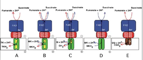
In prokaryotic respiratory chains SQR is also expressed and can be replaced by a similar complex under anaerobic conditions, the quinol:fumarate oxidoreductase (QFR). This protein catalyzes the reverse reaction, i.e., the coupling of quinol oxidation to the reduction of fumarate, termed fumarate respiration [81]. SQR and QFR complexes can be classified in five types, from A to E, based on their hydrophobic domain and heme content [82,83] (Figure 3).
Fig. 3. Distribution of SQR/QFR types in prokaryotic and eukaryotic organisms (from A to E). In common, all enzymes’ hydrophilic domain, which contacts the mitochondrial matrix
14
The mammalian complex II and E. coli SQR assemble a type-C hydrophobic domain, bearing only one b-type heme sandwiched between transmembrane helices of the two integral membrane subunits, SqrC and SqrD [84]. These subunits are also present in type-A domain, being distinguished from the former by the presence of an additional b-type heme group classified as bD-type heme given
its distal location from the hydrophilic domain in comparison with the proximal location of the bP-type heme. SQR type-A domain is present in the archaea
Thermoplasma acidophilum and the bacterium T. thermophilus [85,86]. Two b-type hemes are also present in the b-type-B domain of B. subtilis SQR and Wolinella succinogenes QFR [87,88]. What better distinguishes this domain is the exclusive presence of a single large hydrophobic subunit (C), which probably evolved from the fusion of the genes coding for the smaller subunits C and D [77]. Type-D domain is present in E. coli QFR [89] and contains the two integral membrane subunits but the b-type hemes are absent. In contrast, type-E SQRs lack the typical membrane anchoring subunits as found in the other four types of membrane domains. The proposed type-E domain consists of two polypeptides which lack transmembrane spanning helices and are not sequence-related to SqrC and SqrD, being renamed SqrE and SqrF subunits, respectively, for that reason [90]. Type-E subunits do not contain b-type hemes and SqrE was proposed to function as a monotopic membrane anchor of the enzyme, harboring the quinone binding site (caldariella quinone) and potentially assembling one [4Fe-4S]3+/2+ cluster [91,92].
Besides complex II, three other membrane-bound enzymes are able to deliver electrons to ubiquinone without proton translocation in mammalian mitochondria: i) ETF:ubiquinone oxidoreductase; ii) dihydroorotate and iii) s,n-glycerophosphate dehydrogenases.
15
electron transferring flavoproteins, which in turn accept electrons from FAD-containing dehydrogenases involved in mitochondrial fatty acid β-oxidation and amino acid catabolism, to ubiquinone [93].
Dihydroorotate and s,n-glycerophosphate dehydrogenases bind their substrates from the outer side of the membrane. The former contains a tightly bound FMN and is involved in pyrimidine biosynthesis, catalyzing the oxidation of dihydroorotate to orotate [94], while the latter contains one FAD, at least one [Fe-S] cluster and oxidizes s,n-glycerophosphate.
1.1.3 Quinol:cytochrome
c
oxidoreductase
The electron transfer from quinol to cytochrome c, with concomitant proton translocation, is catalyzed by quinol:cytochrome c oxidoreductase, also known as
bc1 complex or complex III in mammalian mitochondria. This membrane complex
is also found in many species of bacteria and is similar in many aspects to the cytochrome b6f complex of plant thylakoids (see [95] for a review). The
16
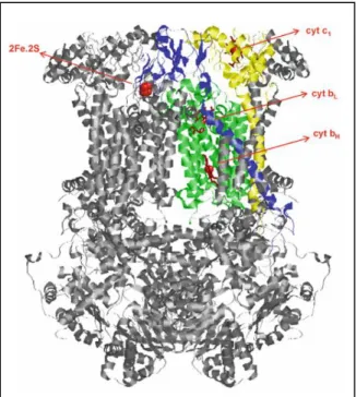
In detail (Figure 4), one subunit (cytochrome b subunit) contains two low spin b-type hemes of lower and higher reduction potentials, bL and bH, respectively. bL is
located towards the P-side while bH faces the N-side of the membrane, where the
binding centres for ubiquinol oxidation and ubiquinone reduction, Qo and Qi, are
respectively located. Another transmembrane subunit (Rieske Fe-S protein) is folded as a globular structure near the P-side of the membrane to which an [2Fe-2S]2+/1+ cluster is attached. The third core catalytic subunit (cytochrome c
1 subunit)
bears a globular domain that carries one c-type heme, to where one electron at a time is transferred in each monomer [98].
The bacterial bc1 complex generally comprises only the three core subunits [99],
of which Rieske Fe-S protein and cytochrome b subunits are conserved between different species. In contrast, the third core catalytic subunit strongly varies between phyla, cytochrome c1-like subunit being only present in α-, β- and γ
-proteobacteria, whilst ε-proteobacteria contain a diheme cytochrome c that belongs to the cytochrome c4 family [100], which is replaced by a tetrahemic
Fig. 4. X-ray crystal structure of
complex III homodimeric assembly from mammalian mitochondria. Each monomer comprises eleven subunits, three of which represent the core catalytic subunits, i) a cytochrome b subunit (green) bearing two b-type hemes of different reduction potentials and the quinone binding centres Qo and Qi, ii) a Rieske Fe-S
protein subunit (blue) containing a binuclear [Fe-S] cluster and iii) a cytochrome c1 subunit (yellow)
17
cytochrome c3, structurally unrelated with cytochrome c1,in the δ-proteobacteria
branch. Moreover, the cytochrome c1-like subunit is replaced by an f-type
cytochrome subunit in cyanobacteria respiratory chains (cytochrome b6f complex)
[101].
Some prokaryotes do not contain the typical bc1 complex, as in the case of the
thermohalophilic bacterium R. marinus, where a multihemic cytochrome bc complex lacking the Rieske center catalyzes the quinol:cytochrome c (HiPIP) oxidoreductase activity [102], hence termed alternative complex III (ACIII). In addition, some archaeal aerobic respiratory chains do not contain the canonical
bc1 complex. For instance, in Sulfolobus acidocaldarius aerobic respiratory chain,
bc1 complex analogs are found within two different supercomplexes, SoxEFGHIM
(SoxM) and SoxABCD, which will be later analyzed in the context of the archaeal aerobic respiratory chain supramolecular assemblies. In common, SoxG and SoxC subunits of both supercomplexes are similar to the cytochrome b subunit from bc1
complex, but the di-heme redox component is exclusively composed of two a-type hemes (AS) [103,104].
Complex III concomitant electron transfer and proton translocation is explained by the protonmotive Q-cycle, first proposed by Peter Mitchell [105] and later refined by Crofts and colleagues (see [106] for a review). The Q-cycle is divided in two steps, each being initiated with the oxidation of a quinol molecule at the Qo
site. In both steps, this reaction releases two electrons that are separated in a bifurcated pathway [107]. One electron is transferred to the [Fe-S] cluster of the Rieske subunit, where it triggers a conformational change of the subunit globular domain that allows the electron to be transferred to cytochrome c1, which in turn
18
higher reduction potential (heme bH). The quinone molecule produced in the
reaction is, in theory, able to freely migrate to the Qi site. There, heme bH reduces
the quinone molecule to a stabilized semiquinone [108]. In the second step, the bifurcated pathway is repeated with the concomitant translocation of two protons. At the Qi site the stabilized semiquinone produced in the first step is reduced by
heme bH, yielding, together with the sequestration of two protons from the N-side
of the membrane, a quinol molecule, which returns to the bulk pool, completing the cycle. The overall reaction catalyzed by complex III involves the oxidation of two quinol molecules in the Qo site, the reduction of one quinone at the Qi site, the
reduction of two cytochromes c1 and consequent transfer of two electrons to the
soluble cytochrome c, the release of four protons at the P-side of the membrane and the uptake of two protons from the N-side of the membrane.
Antimycin A, stigmatellin and myxothiazol are typical inhibitors of complex III and have been essential for the elaboration of the Q-cycle mechanism. Antimycin A acts at the Qi site, preventing the formation of semiquinone. Stigmatellin inhibits
the electron flow from the quinol molecule to the Rieske Fe-S protein and myxothiazol acts at the Qi site [109,110]. Interestingly, the use of antimycin A and
myxothiazol results in the production of superoxide at the Qo site.
It is worth mentioning that complex III monomers do not function independently. The two monomeric units interact with each other through the globular domain of the Rieske Fe-S protein of one monomer with the Qo site and the cytochrome c1
19
1.1.4 Oxygen reductases
The final step of the electron transfer chain in mitochondria and oxygen respiring prokaryotes is accomplished by oxygen reductases. These complexes couple the oxidation of electron carriers, such as cytochrome c or quinol molecules, to the reduction of dioxygen to water. There are three types of oxygen reductases, i) heme-copper oxygen reductases; ii) cytochrome bd oxygen reductases and iii) alternative oxygen reductases (AOX). X-ray crystal structures are not yet available for the cytochrome bd oxygen reductases, whereas the structure of AOX from the trypanosome respiratory chain was recently solved and many have been described for the heme-copper oxygen reductases superfamily, which will be the main focus of this section.
i. Heme-copper oxygen reductases
Heme-copper oxygen reductases superfamily is further classified in three families: A, B and C [112]. The mitochondrial cytochrome c oxygen reductase, or complex IV, belongs to the A-family, which includes also cytochrome c or quinol oxygen reductases from some prokaryotes. The B-family includes oxygen reductases from prokaryotes, such as the ba3-type oxygen reductase from T.
thermophilus [113], while the C-family is represented by cbb3-type oxygen
reductases [114]. X-ray structures from the three families have been elucidating the mechanism by which these complexes operate, which is far from being completely understood [113-117].
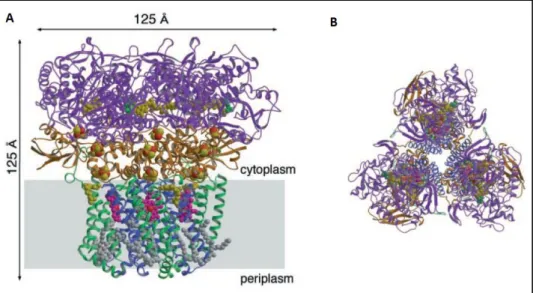
Mitochondrial complex IV catalyzes the sequential transfer of four electrons from the soluble cytochrome c to O2, yielding two water molecules. Concomitantly, four
20
enzyme [119]. Each monomer contains 13 subunits (~205 kDa), in the case of mitochondria, from which only three constitute the catalytic core [120]. Subunits I and II represent the minimal functional unit [121] (Figure 5A), while subunit III is important for the structural integrity of the redox sites and was suggested to participate in the delivery of O2 [122].
In detail, complex IV contains four redox groups, distributed between subunits I and II (Figure 5). Subunit II has two transmembrane helices and a globular domain projected into the P-side of the membrane where a binuclear CuA centre, which is
absent in quinol oxygen reductases, accepts one electron at a time from the soluble cytochrome c. The adjacent subunit I is composed of 12 transmembrane helices and contains one a-type heme and a binuclear complex consisting of a
Fig. 5. Mammalian mitochondrial complex IV architecture and proposed mechanism. A)
21
second a-type heme (designated as a3) together with CuB, to where the electron
transfer sequentially proceeds and the reduction of oxygen takes place [123]. In some prokaryotes, the a-type heme (a3) from the binuclear complex is replaced by
b- or o-type hemes, hence designated as b3 [114] and o3 [124], respectively.
In this region, two hydrogen-bond networks ensure a connection from the reduction of oxygen to the N-side of the membrane, known as D- and K-pathways, where the uptake of four protons from the N-side of the membrane, necessary to produce two water molecules, is achieved (Figure 5B). The translocation of the remaining four protons to the mitochondrial intermembrane space or bacterial periplasm is still debated, although mutations of amino acid residues involved in K- and D- pathways has shown that both pathways contribute for this process [125,126].
22
ii. Cytochrome bd oxygen reductases
Cytochrome bd oxygen reductases are quinol oxygen reductases found in many prokaryotes and generally use ubiquinol or menaquinol as substrates (see [130] for a review). These membrane complexes are composed of two transmembrane subunits (I and II) which harbor three prosthetic groups: i) a low spin heme b558
located in subunit I and ii) a catalytic centre in subunit II, formed by two high spin hemes, b595 and d, suggested to form a binuclear centre similar to the one
described for heme-copper oxygen reductases [131]. Cytochrome bd oxygen reductases do not translocate protons to the periplasm. Their function requires the uptake of four protons from the cytoplasmic side of the membrane for O2 reduction
and the release of two protons in the periplasmic side of the membrane for quinol oxidation with a concomitant transfer of four electrons through heme b558 to the O2
-reducing site. The quinol oxidation is performed, at least partially, in the hydrophilic region of subunit I, termed “Q-loop”, which distinguishes two groups of cytochrome bd oxygen reductases, the ones that harbor a long Q-loop, such as in the E. coli enzymes [132], and those in which an insert in the C-terminal portion is absent, bearing a short Q-loop [133]. The latter feature is observed in cyanide insensitive oxygen reductases, that include also enzymes in which the high spin heme d is replaced by a b-type heme, as in the case of B. subtilis cytochrome bb’ -type quinol oxygen reductase [134].
iii. Alternative oxygen reductases (AOXs)
The AOXs were mainly described in the mitochondria of higher plants, fungi and protists but are also present in some prokaryotes and animal species (see [135,136] for a review). These are homodimeric enzymes insensitive to cyanide that, similarly to cytochrome bd oxygen reductase, do not translocate protons. They contain a non-heme di-iron carboxylate active site for O2 reduction buried in
23
proposed that the protons required for O2 reduction are the ones derived from
quinol oxidation, yet the site where the latter process occurs remains elusive [136]. Nevertheless, since no proton translocation is registered, AOXs allow the presence of partially or completely “uncoupled” pathways. The former pathway relies on the utilization of complex I for the oxidation of NADH, while the latter is observed when the electron transfer from the reducing substrates NADH or succinate to O2 occurs by alternative NADH dehydrogenases or SQR enzymes,
bypassing complexes I, III and IV. This pathway does not contribute to the generation of a proton motive force and therefore the redox energy released during AOX activity is not conserved for the production of ATP, but is dissipated as heat [138]. In plants, this can be of great importance if the main respiratory chain is impaired by factors such as growth inhibition or exposure to subzero temperatures [139]. As for trypanosomes, free-living cells make more use of the standard complexes III and IV, whereas AOX plays a critical role in the survival of the parasite in its bloodstream form, where the organism makes use of the glycolytic pathway as its major source of ATP [140]. The mechanism whereby electrons are distributed between the two electron transport pathways to oxygen is not fully understood, but may depend on the ubiquinone/ubiquinol ratio.
1.1.5 ATP synthase
The transmembrane potential generated by the respiratory chain complexes is used by F1FO-ATP synthases to drive ATP synthesis from ADP and inorganic
phosphate (Pi) [141]. These are membrane complexes present in mitochondria,
bacteria and chloroplasts. Na+-dependent ATP synthases can also be found in
bacteria and are broadly similar to F1FO-ATP synthase with respect to structure
and function, although through the generation of a Na+ ion motive force [142].
Vacuolar-type H+-ATPases (V-ATPases), present in other eukaryotic intracellular
24
F1FO-ATP synthase. ATP synthases have similar structures, irrespectively of their
source. Two major structural domains compose the enzyme, i) a membrane-extrinsic F1 domain, where the catalytic site of the enzyme is located, and ii) a
membrane-intrinsic FO domain, responsible for the uptake of protons from the
P-side of the membrane (Figure 6).
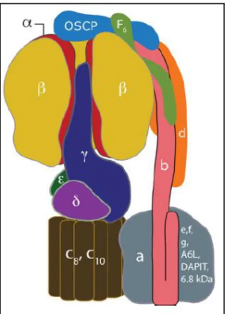
The bovine mitochondrial F1 water-soluble domain is constituted by five different
subunits, α, β, δ, γ and ε, with the stoichiometry α3β3δ1γ1ε1 (~350 kDa). The α3β3γ1
module represents the central stalk, a rotary motor present in all ATP synthases [143] that is linked to the membrane part of the protein by the γ-subunit, which protrudes ~30Ȧ beyond the α3β3 domain, where 3 α- and β-subunits are alternately
arranged [144]. This linkage is reinforced by the δ- (known as ε subunit in bacteria and chloroplasts) and ε-subunits, the latter having no counterpart in bacterial and chloroplast F1FO-ATP synthases [145]. The α3β3 domain is also connected to the
FO domain by the peripheral stalk which, in the case of mitochondria, is composed
of subunits OSCP (oligomycin sensitivity-conferring protein), b, d and F6 [146]. The FO domain contains a c ring, composed of several c-subunits (from 8 in the
Fig. 6. Representation of the mitochondrial F1FO
-ATP synthase. The membrane-extrinsic F1
catalytic domain is located at the upper part of the model and includes the α3β3γ1 rotary motor.
This module connects with FOvia subunit γ and a
peripheral stalk composed of subunits OSCP, b, d and F6. FO domain is responsible for the uptake