Structural and vibrational properties of phase-pure monoclinic
NdLuO
3
interlanthanides synthesized from nanostructured precursors
Júlia C. Soares
a, Kisla P.F. Siqueira
a, Roberto L. Moreira
b, Anderson Dias
a,* aDepartamento de Química, Universidade Federal de Ouro Preto, Campus Morro do Cruzeiro, ICEB II, Ouro Preto, MG, 35400-000, Brazil bDepartamento de Física, ICEx, Universidade Federal de Minas Gerais, C.P. 702, Belo Horizonte, MG, 30123-970, Brazila r t i c l e
i n f o
Article history:
Received 8 December 2015 Received in revised form 17 February 2016 Accepted 30 March 2016 Available online 1 April 2016
Keywords:
Ceramics
Electron microscopy Nanomaterials Raman spectroscopy Rare-earth
a b s t r a c t
Phase-pure monoclinic NdLuO3 interlanthanides were successfully synthesized from hydrothermal-derived precursors. LuO(OH) and Nd(OH)3materials werefirstly obtained by hydrothermal synthesis at 250C, for 24 h. X-ray diffraction, micro-Raman spectroscopy, scanning and transmission electron
microscopies were employed to investigate the thermal behavior of these precursors and NdLuO3 interlanthanide up to 1600 C. It was observed that LuO(OH) and Nd(OH)3were converted to cubic
lanthanide oxides below 1000C. Cubic Nd2O3has changed to the monoclinic structure at 1000C,
followed by reaction with cubic Lu2O3at 1400C to produce monoclinic NdLuO3. Phase-pure monoclinic (C2/mspace group, #12) interlanthanide was then produced at 1600C for thefirst time, as confirmed by
all the employed techniques. Phonon modes were additionally determined and assigned for this monoclinic structure.
©2016 Elsevier B.V. All rights reserved.
1. Introduction
LnLn0O3mixed oxides, known as interlanthanide materials, have
recently received an enormous scientific and technological interest because of their outstandingly complex structures toward prom-ising microwave-related, magnetic, and optical applications[1e5].
These interlanthanides are frequently interpreted as mixtures of simple binary sesquioxides of trivalent lanthanides, which also exhibit intricate crystal chemistry and can be found in at least three different modifications ranging from cubicfluorite-type (C-type), hexagonal (A-type), to monoclinic (B-type) polymorphs, with very remarkable properties[6e11]. In the last decades, many works have
been devoted to investigate a large number of trivalent lanthanide combinations in order to obtain potentially ordered ternary mixed-lanthanide phases by systematically substituting smaller ions into lower coordination sites and larger ions into higher coordination sites[1e4,12,13]. These attempts revealed a systematic occurrence
of order-disorder phenomena in many ternary interlanthanide combinations, which can be advantageous from the perspective of doping (in disordered materials) or from the point of view of the
polymorphism (in ordered materials). However, all compositions deserve investigation towards a complete assessment of structure-property relationships, where different lanthanide ions lead to diverse crystal structures.
The present work focuses the ternary lanthanide oxide NdLuO3,firstly studied by Schneider and Roth[14]and again re-ported by Berndt et al.[15]as a new materialfifteen years later. Both papers did not presented any x-ray diffraction (XRD) data nor further evidence for their findings, only reporting either a mixture of monoclinic and cubic phases after heat treatments at 1650C for 6 h[14]or a mixture of 50% of the perovskite phase
(orthorhombic) in combination with monoclinic and cubic phases after solid-state reactions of coprecipitated hydroxides at 1250C
[15]. Su et al. [16] proposed the synthesis of many ternary lanthanide oxides, including NdLuO3, by using for the first time high pressure and high temperature methods. Although no XRD was presented, the authors claimed a phase-pure NdLuO3 in a perovskite form at 1200 C and 40 kbar. More recently, Dubok
et al. [13] reported the electrophysical properties of inter-lanthanide oxides, and NdLuO3appeared as belonging to a C-type structure after coprecipitation of hydroxides followed by thermal decomposition and sintering at high temperatures. Bharathy et al.
[12] presented a nice structure map for all known ternary
lanthanide oxides in the literature, which shows that NdLuO3is *Corresponding author.
E-mail address:[email protected](A. Dias).
Contents lists available atScienceDirect
Journal of Alloys and Compounds
j o u r n a l h o m e p a g e :h t t p : / / w w w . e l s e v i e r . c o m / l o c a t e / j a l c o m
known, but its structure has not been reported by X-ray or neutron diffraction techniques. In fact, in their introduction, Barathy et al.[12]mentioned NdLuO3as studied by Berndt et al. [15], Ito et al.[3]and Su et al.[16]. Unfortunately, Ito et al.[3]did not presented any result for this interlanthanide.
The synthesis procedures employed to produce binary and even ternary lanthanide oxides vary enormously, either as single crystals, ceramic powders or films. The standard solid-state re-action remains the major processing route for these materials [2,4], although other non-conventional methods are currently applied, such as reverse-strike coprecipitation [17], mechano-chemical [18], thermal decomposition [5,6,13,19e21], sol-gel
processing[22e24], combustion synthesis[25], molecular beam
epitaxy technique[26,27], and hydroxidefluxes[12]. The hydro-thermal technology is currently applied by our research group to synthesize nanostructured materials with technological interest [28e34]. In our previous investigations, crystal chemistry and
morphology are monitored and controlled by choosing the appropriate feed chemistry and processing conditions during hydrothermal syntheses of nanostructured precursors[28e34]. By
using experimental setups with fully-controlled temperature-pressure-time-pH environments, potentially reactive materials can be produced in mild, reliable and reproducible conditions [28e34]. These processing parameters have decisive influence on
the crystal structure, purity, ordering degree, particle size and morphology of the resulting materials [35,36]. It is well known that these features determine the performance of the materials, which lead us to conclude that the investigation of their pro-cessing conditions becomes very important in this context.
In view of that, this work introduces the hydrothermal pro-cessing route for the synthesis of phase-pure monoclinic NdLuO3. Based on the available literature discussed above, it seems that all attempts to produce phase-pure NdLuO3failed. Thus, this paper reports the thermal behavior of nanostructured hydrothermally-derived precursors (hydroxides and oxy-hydroxides), which was monitored by XRD, Raman spectroscopy, scanning and trans-mission electron microscopies. Our procedures allowed us to synthesize, for thefirst time, phase-pure NdLuO3interlanthanide belonging to the monoclinicC2/m(#12) space-group. Moreover, in order to expand the knowledge of this material, the complete set of non-polar phonon modes for the monoclinic NdLuO3was determined and assigned for thefirst time, with appropriate use of polarized micro-Raman scattering tools on sintered samples.
2. Experimental section
Stoichiometric amounts of Nd(NO3)3$6H2O and Lu2O3
(Sigma-Aldrich, purity>99.9%) were used as starting materials. Lutetium
oxide wasfirstly treated with HNO365% to produce a mother so-lution of lutetium nitrate, according to the chemical reaction:
Lu2O3þ6HNO3/2LuðNO3Þ3þ3H2O
: (1)
Neodymium nitrate hydrate was then introduced and mixed to this mother solution followed by adding of NaOH (10 mol/L) until the pH reach values above 13 under vigorous stirring. The resulting solutions were loaded into stainless steel Parr®
autoclaves equip-ped with pressure and temperature controllers. The reaction ves-sels were heated at 10C min 1up to the processing temperature
and its corresponding saturated vapor pressure (250±1 C and
580±2 psi) for 24 h. After synthesis, the obtained precursors were
washed with distilled water in order to remove any remaining Naþ
ion and dried at 70C. The general chemical reaction for the
syn-thesis of the precursors can be written as:
LuðNO3Þ3þNdðNO3Þ3þ6NaOH/LuOðOHÞ þ NdðOHÞ3þ6NaNO3
þH2O:
(2)
Following, the materials were calcined (air atmosphere) in the temperature range 800e1600C forfixed times of 2 h in order to
investigate their thermal behavior. For sintering, cylindrical pucks of about 5 mm height and 12.5 mm diameter were produced by applying a pressure of 150 MPa in samples previously treated at 1200C. The obtained pucks were then sintered in a conventional
oven (air atmosphere) at 1600C, for 8 h.
In the sequence, the structural properties were investigated by x-ray diffraction (XRD) using a Shimadzu D-6000 diffractometer with graphite monochromator and a nickelfilter in the range of 10e602
q
(15 s/step of 0.022q
), operating with FeKa
radiation(
l
¼0.1936 nm), 40 kV and 20 mA (the results were automatically converted to CuKa
radiation for data treatment and manipulation). The lattice parameters were determined by the software MDI Jade 9.0. The vibrational properties were studied by micro-Ramanspectroscopy in back-scattering configuration by using an
Olympus confocal microscope (100 objective) attached to a
Horiba/Jobin-Yvon LABRAM-HR spectrometer (equipped with 600 and 1800 grooves/mm diffraction gratings). The 632.8 nm line of a HeeNe laser (maximum effective power of 6 mW at the surface of
each sample) was used as exciting line and a Peltier-cooled charge coupled device (CCD) detected the scattered light. An edgefilter was employed to stray light rejection (Rayleigh line). In order to avoid any kind of crystallization due to laser exposition, very low intensities were employed, besides lens with low magnification and defocused laser, when possible. Accumulation times of typi-cally 20 collections of 10 s were employed with spectral resolution better than 1 cm 1. The resulting spectra were corrected by the
Bose-Einstein thermal factor besides individual baseline
subtractions.
Morphological and structural features of the samples were also investigated by electron microscopy. A Quanta FEG 3D (FEI)fi eld-emission scanning electron microscope (FESEM) equipped with energy-dispersive spectrometer (EDS) was employed to analyze the morphology and chemical features of the NdLuO3-derived mate-rials (applied voltages varied from 5 to 30 kV). Images were taken in both detection modes, i.e., secondary (SE) and backscattered (BSE) electrons, in order to differentiate the precursors andfinal mate-rials. High-resolution transmission electron microscopy (HRTEM) was carried out to investigate the crystalline aspects of the nano-powders in a Tecnai G2-20 (FEI) transmission microscope (applied voltage of 200 kV). Selected area electron diffraction (SAED) pat-terns were obtained for all samples in order to characterize their polycrystalline nature.
3. Results and discussion
Fig. 1presents XRD pattern and Raman spectrum obtained for a stoichiometric mixture of the precursors hydrothermally synthe-sized at 250C. The results showed that Nd(OH)3 and LuO(OH)
were produced and may be indexed according to the ICDD (Inter-national Committee for Diffraction Data) cards #00-006-0601 and #01-072-0928, respectively (Fig. 1a).Fig. 1b presents the Raman spectrum for this mixture of precursors at room temperature. As it can be seen, a set of relatively broad Raman modes in the region 100e950 cm 1 corresponds to the LuO(OH) and Nd(OH)3
vibra-tions. The bands related to LuO(OH) occur below 200 cm 1and at around 410, 660 and 830 cm 1, while the modes related to
neo-dymium hydroxide occur at 297 and 371 cm 1 [20]. In the
sequence, the thermal evolution aiming to the production of
pure monoclinic NdLuO3 from hydrothermal-derived precursors was studied in the temperature range 800e1600C. Morphological
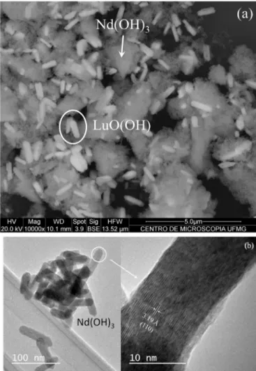
and structural aspects of the precursors were investigated by FESEM and HRTEM.Fig. 2a shows a FESEM image where nanosized, agglomerated particles can be observed for the precursors syn-thesized by hydrothermal processing at 250C, for 24 h. As it can be
seen, two different morphologies are present and could be associ-ated after chemical analyses (EDS) with the precursors obtained after synthesis. LuO(OH) exhibits tabular, faceted particles (easily identified by a circle in Fig. 2a), while Nd(OH)3 shows nano-structured, heavily agglomerated particles (identified by an arrow inFig. 2a).Fig. 2b shows representative HRTEM images obtained in Nd(OH)3precursor hydrothermally synthesized at 250C, in which nanosized, well-dispersed tabular particles can be visualized. For this sample, the interplanar spacing was computed to be about 3.19 Å, which correspond to the (110) plane of theP63/mhexagonal space group.
Fig. 3shows the XRD results for the samples thermally treated for 2 h at selected temperatures (800, 1000, 1200, 1400 and 1600 C). At temperatures below 1000 C, the precursors have
converted to lanthanide oxides according to the ICDD cards
#00-021-0579 (Nd2O3) and #01-072-6366 (Lu2O3). These oxides belong to theIa3 (#206) space group (cubicfluorite-type phase) and can be visualized as phase-pure materials at 800C inFig. 3b with lattice
parametersa¼10.96 Å for Nd2O3anda¼10.39 Å for Lu2O3. At 1000 C, cubic Nd2O3 changed (see Fig. 3b) to monoclinic
neo-dymium oxide,C2/mspace-group (#12), according to the ICDD card #01-076-7416, with lattice parameters a¼14.12 Å, b¼3.63 Å,
c¼8.80 Å and
b
¼100.2. This result was unexpected, since ahexagonal structure (space group P3m1) is frequently found for pure Nd2O3[20,37,38]. At 1200C, a mixture of pure monoclinic Nd2O3and cubic Lu2O3phases can be observed. At this temperature and above,Fig. 3b shows clearly the chemical reaction and crystal phase evolutions, since it focuses on the range 25e352
Q
, where the most important changes in the diffraction patterns ofFig. 3a occur. InFig. 3b, each peak could be identified in agreement with ICDD cards of previously published works[5,17,39,40]. At 1400C,the NdLuO3interlanthanides become visible as a monoclinic phase (C2/m space group, #12) together with residual cubic lutetium oxide. Finally, heat treatment at 1600 C produced phase-pure
monoclinic NdLuO3, whose identification and assignment could only be accomplished by comparing its XRD pattern with that observed for the LaYO3material (ICDD card#70-3117)[39].
According to the previous literature discussed above, perovskite-like NdLuO3interlanthanides can be formed as a stable
10
15
20
25
30
35
40
45
50
55
60
In
tens
it
y
(
a
rb
. uni
ts
)
2 Theta (°)
Nd(OH)3LuO(OH)
(a)
150 300 450 600 750 900
LuO(OH)
Nd(OH)3
(b)
Raman i
n
tensity (arb. units)
Wavenumber (cm
-1)
Fig. 1.(a) XRD pattern and (b) Raman spectrum for an equimolar mixture of the hydrothermally-synthesized Nd(OH)3and LuO(OH) precursors. In (b), the main bands for each phase are indicated in different colors.(For interpretation of the references to colour in thisfigure legend, the reader is referred to the web version of this article.)
Fig. 2.(a) FESEM image for the precursors obtained by hydrothermal synthesis at 250C, for 24 h. LuO(OH) tabular particles are identified by a circle, while Nd(OH)3
particles are indicated by an arrow; (b) HRTEM images for the Nd(OH)3. Tabular, faceted particles are shown (left image) with interplanar spacing at 3.19 Å, corre-sponding to the (110) plane of theP63/mhexagonal space group (right image).
phase [15,16]. In fact, the structural stability and formability of perovskite materials are based on the so-called tolerance factor (t), a criterion proposed by Goldschmidt through the relationship[41]:
t¼ ðffiffiffiRAþROÞ
2 p
ðRBþROÞ
; (3)
where RA, RBand ROare the ionic radii of A, B and oxygen in the general formula ABO3. In an ideal cubic perovskite structure, the tolerance factor is equal to the unity. Lowertvalues could occur, which means that distorted structures would be present. It is
currently accepted that almost all perovskites havetvalues ranging from 0.75 to 1.00. However, the tolerance factor is a necessary but not a sufficient condition for the formation of perovskite structures. The Goldschmidt's criterion neglects the influence of the coordi-nation number on the effective ionic radii, and was debated by the literature by years without a definitive solution. For NdLuO3, the tolerance factor could be calculated as 0.7847, which indicates that this material could be produced in a (highly distorted) perovskite phase, even if one considers this value in the border for the tradi-tional perovskite atomic arrangement. In this work, the phase behavior followed by our NdLuO3was quite different: the hydro-thermal precursors were converted to monoclinic Nd2O3and cubic Lu2O3at 1200C, followed by a chemical reaction at higher tem-peratures allowing the production of phase-pure monoclinic (C2/m, #12) materials at 1600C.Fig. S1(Supplementary Material)
pre-sents XRD data for the phase-pure monoclinic NdLuO3 together with its Miller indexes for the monoclinicC2/mspace group. In a
10 20 30 40 50 60
2 Theta (°)
800°C 1000°C
In
te
ns
ity
(a
rb
. un
its
)
1200°C 1400°C 1600°C
(a)
26 28 30 32 34
cubic Lu O
cubic Nd O
NdLuO
monoclinic Nd O
2 Theta (°)
800°C 1000°C
Intensity (arb. units)
1200°C 1400°C 1600°C
(b)
Fig. 3.XRD patterns exhibiting the thermal behavior for samples produced in the temperature range 800e1600C (from bottom to top). (a) General XRD patterns in the 10e602Q range, and (b) particular view in the region 25e352Qwith identification of all observed phases for better visualization.
150 300 450 600 750 900
1600°C
1400°C
1000°C
800°C cubic Lu2O3
Wavenumber (cm
-1)
cubic Nd2O3
Raman Intensity (arb. units)
monoclinic Nd2O3 NdLuO3
Fig. 4. Raman spectra (100e1000 cm1 range) showing the thermal evolution (800e1600C) of the samples produced from hydrothermally-processed precursors at 250C. The main bands for each phase are indicated in different colors.(For inter-pretation of the references to colour in thisfigure legend, the reader is referred to the web version of this article.)
200 400 600 800 1000 1200
Raman intensity (arb. units)
Wavenumber (cm
-1)
Fig. 5.Micro-Raman spectra for the NdLuO3 sample in the spectral region 50-1200 cm 1. Experimental data are in open circles, whereas thefitting curve is repre-sented by a red line. Green lines represent the phonon modes adjusted by Lorentzian curves. (For interpretation of the references to colour in thisfigure legend, the reader is referred to the web version of this article.)
previous publication[39], Yamamura et al. studied the synthesis of monoclinic LaYO3and their results were employed in the present work to indexing our NdLuO3samples, since no XRD pattern for this material in the monoclinic phase could be found in the literature. The lattice parameters were determined asa¼14.08 Å,b¼3.54 Å,
c¼8.67 Å and
b
¼101.3for the monoclinicC2/m space group,according to the equation[42]:
1
d2¼
1
sen2
b
h2
a2þ k2sen2
b
b2 þ l2 c2
2hlcos
b
ac
: (4)
It is important to highlight that conventional solid-state reac-tion using neodymium oxide with lutetium oxide at 1600C did not
result in a phase-pure monoclinic NdLuO3.Fig. S1 also presents XRD results for conventionally-synthesized materials by solid-state reaction. We can note the presence of both unreacted Lu2O3and Nd2O3 in the final product obtained throughout solid-state reaction.
The thermal behavior of hydrothermally treated precursors for
the synthesis of NdLuO3 was also monitored by micro-Raman
scattering. Fig. 4 presents selected spectra for the samples sequentially treated at 800C, 1000C, 1400C and 1600C. The
spectrum for the samples heat treated at 800C is quite similar to
those obtained by Ubaldini and Carnasciali[20]for lutetium and neodymium cubic oxides. The strongest bands for these materials are located, respectively, at 338 and 390 cm 1. For the samples prepared at 1000C, we can also observe the strongest band related
to the Lu2O3(390 cm 1) together with vibrational modes associated with monoclinic Nd2O3.20These additional modes are also reported for the same crystal structure by Mele et al.[5], for Nd2O3eGd2O3
mixed system, Martel et al.[43], for Sm2O3, and Heiba et al.[22], for Dy2-xHoxO3(x>0.4). At 1400C, monoclinic NdLuO3can now be observed, in accordance with Tompsett et al.[17], particularly in the frequency range 350e500 cm 1. Although some secondary phases
(lanthanide oxides) are still present, as verified by XRD and dis-cussed previously, in the range 350e500 cm 1 a broad group of
Raman modes could be assumed as afingerprint of the monoclinic
C2/m (#12) space group, as previously proposed for the LaGdO3 material[17]and for the (Gd0.5Nd0.5)2O3system[3].
The spectrum for the calcined sample at 1600C (Fig. 4, top)
shows the Raman-active modes for the phase-pure NdLuO3. In this monoclinicC2/mspace group, there are six formula units per uni-tary cell (Z¼6)[5,44]. Four oxygen ions and three lanthanide ions occupy the 4iWyckoff position with Cssymmetry (2Ag4Bg4Au 42Bu), and one oxygen ion sites on an 2bposition with C2h
sym-metry (Au42Bu). Based on these occupation sites, the site-group
method of Rousseau et al. [45] was applied to obtain the
following representation of the Raman-active modes (gerade) at the Brillouin-zone center:
G
RAMAN¼14Agðxx;yy;zz;xyÞ47Bgðxz;yzÞ: (5)According to this model, one would expect to observe up to 21
first-order Raman modes in the spectrum of NdLuO3 sample
treated at 1600C (beside 24 infrared bands, 8Au(y)416Bu(x,z)).
However, the spectrum of this sample showed inFig. 4 presents relatively broad peaks and does not allow one to visualize more than 10 bands. Therefore, in order to resolve the superimposed phonon modes, peakfitting procedures were required. The Raman spectrum for this sample was thenfitted by Lorentzian curves and the results are presented inFig. 5. The experimental data are in open circles and thefitting curve is presented by a red line. The results showed that 21 Raman modes were required for thefitting (green curves), in perfect agreement with the theoretical pre-dictions. Based on this fitted spectrum, the frequencies and full-width at half maxima (FWHM) for all bands were determined for our phase-pure monoclinic NdLuO3and are presented inTable 1.
The Raman spectrum depicted in Fig. 5 could represent a
fingerprint of the NdLuO3interlanthanides with monoclinicC2/m structure. In addition, as already mentioned, 21 Raman-active modes were expected, with the following distribution in terms of the irreducible representations (i.r.) of theC32hpoint group: 14Ag4
7Bg[2,22,44]. In order to corroborate ourfindings and assign these non-polargerademodes to their respective i.r., additional experi-ments were carried out by using polarized micro-Raman spec-troscopy. For this study, a ceramic sample was sintered at 1600C
for 8 h. These processing conditions were employed because the goal was to obtain ceramics with large grains in order to get suit-able polarized micro-Raman spectra. It is well known that the in-elastic scattered light intensities due to the Raman effect are Table 1
Phonon frequencies, FWHM and assignment of thegerademodes determined from the adjustment of the Raman experimental data by Lorentzian curves, for the phase-pure monoclinic NdLuO3.
Band Frequency (cm 1) FWHM (cm 1) Assignment
1 65.7 5 Ag
2 96.5 19 Ag
3 110.5 6 Bg
4 123.6 14 Ag
5 168.4 11 Ag
6 181.1 16 Ag
7 200.2 19 Ag
8 218.4 18 Bg
9 265.9 43 Bg
10 306.0 52 Ag
11 338.5 47 Bg
12 381.7 43 Bg
13 408.7 42 Ag
14 439.9 27 Ag
15 486.9 9 Bg
16 572.2 36 Ag
17 595.2 29 Bg
18 702.3 56 Ag
19 815.2 48 Ag
20 840.0 40 Ag
21 953.4 17 Ag
200 400 600 800 1000 1200
B B B B B B
Raman intensi
ty (arb. unit
s
)
Wavenumber (cm
-1)
B//
T
Fig. 6.Polarized micro-Raman scattering for the sintered NdLuO3. Parallel (//) and cross-polarized (t
) configurations are indicated in red and blue lines, respectively. The relative strengthening of the Agmodes in the parallel configuration are accompanied by the relative weakening of the Bgmodes (indicated in blue). Conversely, Bgmodes are favored by crossed light configuration, for the same sample position (particular grain size orientation). (For interpretation of the references to colour in thisfigure legend, the reader is referred to the web version of this article.)
proportional to the square of the elements of the polarizability tensor (second-order tensor). Then, the base functions of the i.r. that are Raman-active have quadratic forms, i.e., they transform like the product of the Cartesian coordinates (e.g.,xx,xy,xz,yy,etc). For single crystals, we take benefit of the crystal symmetry to assign the lattice vibrations to the different i.r.[45,46]. However, in the case of the ceramics, although the group-theory predictions remain valid, the symmetries of the modes are generally mixed up due to the random orientation of the crystalline grains. In this work, we have used a confocal microscope with an objective of magnification of 100, which allows the selection of observation regions smaller
than the grain sizes (scattering volumes ofcirca1
m
m3). However, we do not know anything about the crystallographic axes of these grains, which have also random orientation throughout the sample. By measuring the micro-Raman spectra of NdLuO3sintered sample with polarized light (Fig. 6), we observed that for some grains the spectra of parallel (red line) and crossed light (blue line) become quite different. We can observed the relative strengthening of the totally symmetric 14Ag modes in the parallel configuration (because these modes are particularly observed forxx,yyandzzconfigurations of polarizer and analyzer), accompanied by the relative weakening of the 7Bgmodes, which are favored by crossed Fig. 7.FESEM images for samples obtained at different conditions. (a) 800C, (b) 900C, (c) 1000C, (d) 1400C and (e) 1600C.
light configuration (selected only for xz and yz configurations). These crossed-polarized Bgmodes are indicated in blue inFig. 6. Therefore, we could assign all the 21 Raman-activegerademodes as belonging to the Agand Bgsymmetries, as also presented inTable 1. These results are in complete agreement for the proposed mono-clinicC2/m(#12) space group for our phase-pure NdLuO3.
The thermal behavior of all samples was examined in their morphological, chemical and structural aspects by FESEM and HRTEM.Fig. 7presents a sequence of FESEM images (backscattered electrons) for samples obtained under different conditions. Fig. 7aee shows representative FESEM images for samples obtained
at 800, 900, 1000, 1400 and 1600C. It was observed a general
increase in their particle sizes; in particular, the morphologies due to LuO(OH) and Nd(OH)3 precursors presented and discussed in Fig. 2were changed above 800C (Fig. 7aec) due to their
conver-sion into lanthanide oxides. EDS analyses showed that Lu and Nd are still in opposite corners, which means that a chemical reaction
between these precursors was not in progress. Above 900 C
(Fig. 7b and c), it is expected that some changes can occur since XRD data verified a phase transformation for Nd2O3, from cubic to monoclinic. However, the morphology had no significant changes above 900C, except for a further increase in the particle size.
Fig. 7d and e presents the morphology of the samples obtained at 1400C and 1600C, which exhibit features of materials where
some solid-state reaction cannot be neglected. Hard, compacted agglomerates can be seen, which indicate that a chemical reaction between the hydrothermally-derived precursors have now occurred. In fact, EDS analyses indicate that it is no longer possible to discern between lutetium and neodymium along the samples.
Fig. 8presents HRTEM images for the phase-pure monoclinic NdLuO3produced at 1600C. It was observed that thick samples dominate the scenario, which were the result of the solid-state reactions that occurred at this temperature (left image). The interplanar spacing was determined to be about 3.13 Å, corre-sponding to the (111) plane of the monoclinicC2/m space group (right image). The inset inFig. 8shows the SAED pattern for the
sample obtained at 1600 C, in which spots observed in
well-aligned samples confirm their monoclinic structure.
4. Conclusions
In this work, phase-pure monoclinic NdLuO3was synthesized
for the first time from nanostructured hydrothermally-derived precursors. Aqueous solutions of lutetium and neodymium ni-trates were hydrothermally treated at 250C for 24 h in order to
produce nanostructured LuO(OH) and Nd(OH)3 precursors. The
thermal behavior of this interlanthanide material was investigated by XRD, micro-Raman spectroscopy and electron microscopy (FESEM and HRTEM). It was observed that the starting materials were converted to cubic lanthanide oxides below 1000C. Cubic
Nd2O3has changed to the monoclinic structure at 1000C, followed by reaction with Lu2O3at 1400C to produce monoclinic NdLuO3. Phase-pure monoclinic C2/m space group was obtained only at 1600C, as verified by XRD, Raman spectroscopy and HRTEM. In
order to validate our results, polarized micro-Raman scattering was also employed to probe the phonon modes in sintered NdLuO3. All the 21 non-polar vibrational modes predicted by group theory calculations for theC2/mspace group were identified and assigned,
which also confirmed the production of phase-pure NdLuO3
interlanthanides at 1600C.
Acknowledgements
The authors acknowledge the financial support from CAPES,
CNPq, FINEP and FAPEMIG. The Center of Microscopy at the Uni-versidade Federal de Minas Gerais (http://www.microscopia.ufmg. br) is also acknowledged for providing the equipment and technical support for experiments involving electron microscopy.
Appendix A. Supplementary data
Supplementary data related to this article can be found athttp:// dx.doi.org/10.1016/j.jallcom.2016.03.291.
References
[1] R.L. Moreira, A. Feteira, A. Dias, Raman and infrared spectroscopic investiga-tion on the crystal structure and phonon modes f LaYbO3ceramics, J. Phys. Condens. Matter 17 (2005) 2775e2781.
[2] Y. Sharma, S. Sahoo, A.K. Mishra, P. Misra, S.P. Pavunny, A. Dwivedi, S.M. Sharma, R.S. Katiyar, Structural phase transition of ternary dielectric SmGdO3: evidence from angle-dispersive X-ray diffraction and Raman spec-troscopic studies, J. Appl. Phys. 117 (2015) 094101.
[3] K. Ito, K. Tezuka, Y. Hinatsu, Preparation, magnetic susceptibility, and specific heat on interlanthanide perovskites ABO3(A¼La-Nd, B¼Dy-Lu), J. Solid State Chem. 157 (2001) 173e179.
Fig. 8.HRTEM images for the phase-pure monoclinic NdLuO3produced at 1600C. Thick samples were the result of the solid-state reactions that occurred at this temperature (left
image). Interplanar spacing of 3.13 Å corresponds to the (111) plane of the monoclinicC2/mspace group (right image). The inset shows the SAED pattern for this sample, in which the aligned spots confirm its monoclinic structure.
[4] J. Blanusa, N. Jovic, T. Dzomic, B. Antic, A. Kremenovic, M. Mitric, V. Spasojevic, Magnetic susceptibility and ordering of Yb and Er in phosphors Yb,Er:Lu2O3, Opt. Mater 30 (2008) 1153e1156.
[5] P. Mele, C. Artini, A. Ubaldini, G.A. Costa, M.M. Carnasciali, R. Masini, Synthesis, structure and magnetic properties in the Nd2O3-Gd2O3mixed system syn-thesized at 1200C, J. Phys. Chem. Solids 70 (2009) 276e280.
[6] P. Mele, C. Artini, R. Masini, G.A. Costa, A. Hu, N. Chikumoto, M. Murakami, Synthesis and superconductive characterisation of RuSr2NdxGd1 xCu2O8 compounds (x¼0, 0.09, 0.18, 0.35), Phys. C 391 (2003) 49e54.
[7] D.Y. Chung, E.H. Lee, Microwave-induced combustion synthesis of Ce1 xSmxO2 x/2powder and its characterization, J. Alloys Compd. 374 (2004) 69e73.
[8] C. Peng, Z. Zhang, Nitrateecitrate combustion synthesis of Ce1 xGdxO2 x/2
powder and its characterization, Ceram. Int. 33 (2007) 1133e1136. [9] H. Yang, H. Wang, H.M. Luo, D.M. Feldman, P.C. Dowden, R.F. Depaula, Q.X. Jia,
Structural and dielectric properties of epitaxial Sm2O3 thinfilms, Appl. Phys. Lett. 92 (2008) 062905.
[10] J. Hao, Y.W. Li, J.S. Wang, C.L. Ma, L.Y. Huang, R. Liu, Q.L. Cui, G.T. Zou, J. Liu, X.D. Li, Experimental determinations of the high-pressure crystal structures of Ca3N2, J. Phys. Chem. C 114 (2010) 16750e16755.
[11] G. Azimi, R. Dhiman, H. Kwon, A. Paxson, K. Varanasi, Hydrophobicity of rare-earth oxide ceramics, Nat. Mater 12 (2013) 315e320.
[12] M. Bharathy, A.H. Fox, S.J. Mugavero, H.eC. zur Loye, Crystal growth of inter
-lanthanide LaLn0O3 (Ln0¼Y, Ho-Lu) perovskites from hydroxidefluxes, Solid State Sci. 11 (2009) 651e654.
[13] V.A. Dubok, V.V. Lashneva, Y.N. Kryuchkov, Electrophysical properties of oxide interlanthanides and solid solutions based on them, Glass Ceram 60 (2003) 115e117.
[14] S.J. Schneider, R.S. Roth, Phase equilibria in systems involving the rare-earth oxides. Part II. Solid state reactions in trivalent rare-earth oxide systems, J. Res. NBS 64A (1960) 317e332.
[15] U. Berndt, D. Maier, C. Keller, New AIIIBIIIO3interlanthanide perovskite com-pounds, J. Solid State Chem. 13 (1975) 131e135.
[16] W. Su, D. Wu, X. Li, X. Ma, J. Zhou, Z. Qian, Y. Wang, W. Liu, Z. Ge, An inves-tigation using high-pressure synthesis of double-rare-earth oxides of ABO3-composition, Physica 139&140B (1986) 658e660.
[17] G.A. Tompsett, R.J. Philips, N.M. Sammes, A.M. Cartner, Characterisation of LaGdO3 by X-ray powder diffraction and Raman spectroscopy, Solid State Commun. 108 (1998) 655e660.
[18] T. Tsuzuki, W.T.A. Harrison, P.G.J. McCormick, Synthesis of ultrafine gadolin-ium oxide powder by mechanochemical processing, J. Alloys Compd. 281 (1998) 146e151.
[19] C. Artini, G.A. Costa, R. Masini, Study of the formation temperature of mixed LaREO3(RE¼Dy,Ho,Er,Tm,Yb,Lu) and NdGdO3oxides, J. Therm. Anal. Calorim. 103 (2011) 17e21.
[20] A. Ubaldini, M.M. Carnasciali, Raman characterization of powder of cubic RE2O3(RE¼Nd,Gd,Dy,Tm, and Lu), Sc2O3 and Y2O3, J. Alloys Compd. 454 (2008) 374e378.
[21] B. Antic, A. Kremenovic, M. Vucinic-Vasic, Z. Dohcevic-Mitrovic, A.S. Nikolic, M. Gruden-Pavlovic, B. Lancar, A. Meden, Composition related properties of (Yb,Y)2O3nanoparticles synthesized by controlled thermal degradation of AA complexes, Mater. Chem. Phys. 122 (2010) 386e391.
[22] Z.K. Heiba, M.B. Mohamed, H. Fuess, XRD, IR, and Raman investigations of structural properties of Dy2-xHoxO3prepared by sol gel procedure, Cryst. Res. Technol. 47 (2012) 535e540.
[23] Z.K. Heiba, L. Arda, Y.S. Hascicek, Structure and microstructure characteriza-tion of the mixed sesquioxides (Gd1-xYbx)2O3and (Gd1-xHox)2O3prepared by sol-gel process, J. Appl. Cryst. 38 (2005) 306e310.
[24] Z.K. Heiba, Y. Akin, W. Sigmund, Y.S. Hascicek, X-ray structure and
microstructure determination of the mixed sesquioxides (Eu1-xYbx)2O3 pre-pared by a sol-gel process, J. Appl. Cryst. 36 (2003) 1411e1416.
[25] J.C. Panitz, J.C. Mayor, B. Grob, W. Durish, A Raman spectroscopic study of rare earth mixed oxides, J. Alloys Compd. 303e304 (2000) 340e344.
[26] J. Wang, T. Liu, Z. Wang, E. Bugiel, A. Laha, T. Watahiki, R. Shayduk, W. Braun, A. Fissel, H.J. Osten, Epitaxial multi-component rare-earth oxide: a high-k material with ultralow mismatch to Si, Mater. Lett. 64 (2010) 866e868. [27] M. Paranthaman, D.F. Lee, A. Goyal, E.D. Specht, P.M. Martin, X. Cui, J.E. Mathis,
R. Feenstra, D.K. Christen, D.M. Kroeger, Growth of biaxially textured RE2O3 buffer layers on rolled-Ni substrates using reactive evaporation for HTS-coated conductors, Supercond. Sci. Technol. 12 (1999) 319e325.
[28] A. Dias, V.S.T. Ciminelli, Electroceramic materials of tailored phase and morphology by hydrothermal technology, Chem. Mater 15 (2003) 1344e1352.
[29] A. Dias, F.M. Matinaga, R.L. Moreira, Raman spectroscopy of (Ba1-xSrx)(Mg1/ 3Nb2/3)O3 solid solutions from microwave-hydrothermal powders. Chem, Mater 19 (2007) 2335e2341.
[30] A. Dias, V.S.T. Ciminelli, F.M. Matinaga, R.L. Moreira, Raman scattering and X-ray diffraction investigations on hydrothermal barium magnesium niobate ceramics, J. Eur. Ceram. Soc. 21 (2001) 2738e2744.
[31] K.P.F. Siqueira, R.L. Moreira, M. Valadares, A. Dias, Microwave-hydrothermal preparation of alkaline-earth-metal tungstates, J. Mater. Sci. 45 (2010) 6083e6093.
[32] K.P.F. Siqueira, A. Dias, Incipient crystallization of transition-metal tungstates under microwaves probed by Raman scattering and transmission electron microscopy, J. Nanopart. Res. 13 (2011) 5927e5933.
[33] A. Dias, F.M. Matinaga, R.L. Moreira, Vibrational spectroscopy and electron-phonon interactions in microwave-hydrothermal synthesized Ba(Mn1/3Nb2/ 3)O3complex perovskites, J. Phys. Chem. B 113 (2009) 9749e9755. [34] A. Dias, R.L. Moreira, Production of Sr-deficient bismuth tantalates from
microwave-hydrothermal derived precursors: structural and dielectric prop-erties, J. Phys. Chem. Solids 68 (2007) 645e649.
[35] M. Yoshimura, K. Byrappa, Hydrothermal processing of materials: past, pre-sent and future, J. Mater. Sci. 43 (2008) 2085e2103.
[36] K. Byrappa, M. Yoshimura, Handbook of Hydrothermal Technology, William Andrew Publishing, New York, 2001.
[37] M. Zinkevich, Thermodynamics of rare earth sesquioxides, Prog. Mater. Sci. 52 (2007) 597e647.
[38] L. Eyring, in: K.A. Gschneider (Ed.), Handbook on the Physics and Chemistry of Rare Earths, 3, 1979, p. 337. Amsterdam, North Holland.
[39] H. Yamamura, K. Yamazaki, K. Kakinuma, K. Nomura, The relationship be-tween crystal structure and electrical conductivity in the LaY1-xInxO3(x¼ 0,0-0,7), Solid State Ionics 150 (2002) 255e261.
[40] N. Okita, A. Higashide, M. Saito, H. Yamamura, Structure transformation be-tween perovskite-type and B-type rare earth structures, Procedia Eng. 36 (2012) 2e6.
[41] H. Zhang, N. Li, K. Li, D. Xue, Structural stability and formability of ABO3-type perovskite compounds, Acta Cryst. B63 (2007) 812e818.
[42] S.R. Bathe, P.S. Patil, Electrochromic characteristics of pulsed spray pyrolyzed polycrystalline WO3thinfilms, Smart Mater. Struct. 18 (2009) 025004. [43] J.eF. Martel, S. Jandl, A.M. Lejus, B. Viana, D. Vivien, Optical crystalfield study
of Sm2O3(C- and B-type), J. Alloys Compd. 275e277 (1998) 353e355. [44] J. Gouteron, D. Michel, A.M. Lejus, J. Zarembowitch, Raman spectra of
lanthanide sesquioxide single crystals: correlation between A and B-type structures, J. Solid State Chem. 38 (1981) 288e296.
[45] W. Hayes, R. Loudon, Scattering of Light by Crystals, Wiley, New York, 1978. [46] D.L. Rousseau, R.P. Bauman, S.P.S. Porto, Normal mode determination in
crystals, J. Raman Spectrosc. 10 (1981) 253e290.